Yaorun Zhang, Xu Sun, Yifan Yang, Juanfen Xu, Qingfeng Wang, Jiang Wu (2025). Motion sickness detection in autonomous vehicle with biosensors: A review. In: Advances in Human Factors of Transportation. AHFE Open Access, vol. 186, pp. 259–268. https://doi.org/10.54941/ahfe1006515
The advent of autonomous driving reduces human control and situational awareness on the road, significantly increasing the likelihood of motion sickness (MS) among passengers. This review explores key physiological measurement methods for detecting MS in autonomous vehicle environments, emphasizing the effectiveness of various biosensors—such as those monitoring body motion, brain activity, eye response, and heart rate. It highlights the potential of multi-sensor fusion methods to enhance sensor usability and provide comprehensive, real-time MS detection, ultimately improving passenger comfort.
Traditional motion sickness (MS) assessment methods primarily rely on subjective questionnaires, such as the Motion Sickness Assessment Questionnaire and the Fast Motion Sickness Scale (Reinhard et al., 2017). While useful for statistical analysis, these tools are limited by their inability to capture real-time physiological changes and dependence on self-reporting. This subjectivity hampers accurate, continuous MS detection in autonomous vehicle settings. In contrast, biosensor technology presents a promising alternative, enabling objective measurement of physiological responses for continuous, real-time monitoring, which could enhance MS detection and passenger comfort (Wang, Liang, Monteiro, Xu, & Xiao, 2023).
The advancements in biosensor technology allow for real-time detection of MS by measuring objective physiological responses across various biological systems, including the digestive, central nervous, and autonomic systems (Koohestani et al., 2019). Biosensors convert biological responses—such as pulse rate, blood pressure, and gastrointestinal signals—into quantifiable data, offering a viable alternative to subjective MS assessments (Iskander
et al., 2019). This review synthesizes experimental findings from the past decade to evaluate the effectiveness of physiological biosensors for MS detection in autonomous vehicles. It underscores the potential of multi-sensor fusion methods to provide a comprehensive, accurate framework for real- time MS monitoring, addressing individual and environmental variability to enhance passenger comfort.
METHODOLOGY
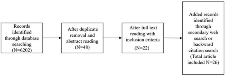
This study reviews research conducted over the past decade on MS detection using biosensors in vehicle environment. Electronic databases were searched, including SAGE Journals, IEEE Explore, Elsevier Science Direct and ACM Digital Library. Keywords used include motion sickness, detection, biosensors and autonomous vehicle. Duplicate articles were removed, articles from same author were compared and 26 most relevant articles were finally selected from 6202 for detailed review. The major inclusion criteria are:
The study must investigate the use of biosensors in detecting motion sickness within the context of autonomous vehicles, focusing on physiological measurement methods.
It should identify and analyse key physiological signals that contribute to real-time and objective motion sickness detection.
The study should present findings that enhance the understanding of biosensor usability or address challenges such as individual variability and environmental factors in motion sickness monitoring.
Figure 1: Flow chart for article selection process.
RESULT
The sensors involved in detecting MS are categorized according to the type of biological signal measured, including: Body Motion Detection, Brain Activity Detection, Stomach/Gastric Response Detection, Eye Response Detection, Heart/Cardiac Response Detection, Skin Response Detection, Body Temperature Detection.
Body motion sensors track postural instability linked to motion sickness (Diels & Bos, 2016). Techniques such as motion capture suits combine neural network analysis demonstrate the significant role of body movement in the onset and severity of motion sickness symptoms. Electroencephalography
Motion Sickness Detection in Autonomous Vehicle With Biosensors: A Review 261
(EEG) and Functional Near-Infrared Spectroscopy (fNIRS) detect brain signals during motion sickness, highlighting affected regions like the left frontal cortex (Zhang, Li, Li, Li, & Nie, 2020). Machine learning applied to EEG data shows promise in accurately predicting symptoms. Gastric activity measured by electrogastrogram (EGG) links nausea and dysrhythmia to motion sickness (Jakus, Sodnik, & Miljkovic, 2022). Although non- invasive, EGG data may vary with posture, limiting its real-time application in autonomous vehicles. Eye-tracking and pupillometry monitor indicators like blink rate and pupil size, which are associated with motion sickness (Adachi et al., 2014). While effective, these methods can be costly and face challenges with calibration and adaptability in vehicle environments. Heart rate (HR) and variability (HRV) are commonly measured using electrocar- diography (ECG) or photoplethysmography (PPG) to capture autonomic responses to motion sickness. While wearable PPG devices offer convenience, inconsistent HR data suggests that further research is needed to establish reliable markers (Rauterberg, Delbressine, Terken, Md Yusof, & Karjanto, 2022). Electrodermal activity (EDA) monitors sweat response during motion sickness. Increases in EDA generally correlate with symptoms, but individual variations indicate the need for a more refined understanding of these responses (Smyth, Birrell, Woodman, & Jennings, 2021). Changes in facial and body temperature, detected via wristband or thermal imaging, have proven inconsistent as motion sickness indicators. External factors, such as climate control, can affect results, necessitating further study for reliability. Additional signals, such as respiratory rate (Keshavarz, Peck, Rezaei, & Taati, 2022) and myogenic potential changes, are also linked to motion sickness. These findings show the complexity of physiological responses and supports the integration of multiple bio-signals for comprehensive detection. The results also outline various physiological and sensor-based indicators associated with MS, detailing the increase or decrease of each indicator under MS conditions, along with relevant studies, see Table 1.
Table 1: Biosensor detection results from selected articles.
Table available in the original PDF paper.
Measurement Indicator Result Reference Increase (Karjanto et al., 2018) Body Motion Motion Sickness Dose
Values (MSDV) Head rotation variation Increase (Brietzke, Xuan, Dettmann, &
Bullinger, 2021) No Change (Irmak, Pool, & Happee, 2021)
Alpha power Increase (Henry et al., 2021; Huang et al., 2021) Beta power Most effect (Recenti et al., 2021)
Increase (Tan et al., 2022) fNIR Oxyhemoglobin Decrease Decrease (O2Hb): Left prefrontal cortex (TX1,TX2) Left prefrontal cortex (TX3,TX4) Right prefrontal cortex (TX1,TX2,TC3,TX4)
Deoxyhemoglobin Increase
(Tan et al., 2022) Decrease Decrease
(HHb): Left prefrontal cortex (TX1) Left prefrontal cortex (TX2,TX3,TX4) Right prefrontal cortex (TX1,TX2,TC3,TX4) Total hemoglobin (tHb): Increase
(Tan et al., 2022) Decrease Decrease
Left prefrontal cortex (TX1) Left prefrontal cortex (TX2,TX3,TX4) Right prefrontal cortex (TX1,TX2,TC3,TX4) Oxyhemoglobin Increase
(Tan et al., 2022) Decrease Decrease
difference (HbDiff): Left prefrontal cortex (TX1,TX2) Left prefrontal cortex (TX3,TX4) Right prefrontal cortex (TX1,TX2,TC3,TX4)
Continued
Motion Sickness Detection in Autonomous Vehicle With Biosensors: A Review 263
Table 1: Continued Measurement Indicator Result Reference
Table available in the original PDF paper.
Cerebral oxygen
(Zhang et al., 2020) Highest (Striaght driving) Highest (Curved driving)
exchange activity – (COS): The visual cortex of the occipital lobe (BA17 and BA18) and the frontal cortex (BA6) The visual cortex of the occipital lobe (BA17 and BA18) and the prefrontal cortex (BA10) Increase (Adachi et al., 2014) Eye Response The resting potential of the retina
Pupil diameter Increase (Beggiato et al., 2018; Matthias Beggiato, Hartwich, & Krems, 2019; Niermann et al., 2021)
Interblink time Increase (Beggiato et al., 2018; Matthias Beggiato et al., 2019)
Eye blink Decrease (Beggiato et al., 2018; Matthias Beggiato et al., 2019)
Eye fixation No change (Brietzke et al., 2021)
No change (Popovic et al., 2019) (Gruden EGG Dominant frequency (DF) et al., 2021) Increase, significant
Median frequency (MF) No change
(Popovic et al., 2019) (Gruden et al., 2021) Increase, not significant
Crest factor (CF) No change
(Popovic et al., 2019) (Gruden et al., 2021) Decrease, significant
Root mean square
Increase Increase (Popovic et al., 2019) (Gruden (RMS) et al., 2021) but not significant
Percentage of spectral
No change (Popovic et al., 2019) power in the normogastric range (2–4 cpm)
Percentage of the high Increase,
(Gruden et al., 2021) significant
power spectrum density (FSD) Maximum magnitude of Increase but not
(Gruden et al., 2021) significant
power spectrum density (MFM) Percentage of Increase (Schartmüller & Riener, 2020) tachygastria activity
Percentage of
Increase (Schartmüller & Riener, 2020) arrhythmia activity Continued
Table 1: Continued Measurement Indicator Result Reference
Table available in the original PDF paper.
Dominant frequency in
Increase (Schartmüller & Riener, 2020) contractions per minute (DF cpm)
Maximum power
No effect (Schartmüller & Riener, 2020) spectrum density in decibels (MPSD dB)
Gastric contractions
No effect (Keshavarz et al., 2022) (EGG) (Beggiato et al., 2018;
Heartbeat/Pause HR Decrease Increase
Matthias Beggiato et al., 2019; Karjanto et al., 2018; Schartmüller & Riener, 2020; Schneider et al., 2022) (Gruden et al., 2021; Keshavarz et al., 2022; Kojima, Ohsuga, Kamakura, Hori, & Watanabe, 2022; Tan et al., 2022) IBI Decrease (Keshavarz et al., 2022)
LF Increase (Bando et al., 2021) Skin Conductivity EDA No effect (Beggiato et al., 2018; Matthias Beggiato et al., 2019)
Increase (Gruden et al., 2021; Irmak et al., 2021; Keshavarz et al., 2022; Schneider et al., 2022; Shizuka Bando et al., 2021; Tan et al., 2022)
Skin Temperature Facial temperature Decrease (Keshavarz et al., 2022)
No significant (Bando et al., 2021) changes
Body temperature Increase (Keshavarz et al., 2022) Increase (Schartmüller & Riener, 2020)
No significant (Tan et al., 2022) changes
The table summarizes physiological indicators of motion sickness and their trends across multiple measurement domains. Body motion metrics, such as motion sickness dose values and head movements, predominantly showed increases. Brain activity revealed increases in theta, alpha, and beta power, with hemoglobin changes in fNIR demonstrating regional specificity. Eye metrics, including pupil diameter and inter-blink time, increased, while blink frequency decreased. Gastric responses showed significant increases in spectral power density and tachygastria activity. Heart rate metrics varied, with a general trend of reduced autonomic regulation. Skin conductivity and body temperature generally increased, while facial temperature changes were inconsistent. These findings provide valuable physiological insights for motion sickness detection.
Motion Sickness Detection in Autonomous Vehicle With Biosensors: A Review 265
DISCUSSION
The application of biosensors for detecting MS in transportation settings, particularly in autonomous vehicles, shows significant promise. The results indicate that physiological responses to MS are complex and multifaceted, with different biological signals providing complementary insights. While single-sensor approaches can capture specific aspects of MS, a multimodal fusion strategy integrating multiple biosignals—such as motion data, EEG, HRV, EDA, and gastric responses—could significantly improve detection accuracy.
Despite promising results, several challenges remain. Environmental factors (e.g., climate control affecting skin temperature), individual physiological variations, and real-time signal variability pose limitations to current sensor-based MS detection (Podoprigora, Marusin, Pegin, Karelina, & Akulov, 2022). Additionally, future research should prioritize the development of reliable and resilient multi-sensor fusion techniques that can effectively integrate physiological, vehicular motion, and environmental data for real-time MS monitoring (King et al., 2017). Advances in machine learning and deep learning algorithms can further refine signal interpretation, improving classification accuracy. Moreover, enhancing wearable sensor technology for greater usability (Liu, Zhang, Chen, Liu, & Zhang, 2021), comfort, and reliability will be crucial for practical deployment in autonomous vehicle applications.
CONCLUSION
This review underscores the potential of biosensors for achieving real-time, objective detection of motion sickness in autonomous vehicles. Despite progress, individual variability and environmental factors pose ongoing challenges to sensor reliability. Future research should prioritize sophisticated multi-sensor fusion techniques, integrating diverse physiological and environmental data, to enhance accuracy and usability in MS monitoring, ultimately promoting passenger comfort in autonomous driving contexts.
ACKNOWLEDGMENT
This work was supported by the University of Nottingham Ningbo China 2023 LDS innovation fellowship award - Motion Sickness Detection Application Project (E01231200039).
REFERENCES
Adachi, T., Yonekawa, T., Fuwamoto, Y., Ito, S., Iwazaki, K., & Nagiri, S. (2014).
Simulator Motion Sickness Evaluation Based on Eye Mark Recording during Vestibulo-Ocular Reflex. Paper presented at the SAE Technical Paper Series. Beggiato, M., Hartwich, F., & Krems, J. (2018). Using Smartbands, Pupillometry and Body Motion to Detect Discomfort in Automated Driving. Front Hum Neurosci, 12, 338. doi:10.3389/fnhum.2018.00338.
Beggiato, M., Hartwich, F., & Krems, J. (2019). Physiological correlates of discomfort in automated driving. Transportation Research Part F: Traffic Psychology and Behaviour, 66, 445–458. doi:10.1016/j.trf.2019.09.018. Brietzke, A., Xuan, R. P., Dettmann, A., & Bullinger, A. C. (2021). Concepts
for Vestibular and Visual Stimulation to Mitigate Carsickness in Stop-and- Go-Driving. Paper presented at the 2021 IEEE International Intelligent Transportation Systems Conference (ITSC). Buchheit, B., Schneider, E. N., Alayan, M., Dauth, F., & Strauss, D. J. (2022).
Motion Sickness Prediction in Self-Driving Cars Using the 6DOF-SVC Model. IEEE Transactions on Intelligent Transportation Systems, 23(8), 13582–13591. doi: 10.1109/tits.2021.3125802. Chang, C. H., Chen, F. C., Kung, W. C., & Stoffregen, T. A. (2017). Effects of Physical Driving Experience on Body Movement and Motion Sickness During Virtual Driving. Aerosp Med Hum Perform, 88(11), 985–992. doi:10.3357/ AMHP.4893.2017. Diels, C., & Bos, J. E. (2016). Self-driving carsickness. Appl Ergon, 53 Pt B, 374–382.
doi:10.1016/j.apergo.2015.09.009. Gruden, T., Popovic, N. B., Stojmenova, K., Jakus, G., Miljkovic, N., Tomazic, S., & Sodnik, J. (2021). Electrogastrography in Autonomous Vehicles-An Objective Method for Assessment of Motion Sickness in Simulated Driving Environments. Sensors (Basel), 21(2). doi:10.3390/s21020550. Henry, E. H., Bougard, C., Bourdin, C., & Bringoux, L. (2021). Changes in Electroen-
cephalography Activity of Sensory Areas Linked to Car Sickness in Real Driving Conditions. Front Hum Neurosci, 15, 809714. doi:10.3389/fnhum.2021.809714. Huang, K. C., John, A. R., Jung, T. P., Tsai, W. F., Yu, Y. H., & Lin, C. T.
(2021). Comparing the Differences in Brain Activities and Neural Comodulations Associated With Motion Sickness Between Drivers and Passengers. IEEE Trans Neural Syst Rehabil Eng, 29, 1259–1267. doi:10.1109/TNSRE.2021.3092876. Irmak, T., Pool, D. M., & Happee, R. (2021). Objective and subjective responses to motion sickness: The group and the individual. Exp Brain Res, 239(2), 515–531. doi:10.1007/s00221-020-05986-6. Iskander, J., Attia, M., Saleh, K., Nahavandi, D., Abobakr, A., Mohamed, S.,
Hossny, M. (2019). From car sickness to autonomous car sickness: A review. Transportation Research Part F: Traffic Psychology and Behaviour, 62, 716–726. doi:10.1016/j.trf.2019.02.020. Jakus, G., Sodnik, J., & Miljkovic, N. (2022). Electrogastrogram-Derived Features for Automated Sickness Detection in Driving Simulator. Sensors (Basel), 22(22). doi:10.3390/s22228616. Karjanto, J., Md. Yusof, N., Wang, C., Terken, J., Delbressine, F., & Rauterberg,
M. (2018). The effect of peripheral visual feedforward system in enhancing situation awareness and mitigating motion sickness in fully automated driving. Transportation Research Part F: Traffic Psychology and Behaviour, 58, 678–692. doi:10.1016/j.trf.2018.06.046. Keshavarz, B., Peck, K., Rezaei, S., & Taati, B. (2022). Detecting and predicting visually induced motion sickness with physiological measures in combination with machine learning techniques. Int J Psychophysiol, 176, 14–26. doi:10.1016/ j.ijpsycho.2022.03.006. King, R. C., Villeneuve, E., White, R. J., Sherratt, R. S., Holderbaum, W., &
Harwin, W. S. (2017). Application of data fusion techniques and technologies for wearable health monitoring. Med Eng Phys, 42, 1–12. doi:10.1016/ j.medengphy.2016.12.011.
Motion Sickness Detection in Autonomous Vehicle With Biosensors: A Review 267
Kojima, T., Ohsuga, M., Kamakura, Y., Hori, J., & Watanabe, S. (2022). Assessment of car sickness in passengers using physiological indices. Paper presented at the 2022 IEEE International Conference on Systems, Man, and Cybernetics (SMC). Koohestani, A., Nahavandi, D., Asadi, H., Kebria, P. M., Khosravi, A., Alizadehsani,
R., & Nahavandi, S. (2019). A Knowledge Discovery in Motion Sickness: A Comprehensive Literature Review. IEEE Access, 7, 85755–85770. doi:10.1109/ access.2019.2922993. Liu, C., Zhang, B., Chen, W., Liu, W., & Zhang, S. (2021). Current development of wearable sensors based on nanosheets and applications. TrAC Trends in Analytical Chemistry, 143. doi:10.1016/j.trac.2021.116334. Niermann, D., Trende, A., Ihme, K., Drewitz, U., Hollander, C., & Hartwich, F.
(2021). An Integrated Model for User State Detection of Subjective Discomfort in Autonomous Vehicles. Vehicles, 3(4), 764–777. doi:10.3390/vehicles3040045. Nooij, S. A. E., Bockisch, C. J., Bulthoff, H. H., & Straumann, D. (2021). Beyond sensory conflict: The role of beliefs and perception in motion sickness. PLoS One, 16(1), e0245295. doi:10.1371/journal.pone.0245295. Podoprigora, N. V., Marusin, A. V., Pegin, P. A., Karelina, E. A., & Akulov, A. A.
(2022). Systematic Approach in Information Support of the ≪Road User-Vehicle- Road-External Environment≫System. Paper presented at the 2022 Systems of Signals Generating and Processing in the Field of on Board Communications. Popovic, N. B., Miljkovic, N., Stojmenova, K., Jakus, G., Prodanov, M., & Sodnik, J.
(2019). Lessons Learned: Gastric Motility Assessment During Driving Simulation. Sensors (Basel), 19(14). doi:10.3390/s19143175. Rauterberg, M., Delbressine, F., Terken, J., Md Yusof, N., & Karjanto, J. (2022).
Level of motion sickness based on heart rate variability when reading inside a fully automated vehicle. Mechanical Engineering for Society and Industry, 2(2), 72–81. doi:10.31603/mesi.7083. Recenti, M., Ricciardi, C., Aubonnet, R., Picone, I., Jacob, D., Svansson, H. A. R.,...
Gargiulo, P. (2021). Toward Predicting Motion Sickness Using Virtual Reality and a Moving Platform Assessing Brain, Muscles, and Heart Signals. Front Bioeng Biotechnol, 9, 635661. doi:10.3389/fbioe.2021.635661. Reinhard, R., Rutrecht, H. M., Hengstenberg, P., Tutulmaz, E., Geissler, B., Hecht, H., & Muttray, A. (2017). The best way to assess visually induced motion sickness in a fixed-base driving simulator. Transportation Research Part F: Traffic Psychology and Behaviour, 48, 74–88. doi:10.1016/j.trf.2017.05.005 Schartmüller, C., & Riener, A. (2020). Sick of Scents: Investigating Non-invasive
Olfactory Motion Sickness Mitigation in Automated Driving. Paper presented at the 12th International Conference on Automotive User Interfaces and Interactive Vehicular Applications. Schneider, E. N., Buchheit, B., Flotho, P., Bhamborae, M. J., Corona-Strauss, F. I., Dauth, F.,... Strauss, D. J. (2022). Electrodermal Responses to Driving Maneuvers in a Motion Sickness Inducing Real-World Driving Scenario. IEEE Transactions on Human-Machine Systems, 52(5), 994–1003. doi:10.1109/ thms.2022.3188924. Shizuka Bando, Yuri Shiogai, & Hirao, A. (2021). Development of evaluating
methods for passenger’s motion sickness in real driving environment. International journal of automotive engineering, 12, 72–77. doi: https:// doi.org/10.20485/jsaeijae.12.2_72. Smyth, J., Birrell, S., Woodman, R., & Jennings, P. (2021). Exploring the utility of EDA and skin temperature as individual physiological correlates of motion sickness. Appl Ergon, 92, 103315. doi:10.1016/j.apergo.2020.103315.
Tan, R., Li, W., Hu, F., Xiao, X., Li, S., Xing, Y.,... Cao, D. (2022). Motion Sickness Detection for Intelligent Vehicles: A Wearable-Device-Based Approach. Paper presented at the 2022 IEEE 25th International Conference on Intelligent Transportation Systems (ITSC). Wada, T., Fujisawa, S., & Doi, S. (2018). Analysis of driver’s head tilt using
a mathematical model of motion sickness. International Journal of Industrial Ergonomics, 63, 89–97. doi:10.1016/j.ergon.2016.11.003. Wang, J., Liang, H.-N., Monteiro, D., Xu, W., & Xiao, J. (2023). Real-Time Prediction of Simulator Sickness in Virtual Reality Games. IEEE Transactions on Games, 15(2), 252–261. doi:10.1109/tg.2022.3178539. Zhang, C., Li, S., Li, Y., Li, S. E., & Nie, B. (2020). Analysis of Motion Sickness
Associated Brain Activity Using fNIRS: A Driving Simulator Study. IEEE Access, 8, 207415–207425. doi:10.1109/access.2020.3038039.
LISTED IN THE AHFE DIGITAL BOOKSTORE CATALOGUE
Explore the complete digital volume
Advances in Human Factors of Transportation
Read this individual research paper free through the original publisher, or explore the full digital proceedings book.
ISBN 978-1-964867-62-5
Explore this ISBN in the AHFE Bookstore ↗Listed in the July 2026 bookstore catalogue; search by ISBN to confirm current availability · Digital book purchase optional