Eduardo Pinos, María Cordero-Mendieta, Roberto Coronel-Berrezueta (2022). Methods for automatic glaucoma detection and feature extraction by different techniques at the pre-processing stage in fundus images. In: Applied Human Factors Research. AHFE Open Access, vol. 67, pp. 130–139. https://doi.org/10.54941/ahfe1002691
2 GIIATA, Research Group on Artificial Intelligence and Assistive Technologies, GIBI Research Group of Bietics in Research Universidad Politécnica Salesiana, Cuenca 010105, Ecuador ↗
3 GIIATA, Research Group on Artificial Intelligence and Assistive Technologies, GIBI, Research Group of Bietics in Research Universidad Politécnica Salesiana, Cuenca, 010105, Ecuador ↗
Glaucoma is a neurodegenerative, progressive and silent disease that affects the optic nerve, characterized by an increase in intraocular pressure causing irreversible damage to the optic nerve. The difficulty in early diagnosis of glaucoma has posed challenges at the technological and medical level, since it requires not only several years of study, but also experience on the part of medical specialists to examine the images and make a timely diagnosis.During this pandemic of COVID-19 many patients with this pathology have suffered constant changes and intraocular pressure has increased considerably, so early detection is of vital importance, in addition to providing appropriate and timely treatment so that the patient does not lose vision in its entirety and mitigate the effects that COVID-19 has caused in these patients.In this article we present the different techniques for the diagnosis of glaucoma and the automatic detection methods, as well as the analysis of image processing and the results obtained in the preprocessing stage, the characterization of this disease according to different points of view, we also present a thorough analysis of each of the methods proposed for the support of medical diagnosis, the characteristics of each of the classifiers and data of great relevance for future work.
Glaucoma is a silent, progressive, neurodegenerative disease that affects the optic nerve due to increased intraocular pressure (IOP) inside the eyeball. A glaucomatous eye presents obstructions in the eye’s natural drainage system, which causes the fluid inside the eye to accumulate and the IOP to rise. As the IOP rises, the sensitive nerve fibers slowly begin to die, the optic disc begins to hollow out, the optic nerve is compressed forming a cup and as the optic nerve is very sensitive it atrophies causing blurred vision in the individual
Methods for Automatic Glaucoma Detection and Feature Extraction 131
without presenting any symptoms beforehand. (C. Del Portillo et al., 2007) (PJ Foster et al., 2002) (O. Maklad et al., 2018) This pathology is also called “the silent thief of sight” since there are no symptoms at an early stage.
Several studies (PJ Foster et al., 2002) (Bright Focus Foundation, 2021) (BN Kumar et al., 2020) have determined that any person can suffer from this pathology regardless of age; however, people with a higher risk of pre- senting this pathology are: patients over 60 years of age, people of African descent and Caucasian Hispanics, people with a family history of glaucoma, patients with high IOP, diabetics, patients with severe myopia, uveitis, ocu- lar trauma, retinal thrombosis, and trauma (C. Del Portillo et al., 2007) (V. Cortés-González et al., 2015). Statistical studies conducted by the World Health Organization (WHO) reveals in its report for the year 2021 that accor- ding to data collection conducted during the period 2014 - 2020 there are in approximate estimate about 64 million people worldwide with glaucoma of which it has been recorded that 13% of this population presents blindness due to the mentioned disease and it is projected that by the year 2022 this statistic will increase to 80 million people suffering from glaucoma. (OMS et al., 2020) (D. M. E Gilbert et al., 2020) Most people with glaucoma have no symptoms until the disease is in an advanced stage because the visual field is compromised (Hariharan L et al.,2018). Therefore, it is necessary to perform eye examinations periodically and thus detect at an early stage if a patient has glaucoma, the specialist asks to perform tests that allow to know the range of IOP of the patient, usually glaucoma is diagnosed when the IOP is greater than 20 mm Hg, (C. Del Portillo et al., 2007) (U.S. Department of Health and Human Services, 2013) (S. Kavitha et al., 2010).
TECHNIQUES FOR GLAUCOMA DETECTION The difficulty in the early diagnosis of glaucoma has posed technological and medical challenges, because it requires not only several years of study, but also experience on the part of medical specialists to examine the images and make a timely diagnosis (V. Moreno Londoño et al., 2014).
Taking into account the difficulties in the detection of glaucoma, medi- cal specialists consider the monitoring of abnormal changes that may occur in the internal structure of the eye vital for the detection and treatment of this pathology. In the last decades, technology has advanced and thanks to this there are several methods that allow obtaining high resolution fundus images such as Heidelberg retinal tomography (HTR), nerve fiber analyzer (GDx), optical coherence tomography (OCT), ultrabiomicroscopy (UBM), among the best known, from these we can obtain internal structural and non-structural information of the eye, (C. Del Portillo et al., 2007).
Since there are several types of glaucoma, each of the authors mentioned above present different techniques for the detection of glaucoma, as in the case of M. V. Moreno Londoño et al., which analyzes pigmentary glaucoma and the technique of ultrabiomicroscopy, showing a study of high interest since they determine that the population with this type of glaucoma is betw- een 1% and 1.5% of all cases diagnosed with glaucoma, of this percentage 78-93% of people suffering from pigmentary glaucoma are men of any age,
132 Pinos-Vélez1 et al.
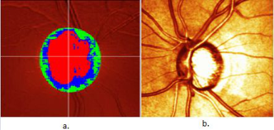
Figure 1: a. Topography take from HTR and the color maps that can be obtained with information from features fundus b. Refractive image f optic disc and neurorretiniano ring.
while women affected are a minority percentage and more frequently in adva- nced age. (V. Moreno Londoño et al.,2014) (Dada T et al., 2011) (M. C. González González et al., 2014).
Through the use of UBM, important characteristic features of this patho- logy have been found, where there is an open angle with posterior widening of the peripheral iris also known as concave iris or pupillary block, that is, thanks to the high resolution of images provided by this technique, it is pos- sible to determine characteristics of the anterior segment of the eye as well as the configuration in which the iris is located and to know how close the anterior chamber of the crystalline lens and the iris are located, it is also pos- sible to determine the permeability in light and dark, therefore concluding that this is a very useful technique for the detection of glaucoma, especially for the diagnosis of pigmentary glaucoma (V. Moreno Londoño et al.,2014).
Another well-known and very effective technique is the Heidelberg retinal tomography because through this we can obtain topographic measurements of both the parapapillary retina and the optic disc, this technique uses scanning technology through laser scanning, which allows us to make a quan- titative study of the optic nerve and the changes it may present over time, in addition, the images obtained through HTR provides focal depth planes cor- responding to the retina and its location that create a three-dimensional image of the optic nerve head for the thorough study of specialists (F. Argones et al., 2021). By means of HTR is possible to identify very precisely the stru- cture state of the optic disc, through three types of images provided by the HTR, Figure 1 a. allows us to perform an analysis of the topographic chan- ges, the color of maps indicate different characteristics, the red color map indicate progression of the excavation, decrease of the neuroretinal ring, the green color map allows visualization of excavation and neuroretinal ring fea- tures and the blue color map allows visualization of the inclination. Figure 1 b. is a reflective image with Moorfield classification and the third image is the three-dimensional reconstruction obtained form 147,456 consecutive acquires imagines.
Through the integration of between 131,072 and 786,432 data points, OCT can performer a tomographic reconstruction of the retina anatomy, additionally, can perform a quantitative analysis of the proprieties of the cup and neurorretiniano ring, define the optic disc margin, scan the thickness
Methods for Automatic Glaucoma Detection and Feature Extraction 133
of the neuroretinal fiber layer (NRFL) peripapillary centered in the optic nerve and allow the classification as normal, borderline or out-of-bounds, depending on age (L Fernández et al., 2009) (D F Ciara et al., 2008).
Automatic Glaucoma Detection Methods Taking in consideration the complexity that the specialist faces every day when diagnosing a patient, several proposals have been put forward to support the treating physician by means of tools that allow the automatic detection of glaucoma. L. Abdel-Hamid made several studies of structural detections based on segmenting the optic disc and the cup through structu- ral calculation, this algorithm allow the segmentation are very complex and require high computational resources a long training time so they are not viable for the real time diagnostic.
On the other hand (L. Abdel-Hamid et al., 2020) emphasizes that there are a few studies have performer features extractions for Wavelet decompo- sition of retinal images, the difficulty of this method is that they are meanly based on a limited number of statistical features as a mean and energy, L. Abdel-Hamid et al., raises a new algorithm based on Wavelet by extracting statistical and texture features. Taking into consideration that the mean fea- tures of the optic disc does looks bring yellow and the retina reddish orange, its segment the images into RGB color maps, the red channel allow to clearly observe the optic disc, while the blue and green channel let to see in higher contrast the cup-disc contrast, in other words, the red channel was used for optical detection while the blue and green channels features extraction; extract 10 features that considers textually relevant for the classification of images among then this: contrast, energy, entropy, dissimilarity, correlation, group shadow, group prominence, difference variance and correlation mean of information, as a statistics features: mean, variance, skewness, kurtosis, energy, entropy, and super pixels. The result obtained from this study has an efficiency of 89.4% and 96.7% using a kNN classifier with a 5-fold cross- variance while the wavelet-based feature detection algorithm offers superior results in high resolution image classification spacing. The presented algori- thm demonstrates reliable results for glaucoma detection and in efficient that it does not represent a high computational expense and requires little time for processing and analysis of high-resolution images (L. Abdel-Hamid et al., 2020). C. Bowd et al., proposes a comparison study of neural networks and linear discriminant functions (LDF) for glaucoma detection, for it takes as input parameters 83 features taken from the topography obtained from HTR, most researchers opt for multilayer perceptron classifiers (MLP) with backpropa- gation, this traditional method differs from statistical techniques such as LDF as these are able to adapt to the distribution of data, instead neural networks tend to assume behaviors based on training for classification. It uses three dif- ferent types of neural networks for comparison, the first one is a MLP type neural network with hyperbolic tangent functions, the second neural netw- ork used is a Gaussian Kernel support vector machine (Gaussian SVM) and a linear support vector machine (Linear SVM) where each of them is different
134 Pinos-Vélez1 et al.
because Gaussian SVM assumes various data distributions in the input and an unknown nonlinear mapping, while, the linear SVM uses a linear mapping. For the training of the networks they have taken 108 glaucoma patients and 189 healthy patients, they used 83 topographic parameters. The results obtai- ned present only those analyzed with Gaussian SVM with backpropagation and without backpropagation, as well as LDF likewise with backpropagation and without backpropagation, furthermore, they mention that all the used techniques of neural networks with HTR perform even better than Linear Discriminant Functions, The maximum sensitivity and specificity reported in the present study were 91% and 90%.
D. Yadav et al., emphasizes on obtaining greater sensitivity and specificity in the processing of medical images obtained by OCT and HTR, for this he proposes a classifier that allows the extraction of texture features using a gray level co-occurrence matrix (GLCM), this method is very useful in the analysis of spatial distribution of gray levels of the image. In image classification they use the adaptive resonance theory method since this method does not need supervision, i.e. it learns automatically from its resonance states and consists of a recognition field conformed a comparison field, a surveillance field and a reset module. Each of these fields has its specific function where the recogni- tion field gives a negative signal to match the quality of the input neuron and gives way to the watchdog parameter through a reset module and compa- res the recognition strength between the recognition field and the watchdog field, the higher watchdog parameter produces a very detailed memory. This method offers 72% accuracy in the classifier and the extracted features are from localized areas of the fundus image near the optic disc, which makes it a promising classifier, however, having 72% accuracy at the time of glaucoma prediction makes this method not feasible (D. Yadav et al., 2014).
On the other hand, X. Chen et al., proposes to perform an automatic classifier based on a deep learning architecture using a convolutional neu- ral network (CNN) because this type of neural networks can successfully perform tasks of segmentation and classification of images, to achieve this purpose, they make a reprocessing stage of the image to reduce and eliminate the bright areas that does not allow the segmentation and cropping of the areas of interest; At the entrance of the deep learning network images are placed with the extraction of parameters of interest in such a way that it has smaller dimensions than those obtained from ophthalmological analysis and thus better optimize the time and the cup-disc characteristics, in addition, they use two layers of max pooling interconnected to the outputs a soft-max classifier. The results , the proposed classifier can detect the presence of glau- coma in the patient, which shows that the method has a good stability in the system and is robust, being able to take the features of importances to bet- ter characterize the patterns that make possible the automatic detection of glaucoma through this deep learning network. One problem encountered in this study is overfitting, which they managed to solve by adapting response normalization layers and overlapping clustering layers, in addition to data augmentation using the random patch station. A. Sevastopolsky in his study (A. Sevastopolsky, 2017) also proposes a cup-disk relationship study using
Methods for Automatic Glaucoma Detection and Feature Extraction 135
a deep learning based convolutional neural network which I could say is a start of the work done in (X. Chen et al., 2015).
K Prasad et al., in the system they propose make as an initial stage the processing of the images with a conversion from RGB to grayscale to pass to a normalization stage, necessary for the elimination of components without relevance in the image and minimize redundancies and elimination of noise in the images, causes a degradation of performance through a medium adaptive filter that allows to eliminate impulsive noise, reduce distortion and smooth other types of noise giving a better contrast to the images improving signifi- cantly the quality of the image thus minimizing the computational expense. For the optic disc segmentation, they used the morphological algorithm of Hough transform and the calculation of the cup-to-disc ratio as a function of the optic disc and the optic cup with a normal value of 0.3. In the characteri- zation process, they select the best features for classification with the lowest error rate and Naive Bayes and k nearest neighbor classifiers are used (kNN) (K Prasad et al., 2018) (P. Constante et al., 2016). The classification stage integrates the Naive Bayes classifier which assumes a conditional independe- nce simplifier that does not influence the accuracy of the classifier and the k nearest neighbor classifier which is a deep learning algorithm that allows taking the input features to search for the nearest neighbor. Figure 6 shows the architecture of the hybrid classifier where according to the classification the values of the disk cup ratio are calculated (normal <= 0.3, mild<=0.5, moderate <=0.7, severe<=0.8) .
S. Pathan et al., in their work proposes a comparative study between an artificial neural network classifier and an SVM classifier by extracting color and texture features from fundus images painted from blood vessels that avoids segmentation of retinal anatomical structures. In the first instance they propose to paint out the blood vessels present in the fundus images as these hinder the identification of the optic disc and cup. In the first place, they make the pigmentation of blood vessels as these are considered as noise that hinder the process of feature extraction and cause difficulties in the data entry for training the network, also considers the segmentation of RGB channels and smoothing the image with the Laplacian filter, the filtering of each channel makes possible the pigmentation of blood vessels and applying the gray level co-occurrence matrix. The results obtained from the classifiers with Artificial Neural Networks obtained a sensitivity 100%, specificity 87.5%, accuracy 94.7% while with SVM obtained a sensitivity 87%, specificity 100% and accuracy 92%. (S. Patha et al., 2018)
DISCUSSION
Information has been collected from several very promising works for the automatic diagnosis of glaucoma using different types of classifiers, image processing and characterization of parametric such as D. Yadav et al., who use the adaptive resonance theory method that allows automatic learning of each of its resonance states and a recognition field consisting of a compari- son field, a surveillance field and a reset module; and although this method is innovative, it still has a long way to go in research and improvement of
136 Pinos-Vélez1 et al.
Table 1. Characteristics used by each author.
Table available in the original PDF paper.
Features L. Abdel- C. Bowd D. Yadav
X. Chen K Prasad
S. Pathan D. W. K.
Hamid et al.
et al.
et al.
A. Seva- stopolsky et al.
et al.
Wong
et al.
et al.
X X X
Age X X Color X X X X Texture X X X X X X X Statistics X X X Energy X X Entropy X X Contrast X X X X X X Cup-to-disc ratio
Table 2. Classifiers used and percentage pressure of each of them.
Table available in the original PDF paper.
C. Bowd D. Yadav X. Chen
K Prasad S. Pathan D. W. K.
Classifier L. Abdelet al.
et al.
et al.
A. Seva- stopolsky et al.
et al.
Wong
Hamid
et al.
et al.
X 94.7 87
SVM 91 92 92.5 kNN 96.7 X Neural network
72
LDF 90 Adaptive resonance
CNN X X
the proposal since it presents an accuracy of 72% in the classifier and the extracted features are from localized areas of the fundus image close to the optic disc, which makes it a promising classifier, however, having 72% accu- racy at the time of glaucoma prediction makes this method not viable. S. Pathan et al., raises an interesting proposal from the point of view of featu- res, they propose in their work a new approach that allows the classification of images by removing and segmenting retinal structures and the features that support this study are the color and texture of fundus images. As in the work of K Prasad et al., they do the removal of blood vessels conside- red as noise at the time of characterization. The proposed study shows an accuracy of 94% with the neural network classifier while the vector machine classification has an accuracy of 92%. If we make a comparison with the work done by C. Bowd et al., which also makes a study with VMS and addi- tionally LDF we observe that, the results obtained with Gaussian SVM with backpropagation and without backpropagation, as well as DLF likewise with backpropagation and without backpropagation, they also mention that all the used techniques of neural networks with HTR perform even better than the Linear Discriminant Functions. It has been noted that within this work there are limitations such as the lack of independent samples to develop the
Methods for Automatic Glaucoma Detection and Feature Extraction 137
LDF techniques and the difference in ages of the healthy and glaucomatous patients, i.e. when the ages were included as one of the characteristics within the classifier, it started the diagnosis taking into consideration only the ages of the patients so this diagnosis is not feasible and therefore unreliable. The ARGLI method shows results obtained through the neural network is 87% and SVM is 92.5% and has the particularity of improving considerably by implementing fusion networks to detect the cup-disc relationship, so far the network that presents a better performance is the SVM compared to that of the neural network for the data of this system. It should be noted that each of these authors took as input features different parameters and characterizati- ons extracted from fundus images, in the case of L. Abdel-Hamid et al. used a greater variety when entering the training data of the network. In Table 1 we can see the results obtained by each of the authors proposed in this study and the methods used by each of them for better analysis and we can realize that the features that in most cases texture, color and contrast features are used. Table 2 shows the types of classifiers and the accuracy of each of them according to the results presented by each of the authors mentioned in the tables.
FUTURE WORKS A support tool is being developed for the presumptive diagnosis of glaucoma through neural networks with a pre-processing stage for noise elimination than the images obtained from the OTC equipment and the possibility of developing an automatic glaucoma detection system by obtaining high resolution HTR images and the implementation of a fuzzy system.
REFERENCES
Bright Focus Foundation: Glaucoma: facts & figures. Available at: https://www.brig htfocus.org/glaucoma/article/glaucoma-factsfigures. Accessed May 2018. C. Bowd, K. Cban, L.M. Zangwill, M.H. Goldbaum, Te-Won Lee, T. J. Sejnow-
ski, and R. N. Weinreb,≪Comparing Neural Networks and Linear Discriminant Functions for Glaucoma Detection Using Confocal Scanning Laser Ophthalmo- scopy of the Optic Disc≫, vol. 43, no 11, p. 11, 2002. C. Del Portillo y D. Calkins, Como entender y vivir con glaucoma, San Francisco, CA 94108: Glaucoma Research Fundation, 2007. Ciara D F, Garway-Heath DP. Evaluating the effect of the new alignment algo-
rithm for longitudinal series of Heidelberg retina tomography images. Acta Ophthalmologica. 2008 March; 86(2):207–214. D. K Prasad, L. Vibha, y K. R. Venugopal, ≪IMPROVED AUTOMATIC DETE- CTION OF GLAUCOMA USING CUP-TO-DISK RATIO AND HYBRID CLASSIFIERS≫, ICTACT J. Image Video Process., vol. 9, n.o 2, pp. 1901–1910, nov. 2018, doi: 10.21917/ijivp.2018.0270. D. M. E. Gilbert-Lucido, D. M. García-Huerta, D. N. Ruiz-Quintero, D. F. Gil-
Carrasco, D. A. García-López, y D. H. Casab-Rueda, ≪Estudio epidemiológico de glaucoma en población mexicana≫, p. 5. D. Yadav, M. P. Sarathi, y M. K. Dutta, ≪Classification of glaucoma based on texture features using neural networks≫, en 2014 Seventh International Conference on Contemporary Computing (IC3), Noida, India, ago. 2014, pp. 109–112, doi: 10.1109/IC3.2014.6897157.
138 Pinos-Vélez1 et al.
Dada T, Gadia R, Sharma A,et al. Ultrasound biomicroscopy in glaucoma. Surv Ophthalmol. 2011;56:433-50. E. C. P. Molina y L. L. Veitía, ≪La fotografía de fondo de ojo como método de
diagnóstico en el glaucoma≫, n.o 2017, p. 8, 2017. Fernández Argones L., Piloto Díaz I., Coba Peña M. J., Pérez Tamayo B., Domínguez R. M., Trujillo Fonseca K. Sistemas de análisis digital de imágenes en el glaucoma. Rev Cubana Invest Bioméd [Internet]. 2009 Sep [citado 2021 Abr 27]; 28(3): 102–111. Disponible en: http://scielo.sld.cu/scielo.php?script=sci_arttext& pid=S0864-03002009000300009&lng=es. Foster PJ, Buhrmann R, Quigley HA, Johnson GJ. The definition and classifi-
cation of glaucoma in prevalence surveys. Br J Ophthalmol. 2002; 86(2): 238–42. Hariharan L, Gilbert CE, Quinn GE, Barg FK, Lomuto C, Quiroga A, et al. Reducing blindness from retinopathy of prematurity (ROP) in Argentina through collaboration, advocacy and policy implementation. Health Policy and Planning. 2018;33(5):654–65. Informe mundial sobre la visión [World report on vision]. Ginebra: Organización
Mundial de la Salud; 2020. Licencia: CC BY-NC-SA 3.0 IGO. J. J. Gómez-Valverde A. Antón, G. Fatti, B. Liefers, A. Herranz, A. Santos, C .I. Sánchez, and M. J. Ledesma-Carbayo., ≪Automatic glaucoma classification using color fundus images based on convolutional neural networks and tran- sfer learning≫, Biomed. Opt. Express, vol. 10, n.o 2, p. 892, feb. 2019, doi: 10.1364/BOE.10.000892. Kumar BN, Chauhan RP, Dahiya N: Detection of glaucoma using image processing
techniques:A critique. Semin Ophthalmol 33(2): 275–228, 2018. L. Abdel-Hamid, ≪Glaucoma Detection from Retinal Images Using Statistical and Textural Wavelet Features≫, J. Digit. Imaging, vol. 33, n.o 1, pp. 151–158, feb. 2020, doi: 10.1007/s10278-019-00189-0. M. C. González González, M. V. Moreno Londoño, M. Takane Imay, W. Koga
Nakamura, G. Islas de La Vega, y L. M. Rodríguez, ≪Glaucoma facomórfico y ultrabiomicroscopia≫, Rev. Mex. Oftalmol., vol. 89, n.o 1, pp. 52–58, ene. 2015, doi: 10.1016/j.mexoft.2014.08.004. O. Maklad, V. Theofilis, y A. Elsheikh, ≪Role of impinging jets in the biomechanical correction of the intraocular pressure (IOP) measurement≫, p. 9, 2018. P. Constante, A. Gordon, O. Chang, E. Pruna, y I. Escobar, ≪Neural networks for
optic nerve detection in digital optic fundus images≫, en 2016 IEEE International Conference on Automatica (ICA-ACCA), Curicó, Chile, oct. 2016, pp. 1–5, doi: 10.1109/ICA-ACCA.2016.7778415. S. Kavitha, S. Kaethikeyan, Dr.K.Duraiswamy. 2010. Early Detection of Glaucoma in Retinal Images Using Cup to Disc Ratio. Second International conference on Computing, Communicationand Networking Technologies. S. Pathan, P. Kumar, y R. M. Pai, ≪The Role of Color and Texture Features in Glau-
coma Detection≫, en 2018 International Conference on Advances in Computing, Communications and Informatics (ICACCI), Bangalore, sep. 2018, pp. 526–530, doi: 10.1109/ICACCI.2018.8554854. U.S. Department of Health and Human Services, El Glaucoma, National Institutes of Health, National Eye Institute, NIH Publication No: 03-6515[13] Khan, Ubaid Ullah Yasin, Usman Qamar. Detection of Glaucoma using Retinal FundusImages. BMEICON –2013.
Methods for Automatic Glaucoma Detection and Feature Extraction 139
V. Cortés-González y C. Villanueva-Mendoza, ≪Epidemiología del glaucoma congénito en la Asociación para Evitar la Ceguera en México, revisión de 5 años≫, Rev. Mex. Oftalmol., vol. 89, n.o 3, pp. 150–153, jul. 2015, doi: 10.1016/j.mexoft.2015.01.003. V. Moreno Londoño, Ma. Cristina González González, M. Takane Imay, W. Koga
Nakamura, L. M. Rodríguez López, y G. Islas de la Vega, ≪Glaucoma pigmentario y ultrabiomicroscopía≫, Rev. Mex. Oftalmol., vol. 88, n.o 3, pp. 124–127, jul. 2014, doi: 10.1016/j.mexoft.2014.03.003. X. Chen, Y. Xu, D. W. Kee Wong, T. Y. Wong, y J. Liu, ≪Glaucoma detection based on deep convolutional neural network≫, en 2015 37th Annual International Con- ference of the IEEE Engineering in Medicine and Biology Society (EMBC), Milan, ago. 2015, pp. 715–718, doi: 10.1109/EMBC.2015.7318462.
LISTED IN THE AHFE DIGITAL BOOKSTORE CATALOGUE
Explore the complete digital volume
Applied Human Factors Research
Read this individual research paper free through the original publisher, or explore the full digital proceedings book.
ISBN 978-1-958651-43-8
Explore this ISBN in the AHFE Bookstore ↗Listed in the July 2026 bookstore catalogue; search by ISBN to confirm current availability · Digital book purchase optional