Fan Yang, Qian Mao, Menghan Shi, Fangling Xie, Ka Wei Eric Cheng (2024). Enhancing Ultrasound Imaging through Convolutional Neural Networks: A Health Informatics Approach. In: Health Informatics and Biomedical Engineering Applications. AHFE Open Access, vol. 142, pp. 82–89. https://doi.org/10.54941/ahfe1005073
Ultrasound imaging, a linchpin in diagnostic medicine, delivers invaluable non-invasive insights into anatomical structures and physiological processes. Despite its widespread application, challenges persist in interpreting ultrasound images due to inherent noise, artifacts, and variations in acquisition conditions. Traditional ultrasound imaging, while invaluable, faces limitations such as lower spatial resolution, susceptibility to noise interference, and challenges in distinguishing subtle abnormalities. The research introduces an innovative approach in health informatics, harnessing the transformative potential of Convolutional Neural Networks (CNNs) to profoundly elevate the clarity and diagnostic utility of ultrasound imaging. The principal objective of this study is to systematically address existing challenges in traditional ultrasound imaging by leveraging deep learning, specifically CNNs. Our approach deploys advanced image processing techniques to significantly enhance the accuracy, resolution, and overall interpretability of ultrasound scans. To achieve this, we propose the implementation of a robust CNN architecture meticulously trained on a diverse dataset of ultrasound images. This architectural design not only enables the CNN to learn intricate patterns and features inherent in ultrasound images but also facilitates intelligent denoising, artifact reduction, and enhancement of anatomical structure visualization. Transfer learning techniques are strategically explored to optimize model performance across different imaging modalities and patient demographics, ensuring versatility and widespread applicability. Moreover, this adaptability has the potential to alleviate the computational burden associated with training large AI models. The initial focus is on denoising, where the CNN is trained to intelligently filter out noise, resulting in clearer and diagnostically valuable ultrasound images. Simultaneously, the model is trained to identify and mitigate common artifacts, such as shadowing and reverberation, significantly enhancing image fidelity. The CNN's capacity for learning hierarchical representations is harnessed to improve the spatial resolution of ultrasound scans. This enhancement proves crucial in aiding the detection of subtle abnormalities, thereby elevating diagnostic accuracy to new heights. Furthermore, the proposed CNN architecture is meticulously designed for adaptability across various ultrasound machines, ensuring seamless integration into diverse clinical settings. This adaptability reinforces its potential to become a standard tool in routine clinical practices. This research envisions the development of an advanced ultrasound imaging tool that seamlessly integrates into existing clinical workflows. The CNN-enhanced ultrasound images are poised to empower healthcare professionals with clearer, more informative visuals, ultimately leading to improved diagnostic accuracy and enhanced patient outcomes. The integration of CNNs into ultrasound imaging represents a significant leap forward in health informatics and biomedical engineering. This approach has the transformative potential to revolutionize routine clinical practices, making ultrasound diagnostics more accessible, reliable, and conducive to enhanced patient care. The intersection of deep learning and ultrasound imaging presents a paradigm shift, laying the groundwork for a new era in medical diagnostics. In the pursuit of advancing healthcare technology, this study heralds a future where the synergy of artificial intelligence and ultrasound imaging sets unprecedented standards in diagnostic precision and patient care.
Enhancing Ultrasound Imaging Through Convolutional Neural Networks 83
INTRODUCTION
Ultrasound (US) imaging has emerged as the gold standard for diagnosing a wide range of diseases, including cardiac and hepatic conditions, thanks to its exceptional temporal resolution, satisfactory image quality, and non- invasive nature (Wells, 2006). Its versatility and effectiveness have spurred numerous research endeavors aimed at expanding the applications of US imaging to various domains. One notable area of exploration is the develop- ment of portable US imaging systems for emergency care (Chan and Perlas, 2011). The portability of these devices enables rapid assessment and on- the-spot imaging, aiding in the timely diagnosis and management of critical conditions (Ng and Swanevelder, 2011). This innovation has the poten- tial to revolutionize emergency medicine by providing immediate access to crucial diagnostic information, improving patient outcomes, and expedit- ing decision-making processes (Nelson and Pretorius, 1998). Furthermore, researchers have been actively exploring advancements in 3-D imaging using ultrasound. By capturing volumetric data sets, 3-D ultrasound imaging offers enhanced visualization and detailed anatomical information. This technology has proven particularly valuable in obstetrics, where it enables comprehensive examinations of fetal development and facilitates accurate prenatal diagnosis (Aldrich, 2007).
Another exciting avenue of research focuses on ultrafast ultrasound imag- ing techniques (Kremkau and Taylor, 1986). By leveraging advanced signal processing algorithms and high-speed imaging capabilities, ultrafast ultra- sound enables real-time visualization of dynamic processes with exceptional temporal resolution (Jensen et al., 2006). This breakthrough paves the way for dynamic studies of blood flow, tissue perfusion, and cardiovascular dynamics, leading to improved diagnostic accuracy and better understand- ing of physiological phenomena (Bercoff, 2011). The continuous efforts to advance ultrasound imaging are driven by the desire to overcome existing limitations, enhance diagnostic capabilities, and improve patient care across various medical disciplines (Saini et al., 2010). As technology evolves, ultra- sound imaging holds great promise in delivering even more sophisticated imaging modalities, expanding its applications, and further cementing its position as a primary diagnostic tool in modern healthcare (Nelson and Pretorius, 1998).
In the realm of portable, 3-D, and ultrafast ultrasound imaging systems, a growing demand exists for reconstructing high-quality images using a limited number of radiofrequency (RF) measurements, often due to receiver (Rx) or transmitter (Xmit) event subsampling (Deshpande et al., 2010). However, the presence of side-flap artifacts in RF subsampling poses challenges, as stan- dard beamformers tend to generate blurred images with reduced contrast, rendering them unsuitable for diagnostic purposes (Whittaker and Stokes, 2011). Currently available compressed sensing techniques, which aim to address this issue, often necessitate hardware modifications or computa- tionally intensive algorithms, thereby offering only limited improvements in image quality (Jensen, 2002). Consequently, there is a pressing need for inno- vative approaches that can effectively mitigate side-flap artifacts and produce
superior image reconstructions without requiring extensive hardware alter- ations or excessively demanding computational resources (Ortiz et al., 2012). Such advancements would be of immense value, allowing for enhanced diag- nostic accuracy and facilitating the optimal utilization of portable, 3-D, and ultrafast ultrasound imaging systems in clinical settings.
Addressing these challenges remains an active area of research, with scien- tists and engineers striving to develop novel algorithms and methodologies that can overcome the limitations of current techniques (Van Sloun et al., 2019). By harnessing cutting-edge signal processing techniques, advanced image reconstruction algorithms, and machine learning approaches, researchers aim to optimize the image quality, contrast, and resolution obtained from RF subsampling in portable, 3-D, and ultrafast ultrasound imaging systems (Narayanan and Wahidabanu, 2009).
These ongoing efforts will undoubtedly contribute to the advancement of ultrasound imaging technology, enabling healthcare professionals to lever- age the full potential of these systems for accurate diagnosis, comprehensive monitoring, and improved patient care. Further research is needed to explore the potential of incorporating artificial intelligence (AI) techniques for com- bining human body information to assist in medical image diagnosis. The integration of AI methodologies with human body information represents an area that requires in-depth investigation and development.
METHEDOLOGY The success of deep learning can be attributed to its ability to achieve excel- lent learning performance by leveraging a large number of labeled training samples. However, in the field of medical ultrasound image analysis, meeting this requirement becomes challenging due to the high cost of expert labeling and the scarcity of data for certain diseases such as lesions or nodules. Con- sequently, training deep models with limited training samples has emerged as a significant challenge in medical ultrasound image analysis.
The goal of model optimization in CNNs is to improve the performance and generalization capability of the network by adjusting its parameters and hyperparameters (Lu et al., 2021). Model optimization involves employing techniques that aim to improve the generalization performance of the model. This may include regularization methods such as dropout, weight decay, or early stopping. These techniques help prevent the model from excessively fitting the training data, thereby enhancing its ability to generalize well to unseen data. Alternatively, transfer learning, also known as migration learn- ing, provides an alternative pathway to address the issue of limited training samples. Transfer learning involves leveraging pre-trained models on large- scale datasets and adapting them to the target task with limited data. By transferring knowledge learned from the source task, the model can benefit from the general features captured by the pre-trained model, even when the target task has limited training samples. This approach allows the model to leverage the knowledge gained from the source task to improve its perfor- mance on the target task. In summary, when dealing with limited training samples in medical ultrasound image analysis, combating model overfitting
Enhancing Ultrasound Imaging Through Convolutional Neural Networks 85
can be achieved through model optimization techniques or by utilizing trans- fer learning approaches. These strategies help enhance the generalization ability of the models and enable them to perform well despite the scarcity of labeled training data.
However, despite advancements in CNN-based classification of medical images, there is still a gap in incorporating patient informatics into the analysis. The integration of patient informatics with medical image label- ing remains an area that requires further investigation. In our proposed approach, we leverage patient informatics to label the medical images before applying the CNN architecture. We believe that this approach holds promise for enhancing the capabilities of AI-based medical image diagnosis and has the potential to extend to the domain of 3D deep learning in medical ultrasound image analysis.
By incorporating patient informatics, such as clinical history, demographic data, and relevant diagnostic information, we aim to augment the label- ing process of medical images. This additional contextual information can provide valuable insights and aid in the interpretation and classification of medical images. The integration of patient informatics with deep learn- ing models has the potential to improve the accuracy and reliability of the diagnostic process, enabling more personalized and precise medical image analysis.
Figure 1: CNN architectures in conjunction with patient informatics.
The application of CNN architectures in conjunction with patient infor- matics (see Figure 1) can yield several advantages. Firstly, the utilization of patient informatics can contribute to better understanding the underlying fac- tors and patterns that influence disease manifestation and progression. By incorporating relevant patient data, the CNN model can learn to identify subtle yet clinically significant features in medical images that may have oth- erwise been overlooked. This comprehensive approach has the potential to enhance the diagnostic capabilities of AI systems, leading to more accurate and reliable medical image analysis.
Secondly, the integration of patient informatics into the labeling process can facilitate the development of more tailored and patient-specific deep learning models. By considering individual patient characteristics, the CNN model can adapt and learn patient-specific patterns and variations, leading to improved diagnostic performance. This personalized approach has the potential to enhance the precision and effectiveness of medical image analysis, particularly in complex cases where individual patient factors play a crucial role.
In summary, the incorporation of patient informatics into the labeling process of medical images, followed by the application of CNN architec- tures, represents a promising approach in the field of AI-based medical image diagnosis. By leveraging patient-specific data, we can enhance the accuracy, reliability, and personalization of medical image analysis. This approach holds significant potential not only for conventional 2D image classifica- tion but also for advancing 3D deep learning in medical ultrasound image analysis. Further research and exploration are warranted to fully exploit the benefits of integrating patient informatics into CNN-based medical image analysis frameworks.
DATA AND MEDICAL IMAGING LABEL The collection and labeling of ultrasonic (US) scanning data were performed using the NVIDIA GeForce GTX 1650 and torch.2.0.1 frameworks. A total of 90 US images were acquired using ultrasonic transducers with a 5MHz center frequency. These images encompassed the US imaging of both liver and lung, which come from publicly available datasets US-4.
Initially, an automated labeling process was executed, wherein a normal- ization procedure was employed as follows: (1)
Ni = xi −xmin xmax −xmin
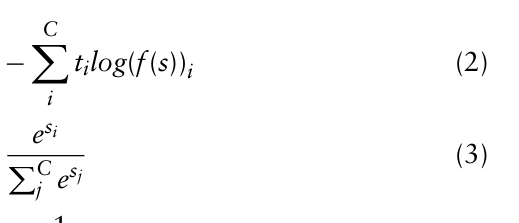
The cross-entropy loss function was defined as Eq. (2) and Eq. (3):
Equation reproduced from the original PDF.
Prior to computing the cross-entropy loss, the scores are subjected to an acti- vation function, namely Sigmoid as Eq. (4). After the application of Softmax, the output yields the most probable possibility.
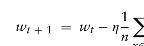
The batch size and learning rate were decided by Eq. (5): X ∇l(x, wt) (5)
Equation reproduced from the original PDF.
The accuracy of the model is calculated using the Eq. (6): Aaccuracy = Predicted labels Correct labels ∗100 (6)
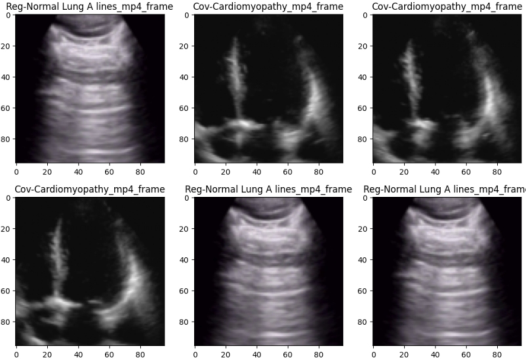
The images were visually represented, accompanied by their corresponding labels, as depicted in Figure 2.
Enhancing Ultrasound Imaging Through Convolutional Neural Networks 87
Figure 2: Visual representation of the images based on the corresponding labels.
The visual representation presented in this study was created through an automated labeling process, which categorizes the images into three dis- tinct types. These types include Cov-Cardiomyopathy, Cov-Consolidation with Air Bronchograms, and Reg-Normal Lung A lines. The training phase consisted of 5 epochs, while the dataset was split into a 70% training sub- set and a 30% testing subset. The CNN model underwent 20 iterations, resulting in an average predicted accuracy of 97.89%. Notably, the ultra- sound images were annotated with detailed health information, leading to improved accuracy levels achieved by the CNN model. This novel architec- ture effectively utilizes a limited training dataset and demonstrates consistent predictive performance, thereby alleviating computational burdens. These findings hold significant academic value, as they highlight the potential of leveraging limited data resources for accurate predictions in the medical imaging domain.
CONCLUSION
The integration of artificial intelligence (AI) for ultrasound imaging analy- sis holds great potential for advancing intelligent medical diagnostics, with CNN-based classification of medical images being a key focus. Our method- ology involves the intelligent labeling of ultrasound images by incorporating human health information, specifically related to the presence or absence of the novel coronavirus. Subsequently, these labeled images are utilized in conjunction with CNN algorithms. Through comprehensive experimenta- tion, our newly proposed architecture demonstrated remarkable robustness, achieving an average accuracy of 97.89% for image classification prediction across 40 iterations. This novel architecture not only reduces the reliance on large training datasets, thanks to the intelligent ultrasound image label- ing process, but also holds the promise of alleviating the diagnostic burden on physicians and reducing the costs associated with patient treatment. By
incorporating human body information into the CNN algorithm, our intel- ligent labeling approach significantly enhances the accuracy of intelligent classification for medical diagnosis.
In future research endeavors, we plan to further refine our methodology. Specifically, we aim to denoise the acquired ultrasound A-scan data and lever- age the Long Short-Term Memory (LSTM) algorithm to assess the quality of ultrasound signals. This will enable the reconstruction of high-quality ultrasound images. Moreover, we intend to integrate the proposed archi- tecture with this denoising and reconstruction process, thereby providing comprehensive assistance for medical diagnosis.
In conclusion, this study contributes to the advancement of AI-enabled medical diagnostics by proposing an innovative approach that combines intelligent ultrasound image labeling with CNN algorithms. The achieved high accuracy rates and the potential to reduce the burden on physicians and patients make our methodology a valuable asset in medical diagnosis. Future research will further refine this approach by incorporating denoising tech- niques and extending its capabilities to assist in various aspects of medical diagnosis.
ACKNOWLEDGMENT
This work was supported by the Hong Kong Research Grants Council, General Research Fund (Grant number: 15225422).
REFERENCES
Aldrich, J. E. 2007. Basic Physics of Ultrasound Imaging. Critical Care Medicine, 35, S131–S137. Bercoff, J. 2011. Ultrafast Ultrasound Imaging. Ultrasound Imaging-Medical Appli-
cations, 3–24. Chan, V. & Perlas, A. 2011. Basics of Ultrasound Imaging. Atlas of Ultrasound- Guided Procedures in Interventional Pain Management, 13–19. Deshpande, N., Needles, A. & Willmann, J. K. 2010. Molecular Ultrasound Imaging:
Current Status and Future Directions. Clinical Radiology, 65, 567–581. Jensen, J. A. 2002. Ultrasound Imaging and its Modeling. Imaging of Complex Media With Acoustic and Seismic Waves. Springer. Jensen, J. A., Nikolov, S. I., Gammelmark, K. L. & Pedersen, M. H. 2006. Synthetic
Aperture Ultrasound Imaging. Ultrasonics, 44, E5–E15. Kremkau, F. W. & Taylor, K. 1986. Artifacts in Ultrasound Imaging. Journal of Ultrasound in Medicine, 5, 227–237. Lu, J., Tan, L. & Jiang, H. 2021. Review on Convolutional Neural Network (Cnn)
Applied to Plant Leaf Disease Classification. Agriculture, 11, 707. Narayanan, S. K. & Wahidabanu, R. 2009. A View on Despeckling in Ultrasound Imaging. International Journal of Signal Processing, Image Processing and Pattern Recognition, 2, 85–98. Nelson, T. R. & Pretorius, D. H. 1998. Three-Dimensional Ultrasound Imaging.
Ultrasound in Medicine & Biology, 24, 1243–1270. Ng, A. & Swanevelder, J. 2011. Resolution in Ultrasound Imaging. Continuing Education in Anaesthesia, Critical Care & Pain, 11, 186–192.
Enhancing Ultrasound Imaging Through Convolutional Neural Networks 89
Ortiz, S. H. C., Chiu, T. & Fox, M. D. 2012. Ultrasound Image Enhancement: A Review. Biomedical Signal Processing and Control, 7, 419–428. Saini, K., Dewal, M. & Rohit, M. 2010. Ultrasound Imaging and Image Segmentation
in the Area of Ultrasound: A Review. International Journal Of Advanced Science And Technology, 24. Van Sloun, R. J., Cohen, R. & Eldar, Y. C. 2019. Deep Learning in Ultrasound Imaging. Proceedings of the Ieee, 108, 11–29. Wells, P. N. 2006. Ultrasound Imaging. Physics in Medicine & Biology, 51, R83. Whittaker, J. L. & Stokes, M. 2011. Ultrasound Imaging and Muscle Function.
Journal of Orthopaedic & Sports Physical Therapy, 41, 572–580.
LISTED IN THE AHFE DIGITAL BOOKSTORE CATALOGUE
Explore the complete digital volume
Health Informatics and Biomedical Engineering Applications
Read this individual research paper free through the original publisher, or explore the full digital proceedings book.
ISBN 978-1-964867-18-2
Explore this ISBN in the AHFE Bookstore ↗Listed in the July 2026 bookstore catalogue; search by ISBN to confirm current availability · Digital book purchase optional