Yvain Tisserand, Elise Prigent, Eva R. Pool, Aline Layachi, Michel-Ange Amorim, David Rudrauf (2026). Integration of Musculoskeletal and Autonomic Ocular Cues in Virtual Humans: Effects on Emotion Perception and Authenticity. In: Intelligent Human Systems Integration (IHSI 2026): Disruptive and Innovative Technologies. AHFE Open Access, vol. 200, pp. 329–339. https://doi.org/10.54941/ahfe1007092
1 Affect Learning and Decisions Lab, University of Geneva, Switzerland | Swiss Center for Affective Sciences, University of Geneva, Switzerland ↗
2 Université Paris-Saclay, CNRS, Laboratoire Interdisciplinaire des Sciences du Numérique, France ↗
3 Affect Learning and Decisions Lab, University of Geneva, Switzerland | Swiss Center for Affective Sciences, University of Geneva, Switzerland | Faculty of Psychology and Educational Sciences, University of Geneva, Switzerland ↗
4 Université Paris Nanterre, Laboratoire des interactions cognition action émotion, France ↗
Virtual human facial emotion expression is typically driven by musculoskeletal control, whereas autonomic physiological cues remain underused. We conducted a virtual reality study with a 5×5 factorial design that crossed five levels of musculoskeletal valence with five ocular states ranging from high parasympathetic activity to high sympathetic activity. Ocular physiology was implemented through parametric control of pupil diameter and scleral redness. Nineteen participants rated each stimulus for perceived valence and authenticity. While musculoskeletal cues were the primary driver of perceived valence, ocular physiology produced significant modulatory effects, even at the resolution of current headsets. This indicates that observers are sensitive to subtle ocular changes. For authenticity there was no direct effect of physiological cues, but there was a significant interaction with musculoskeletal cues, showing that authenticity judgments depend on their combination. Together, these findings suggest that adding physiologically plausible ocular signals enhances the social believability and improves the perception of emotions in virtual humans, with implications for training, serious games, social robotics, and behavioral science.
A large amount of recent work has focused on expressive behavior, and particularly expressive facial behavior, for embodied social agents. This channel is central for human–human interaction, where facial signals regulate turn taking, convey attitudes, and support inferences about the affective state of others. Virtual humans are now deployed as non-player characters, training assistants, and experimental stimuli across games, education, simulation, psychology, and neuroscience. The large majority of the current systems Received October 10, 2025; Revised November 10, 2025; Accepted November 25, 2025; Available online February 1, 2026
generate expressions in virtual humans through musculoskeletal control. Pipelines use pre-recorded motion capture or key-framed animations and, when real-time control is available, they drive blendshapes and joints, often through an abstraction in terms of facial Action Units (AUs). Simple rule sets based on cognitive appraisal variables or annotated conversational streams map each target affective state to a single facial configuration (Niewiadomski et al., 2013; Schröder, 2010; Aylett et al., 2019). This remains largely static and single-level, treating emotions as categorical labels. Critically, this approach also assumes emotional transparency, that is a direct mapping from internal state to visible behavior, which is often violated in social contexts (Aylett et al., 2019). As a result, research on both virtual human expression and human recognition has concentrated on musculoskeletal features such as AUs (Ekman and Friesen, 1976). However, dynamic models argue for direct actuation of expressive modalities and for fluid composition of multiple aspects of the affective experience (Ochs et al., 2010). Multi- stage appraisal architectures run processes on different time scales and naturally connect to the physiological responses that accompany emotion (Scherer, 2005; Levenson, 2014). Although these physiological aspects are well studied through measures of pupil diameter, ocular blood flow, and lacrimation, their visible correlates on the face have been incorporated far less. Examples include pupil dilation and constriction, periocular blood perfusion that changes scleral redness or paleness, and tearing or sweating. These responses reflect activity in the Autonomic Nervous System (ANS) with parasympathetic and sympathetic branches that act in synergy and often in opposition. Increased parasympathetic tone is typically associated with relaxed states, smaller pupils, and redder sclera. Increased sympathetic tone is typically associated with tense states, larger pupils, paler sclera, and sweating. Crucially, these physiological cues are largely involuntary and therefore potentially informative about genuine affect. Nonetheless, they are rarely modeled in real-time virtual human pipelines. This creates a gap between how virtual faces are animated and how people interpret emotion human in their everyday live. The present work addresses this gap with a controlled virtual reality study that quantifies the impact of autonomic ocular cues on emotion recognition and authenticity in virtual humans. We implement a principled model of ocular physiology with parametric control of pupil diameter and scleral redness. We combine this model with musculoskeletal expressions in a factorial design that crosses five levels of musculoskeletal valence, from very negative to very positive, with five ocular states, from high parasympathetic activity to high sympathetic activity. This paper makes three key contributions. First, it introduces an integrated control approach for ocular physiology that complements standard AU-based musculoskeletal pipelines and supports both manual and automatic control. Second, it presents a factorial Virtual Reality (VR) experiment that isolates autonomic- like ocular cues under matched rendering and lighting. Third, it provides behavioral evidence that observers integrate these subtle ocular signals when interpreting virtual human expressions, which informs the design of future virtual humans for training, serious games, social robotics, and behavioral science.
RELATED WORK
Research on facial expression production and perception has centered on musculoskeletal cues. Foundational work on basic emotions and the Facial Action Coding System (FACS) links Action Units to perceived affect (Ekman and Friesen, 1976; Ekman, 1992; 1993; Cohn and Ekman, 2005). Appraisal theories connect cognitive evaluations to temporally layered motor outputs that shape the unfolding of expressions (Scherer, 2005; Scherer and Bänziger, 2010; Scherer et al., 2018). In virtual human systems, high level affect specifications are mapped to low level controllers such as blendshapes, joints, and AU intensities, which enables real time synthesis and systematic manipulation of displays (Ruhland et al., 2015; Vilhjálmsson et al., 2007; Bevacqua et al., 2010). Temporal profiles for onset, peak, and recovery improve perceived naturalness and support multi AU blends (Ochs et al., 2010; Bevacqua et al., 2007; Courgeon et al., 2009). Because musculoskeletal signals can be voluntarily modulated, observers cannot always infer genuine internal states (Aylett et al., 2019; Rehm and André 2005). Perceived naturalness depends on temporal and cross modal coherence in addition to geometry and photometry (Ruhland et al., 2015). Human face perception integrates bottom up evidence with context and task goals, which motivates control layers that combine musculoskeletal and non musculoskeletal facets of expressivity (Calvo and Nummenmaa, 2016; Cunningham et al., 2013; Niewiadomski et al., 2013; Milcent et al., 2019).
A complementary line of work examines how autonomic nervous system processes shape ocular appearance. Iris control is dual innervated, with parasympathetic activity driving constriction and sympathetic activity contributing to dilation (McDougal and Gamlin, 2011; Steinhauer et al., 2004; Bradley et al., 2017). Pupillometry shows that pupil diameter indexes arousal and cognitive effort more than valence (Mathôt 2018; Bradley et al., 2008; Laeng et al., 2012). Conjunctival vasodilation increases perceived scleral redness and reduced perfusion makes the sclera appear paler, and these cues bias judgments of emotion and intensity (Provine et al., 2013). Work on lacrimation documents additional visible changes during intense affect (Botelho, 1964; Vingerhoets and Bylsma, 2016). Because illumination strongly drives the pupillary light reflex, photometric control is essential to isolate Autonomic Nervous System like visual effects from lighting confounds (McDougal and Gamlin, 2011). Ocular cues are largely involuntary and can diverge from posed musculoskeletal displays, which may offer diagnostic information about genuine affect (Levenson, 2014; Kreibig, 2010). Behavioral studies also link tears and redness to greater perceived sincerity and authenticity (Dijk et al., 2011).
Within virtual humans, eye animation has advanced in gaze, blinks, and micro saccades (Ruhland et al., 2015), yet systematic, real time manipulation of ocular physiology remains uncommon in deployed platforms. Surveyed approaches argue for multi channel models where physiological parameters co vary with musculoskeletal expressions under principled rules (Niewiadomski et al., 2013; Milcent et al., 2019). Evidence for incremental benefits of ocular physiology over and above AUs is mixed, likely due to differences in stimulus resolution, illumination control, and temporal dynamics
(Calvo and Nummenmaa, 2016). This motivates controlled factorial designs that jointly vary musculoskeletal and ocular factors, test congruence and conflict, and hold rendering and lighting constant. Our study follows this program by combining parametric ocular physiology with AU driven expressions and measuring effects on valence and authenticity.
METHOD
Overview. We control the virtual human face with two interoperable layers. A musculoskeletal layer maps Action Units using FACS to low level facial actuators, and a physiological layer renders autonomic ocular cues through pupil diameter and scleral redness. Illumination is held constant so that pupillary changes reflect ANS like signals rather than the light reflex. The method is implemented in our authoring tool, part of the Geneva Virtual Humans Toolkit (Tisserand et al., 2020).
Musculoskeletal control. Expression synthesis follows the standard AU abstraction used in virtual human systems (Ekman and Friesen 1978; Ruhland et al., 2015). Each AU [0,1] ja ∈ specifies a component intensity. For example, AU12 with AU6 for joy, and AU1/2, AU5, AU20, AU26 for fear. Each AU drives one or more blendshapes and or joints with calibrated weights and linear blending inside validated ranges. A simple priority and range limiting policy handles antagonistic coactivations. Temporal evolution uses smooth easing from neutral to peak and back (Ruhland et al., 2015). For this study, AU trajectories were time locked to the stimulus and kept identical across ocular conditions to isolate ocular contributions.
Physiological ocular cues. The ocular module implements two visible consequences of ANS activity. The pupil size follows the dual innervation of the iris, with parasympathetic constriction and sympathetic dilation (Steinhauer et al., 2004; Bradley et al., 2017; McDougal and Gamlin 2011). Conjunctival vasodilation modulates scleral redness. We expose two normalized controls: pupil [0,1] p∈ that scales iris aperture with 0.5 p = as neutral, and redness [0,1] r ∈ that blends between baseline and hyperemic appearance. Ocular dynamics use the same smooth easing as AUs. Temporal profiles are synchronized with the musculoskeletal sequence, while only amplitudes vary by condition. Illumination and exposure remain fixed to avoid the light reflex confound.
Table 1: Level to label correspondence for the two experimental factors. The five coded levels (–2 to 2) map to musculoskeletal expression valence and to ocular autonomic state. Parenthetical phrases indicate visible cues used in the stimuli.
Table available in the original PDF paper.
Level Musculoskeletal Control Physiological Control 2 − Very negative (high fear) High sympathetic (large pupil, pale sclera)
2 Very positive (high joy) High parasympathetic (small pupil, red sclera)
Integration of Musculoskeletal and Autonomic Ocular Cues in Virtual Humans 333 Implementation. We built a Unity 2022.3 application that schedules stimuli, renders the avatar, and collects ratings in VR. The system logs csv files per trial with participant ID, factor levels, timing, ratings, and head pose, and uses a single configuration file for AU weights, physiology presets, and durations. The application ran at a stable VR frame rate on a Meta Quest Pro via PC VR link and OpenXR. Scenes used fixed illumination and constant exposure and tone mapping.
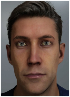
Design and procedure. A 5×5 within-participant factorial design crossed musculoskeletal valence with ocular physiology. Nineteen participants ( 19 N = ) sat across a life-size, eye-level virtual human at about 1.5 m. Each 5 s stimulus displayed a musculoskeletal expression combined with an ocular state. Both factors used five ordered codes ( 2 − to 2): musculoskeletal valence ranged from very negative to very positive, and physiological state from high sympathetic to high parasympathetic activation. Negative valence used fear-like AU programs, positive valence used happiness-like programs, and neutral used baseline activity, yielding 25 expressions per block. Examples are shown in Figure 1. The neutral–neutral condition (musculoskeletal 0, physiological 0) appeared once per block like any other condition. See Table 1 for the exact mapping and visible cues. Two blocks were presented in succession. In the Emotion block, participants rated positivity and negativity after each expression on separate 0–100 sliders. In the Authenticity block, they rated perceived authenticity on a 0–100 slider, defined as the perceived coherence between what the virtual human truly feels and what it displays (0 = not at all authentic, 100 = completely authentic). Each participant viewed every musculoskeletal × physiology combination exactly once per block (25 per block; 50 trials total), with trial order randomized within block; ratings were collected with the VR controllers.
Figure 1: Examples of manipulated musculoskeletal and ocular physiology settings. From left to right: musculoskeletal very negative (−2) with high sympathetic physiology (−2) characterized by dilated pupils and a paler sclera, musculoskeletal neutral (0) with neutral physiology (0), musculoskeletal very positive (+2) with high parasympathetic physiology (+2) characterized by constricted pupils and a redder sclera.
RESULTS
We fit repeated-measures mixed-effects ANOVAs with random intercepts for participants. Both factors (muskulosketal and physiological) were within-participant. Type-II F-tests used Satterthwaite denominator degrees of freedom with sum-to-zero contrasts. Two-sided .05 α = ; Tukey-adjusted pairwise comparisons were obtained with emmeans.
Table 2 : ANOVAs for negativity, positivity, and authenticity ratings. Type-II F-tests with Satterthwaite denominator degrees of freedom (* p<.05, ** p<.01, *** p<.001).
Negativity Positivity Authenticity Effect df F p df F p df F p
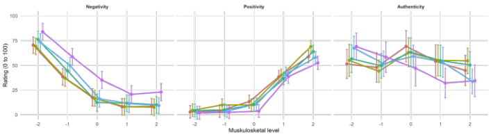
As shown in Table 2, muskulosketal produced a large main effect on both negativity and positivity. Physiological also influenced ratings, clearly for negativity and more modestly for positivity. The muskulosketal × physiological interaction was not significant for either valence outcome, indicating largely additive contributions.
Figure 2: Fused interaction plots (EMMs with 95% CIs) for negativity (left), positivity (center) and authenticity (right) across muskulosketal (x-axis) and ANS driven physiological level.
For authenticity (see Table 2), there was a significant main effect of musculoskeletal configuration, ( ) 4,432 6.04, .001 F p = < , no main effect of physiological state, ( ) 4,432 0.82, .510 F p = = , and a significant Musculoskeletal × Physio interaction, ( ) 16,432 1.79, .030 F p = = , indicating that perceived authenticity depended on the pairing of the facial expression and ocular state (right panel of Figure 2).
Table 3: Tukey all-pairs for physiological level (collapsed across muskulosketal control) for negativity (left) and positivity (right) (* p<.05, ** p<.01, *** p<.001).
Table available in the original PDF paper.
Negativity Positivity Contrast Est. SE df t p Est. SE df t p −2 vs −1 0.537 2.26 432 0.24 0.999 −1.474 1.87 432 −0.79 0.935
Post-hoc tests on physiological collapsed across muskulosketal (Table 3) showed that level 2 elicited higher negativity than levels 1, 0, 1 −, and 2 − (all .001 p < ); other negativity contrasts were not reliable. For positivity, effects were small; only 1 − versus 2 reached significance ( .008 p = ).
Simple-effect tests at musculoskeletal 0 = (Table 4) indicated a physiological effect on negativity ( (4,72) 8.59, .001) F p = < : level 2 exceeded levels 1, 0, 1 −, and 2 − (Tukey .0012 p ≤ ), while other contrasts were not significant. Physiological did not affect positivity at musculoskeletal 0 = (4,72) 1. ) 5 ( 16, .33 F p = = . Consistent with these results, the fused interaction plot in Figure 2 shows nearly parallel profiles for negativity and positivity across physiological levels and clear nonparallel profiles for authenticity, illustrating the interaction.
DISCUSSION
The data show that ocular autonomic cues, specifically pupil size and scleral coloration, measurably shape perceived emotion. As expected, musculoskeletal configuration was the dominant driver of valence. Physiological cues added a smaller but reliable modulation, stronger for negativity than for positivity. There was no musculoskeletal by physiological interaction for valence. This pattern supports an additive account in which observers integrate facial musculature with ocular physiology to judge intensity and valence (Demos et al., 2008; Kret 2015).
The direction of the physiological effects aligns with known interpretations of autonomic signals. Larger pupils with paler sclera, consistent with higher sympathetic activation, index arousal more than valence (Bradley et al., 2008; Steinhauer et al., 2004). Constricted pupils with redder sclera, consistent with higher parasympathetic tone and conjunctival vasodilation, carried a negative bias. Redness likely acted as a distress cue (Provine et al., 2013), which pushed even neutral faces toward negativity. These results suggest that small changes in the ocular region can tilt judgments without reversing the sign imposed by the facial musculature.
Authenticity judgments showed a different structure: the musculoskeletal by physiological interaction was significant. Participants rated expressions as more genuine when ocular and facial signals were congruent in affective message, and less genuine when they conflicted. This fits accounts that treat involuntary physiological cues as honesty markers whose meaning depends on context. In practice, the same ocular state can boost or penalize authenticity depending on the paired facial expression, which highlights the importance of cross channel coherence for social credibility. This pattern is consistent with evidence that observers privilege low-volitional, hard-to-fake physiological signals when inferring sincerity (Levenson 2014; Dijk et al., 2011). Congruent ocular–facial pairings tend to be read as genuine, whereas mismatches are more often read as acted.
These findings raise two important considerations for designing emotional expressions in virtual humans. First, when targeting valence communication, musculoskeletal control should remain primary, with ocular physiology used as a calibrated modulator. Second, when targeting perceived genuineness, designers should favor congruent pairings of ocular state and facial configuration and avoid mismatches that read as acted.
Several constraints qualify the present findings. Illumination was held constant to avoid the pupillary light reflex, which improves internal validity but limits ecological generality. The sample size was modest and the study used a single avatar identity, which may constrain generalization across faces and display devices. Nevertheless, effects were detected at current head-mounted display resolution, which indicates that the relevant ocular differences are perceptually available in common VR settings.
CONCLUSION
We tested how autonomic ocular cues interact with musculoskeletal facial expressions in VR. Musculoskeletal cues dominated perceived valence. Ocular cues provided consistent modulation without interacting with
Integration of Musculoskeletal and Autonomic Ocular Cues in Virtual Humans 337 musculoskeletal configuration for valence, and they interacted with musculoskeletal configuration for authenticity. Congruent pairings increased perceived genuineness, while incongruent pairings reduced it. Together, these results show that users read eye physiology alongside facial musculature and that they weigh alignment between channels when judging authenticity. This is consistent with the broader tendency to weight relatively uncontrollable physiological cues when judging whether an emotion is truly felt.
These findings argue for multi channel control in virtual humans. Adding physiologically plausible eye signals can increase emotional clarity and social credibility in applications such as training, therapy, and entertainment. A practical guideline is to keep ocular variations within plausible ranges, synchronize their dynamics with the facial motion, and apply simple congruency rules when authoring expressions.
Future work should separate pupil size and scleral coloration to estimate their individual contributions, expand to other visible autonomic cues such as skin color and sweat, and test generality across different identities and face morphologies.
REFERENCES
Aylett, Ruth, Christopher Ritter, Mei Yii Lim, et al., 2019. “An Architecture for Emotional Facial Expressions as Social Signals.” IEEE Transactions on Affective Computing. Bevacqua, Elisabetta, Ken Prepin, Radoslaw Niewiadomski, Etienne de Sevin, and
Catherine Pelachaud. 2010. “Greta: Towards an Interactive Conversational Virtual Companion.” In Close Engagements with Artificial Companions: Key Social, Psychological, Ethical and Design Issues. John Benjamins Publishing Company. Bevacqua, Elisabetta, Maurizio Mancini, Radoslaw Niewiadomski, and Catherine Pelachaud. 2007. “An Expressive ECA Showing Complex Emotions.” Proceedings of the AISB Annual Convention, Newcastle, UK, 208–16. Botelho, Stella Y. 1964. “Tears and the Lacrimal Gland.” Scientific American 211
(4): 78–87. Bradley, Margaret M, Rosemarie G Sapigao, and Peter J Lang. 2017. “Sympathetic ANS Modulation of Pupil Diameter in Emotional Scene Perception: Effects of Hedonic Content, Brightness, and Contrast.” Psychophysiology 54 (10): 1419–35. Bradley, Margaret M., Laura Miccoli, Miguel A. Escrig, and Peter J. Lang.
2008. “The Pupil as a Measure of Emotional Arousal and Autonomic Activation.” Psychophysiology 45 (4): 602–7. https://doi.org/10.1111/j.1469- 8986.2008.00654.x. Calvo, Manuel G., and Lauri Nummenmaa. 2016. “Perceptual and affective mechanisms in facial expression recognition: An integrative review.” Cognition and Emotion, ahead of print. https://doi.org/10.1080/02699931.2015.1049124. Cohn, Jeffrey F, and Paul Ekman. 2005. “Measuring Facial Action.” The New
Handbook of Methods in Nonverbal Behavior Research, 9–64. Courgeon, Matthieu, Céline Clavel, and Jean-Claude Martin. 2009. “Appraising Emotional Events During a Real-Time Interactive Game.” Proceedings of the International Workshop on Affective-Aware Virtual Agents and Social Robots, 1–5. Cunningham, William A., Kristen A. Dunfield, and Paul E. Stillman. 2013. “Emotional
states from affective dynamics.” Emotion Review, ahead of print. https://doi. org/10.1177/1754073913489749.
Demos, Kathryn E, William M Kelley, Sophia L Ryan, F Caroline Davis, and Paul J Whalen. 2008. “Human Amygdala Sensitivity to the Pupil Size of Others.” Cerebral Cortex 18 (12): 2729–34. Dijk, Corine, Bryan Koenig, Tim Ketelaar, and Peter J de Jong. 2011. “Saved by the
Blush: Being Trusted Despite Defecting.” Emotion 11 (2): 313. Ekman, Paul, and Wallace V. Friesen. 1976. “Measuring facial movement.”
Environmental Psychology and Nonverbal Behavior, ahead of print. https://doi. org/10.1007/BF01115465. Ekman, Paul, and Wallace V Friesen. 1978. Facial Action Coding System: Investigator’s Guide. Consulting Psychologists Press. Ekman, Paul. 1992. “Facial Expressions of Emotion: New Findings, New Questions.”
Psychological Science, ahead of print. https://doi.org/10.1111/j.1467-9280.1992. tb00253.x. Ekman, Paul. 1993. “Facial expression and emotion.” American Psychologist, ahead of print. https://doi.org/10.1037/0003-066X.48.4.384. Kreibig, Sylvia D. 2010. “Autonomic Nervous System Activity in Emotion: A Review.”
Biological Psychology 84 (3): 394–421. Kret, Mariska E. 2015. “Emotional Expressions Beyond Facial Muscle Actions. A Call for Studying Autonomic Signals and Their Impact on Social Perception.” Frontiers in Psychology 6: 711. Laeng, Bruno, Sylvain Sirois, and Gustaf Gredebäck. 2012. “Pupillometry: A Window
to the Preconscious?” Perspectives on Psychological Science 7 (1): 18–27. https:// doi.org/10.1177/1745691611427305. Levenson, Robert W. 2014. “The autonomic nervous system and emotion.” Emotion Review, ahead of print. https://doi.org/10.1177/1754073913512003. Mathôt, Sebastiaan. 2018. “Pupillometry: Psychology, Physiology, and Function.”
Journal of Cognition 1 (1): 16. McDougal, David H, and Paul D Gamlin. 2011. “Autonomic Control of the Eye.”
Comprehensive Physiology 5 (1): 439–73. Milcent, Anne-Sophie, Erik Geslin, Abdelmajid Kadri, and Simon Richir. 2019.
“Expressive Virtual Human: Impact of Expressive Wrinkles and Pupillary Size on Emotion Recognition.” Proceedings of the 19th ACM International Conference on Intelligent Virtual Agents, 215–17. Niewiadomski, Radosław, Sylwia Julia Hyniewska, and Catherine Pelachaud. 2013.
“Computational models of expressive behaviors for a virtual agent.” Social Emotions in Nature and Artifact. Ochs, Magalie, Radosław Niewiadomski, and Catherine Pelachaud. 2010. “How a Virtual Agent Should Smile?” International Conference on Intelligent Virtual Agents, 427–40. Provine, Robert R, Jessica Nave-Blodgett, and Marcello O Cabrera. 2013. “The
Emotional Eye: Red Sclera as a Uniquely Human Cue of Emotion.” Ethology 119 (11): 993–98. Rehm, Matthias, and Elisabeth André. 2005. “Catch Me If You Can: Exploring Lying Agents in Social Settings.” Proceedings of the Fourth International Joint Conference on Autonomous Agents and Multiagent Systems, 937–44. Ruhland, Kerstin, Christopher E Peters, Sean Andrist, et al., 2015. “A Review of
Eye Gaze in Virtual Agents, Social Robotics and Hci: Behaviour Generation, User Interaction and Perception.” Computer Graphics Forum 34: 299–326. Scherer, Klaus R, Marcello Mortillaro, Irene Rotondi, Ilaria Sergi, and Stéphanie Trznadel. 2018. “Appraisal-Driven Facial Actions as Building Blocks for Emotion Inference.” Journal of Personality and Social Psychology 114 (3): 358.
Integration of Musculoskeletal and Autonomic Ocular Cues in Virtual Humans 339 Scherer, Klaus R. 2005. “Appraisal Theory.” In Handbook of Cognition and Emotion.
https://doi.org/10.1002/0470013494.ch30. Scherer, Kr, and T Bänziger. 2010. “On the use of actor portrayals in research on emotional expression.” Blueprint for Affective Computing: A Sourcebook. Schröder, Marc. 2010. “The SEMAINE API: Towards a Standards-Based Framework
for Building Emotion-Oriented Systems.” Advances in Human-Computer Interaction 2010. Steinhauer, Stuart R, Greg J Siegle, Ruth Condray, and Misha Pless. 2004.
“Sympathetic and Parasympathetic Innervation of Pupillary Dilation During Sustained Processing.” International Journal of Psychophysiology 52 (1): 77–86. Tisserand, Yvain, Ruth Aylett, Marcello Mortillaro, and David Rudrauf. 2020. “Real- Time Simulation of Virtual Humans’ Emotional Facial Expressions, Harnessing Autonomic Physiological and Musculoskeletal Control.” Proceedings of the 20th ACM International Conference on Intelligent Virtual Agents, 1–8. Vilhjálmsson, Hannes, Nathan Cantelmo, Justine Cassell, et al., 2007. “The Behavior
Markup Language: Recent Developments and Challenges.” International Workshop on Intelligent Virtual Agents, 99–111. Vingerhoets, Ad J. J. M., and Lauren M. Bylsma. 2016. “The Riddle of Human Emotional Crying: A Challenge for Emotion Researchers.” Emotion Review 8 (3): 207–17. https://doi.org/10.1177/1754073915586226.
LISTED IN THE AHFE DIGITAL BOOKSTORE CATALOGUE
Explore the complete digital volume
Intelligent Human Systems Integration (IHSI 2026): Disruptive and Innovative Technologies
Read this individual research paper free through the original publisher, or explore the full digital proceedings book.
ISBN 978-1-964867-76-2
Explore this ISBN in the AHFE Bookstore ↗Listed in the July 2026 bookstore catalogue; search by ISBN to confirm current availability · Digital book purchase optional