Jiayi Hu, Yuki Ban, Shin'ichi Warisawa (2026). Acute Physical Stress Responses in Individuals with Chronic Psychological Stress: A Cold Pressor Test Study. In: Human Systems Engineering and Design (IHSED2026): Future Trends and Applications. AHFE Open Access, vol. 237, pp. 627–637. https://doi.org/10.54941/ahfe1008202
Chronic stress is a major public health concern associated with dysregulated physiological and psychological functioning. While responses to acute physical stressors have been studied in clinical populations such as chronic pain patients and individuals with major depression, limited attention has been paid to how non-clinical individuals experiencing chronic psychological stress respond to such challenges. This study examined whether individuals with chronic psychological stress differ from healthy controls in their behavioral, neuroendocrine, and subjective responses to an acute physical stressor.Forty non-clinical adults were stratified into a chronic stress group (n=20) and a healthy control group (n=20) based on the Quick Inventory of Depressive Symptomatology (QIDS; chronic stress: QIDS ≥ 6, healthy: QIDS ≤ 5). The experimental protocol consisted of a 5-minute initial rest period, followed by saliva sampling (S1), a Cold Pressor Test (CPT) phase in which participants immersed their hand in ice water for up to 180 seconds, a second saliva sampling (S2), a 10-minute guided breathing rest period, and a final saliva sampling (S3). Salivary cortisol concentrations were measured to assess HPA axis reactivity. Subjective ratings of relaxation, stress, and task difficulty were collected across four phases: initial rest, CPT, early recovery, and late recovery.The chronic stress group showed a significantly higher risk of withdrawing from the CPT than healthy controls (median: 59.4s vs. 180.0s; survival analysis, Cox HR=2.538, p=0.028). Salivary cortisol at baseline (S1) did not differ between groups (p=0.50). To account for individual differences in baseline cortisol, S2 and S3 values were normalized to S1 ((S-S1)/S1). The normalized cortisol change (S3-S2 normalized) was significantly correlated with CPT tolerance time in both groups (healthy: r=0.446, p=0.049; stress: r=0.604, p=0.006), with no significant difference between groups (Fisher z-test, all p>0.48). Subjective stress ratings during initial rest were significantly higher in the chronic stress group (p=0.017), whereas CPT ratings showed no group difference (both p>0.24). During late recovery, the chronic stress group showed trends toward lower relaxation (p=0.058) and higher stress (p=0.056).Despite prolonged exposure to psychological stress, the chronic stress group showed cortisol reactivity patterns comparable to healthy controls, suggesting that neuroendocrine responsiveness to acute physical stressors may remain relatively preserved in chronic stress populations. In contrast, although CPT tolerance was shorter in the chronic stress group, perceived stress and difficulty during the task did not differ between groups. Given that the chronic stress group reported higher baseline stress prior to CPT, entering the acute stressor in an already-stressed state may have reduced their capacity to sustain performance, rather than reflecting a more intense in-task experience. During late recovery, the chronic stress group showed trends toward lower relaxation and higher stress, consistent with their elevated baseline stress. This suggests that individuals with chronic stress may experience persistent stress and difficulty relaxing in quiet states, which may contribute to slower recovery following acute stress exposure. These findings reveal selective impairments in behavioral tolerance and psychological recovery, with no clear evidence of impaired neuroendocrine reactivity, advancing our understanding of how chronic psychological stress modulates acute stress responses.
Stress is broadly categorized as acute or chronic (McEwen, 2007). Acute stress is a short-term response to a specific threat that subsides once the stimulus is removed, whereas chronic stress is a prolonged state arising from accumulated daily stressors such as work and interpersonal demands. Highly prevalent in modern society, chronic stress has become a major public health issue (World Health Organization, 2022) and is linked to sleep disturbance, emotional dysregulation, and cognitive decline (McEwen, 2007; Kalmbach
Received June 12, 2026 Revised June 22, 2026 Accepted July 6, 2026 Available online September 25, 2026
et al., 2018), as well as elevated risk of depression, anxiety, and cardiovascular disease (Cohen et al., 2007). Beyond disrupting functioning at rest, an equally important question is whether chronic stress alters how individuals respond to new acute challenges, that is, whether their stress response system shows functional changes relative to healthy individuals. Addressing this is essential for understanding the mechanisms of chronic stress and its consequences in real-world situations.
Acute stress responses are mediated by two pathways: the sympathetic– adrenal–medullary (SAM) axis, which drives rapid responses within seconds, and the hypothalamic–pituitary–adrenal (HPA) axis, which governs slower, more sustained responses through cortisol secretion (Chu et al., 2024). Chronic stress may alter the regulation of both, but because the HPA axis is more persistently affected, it is considered a key system for evaluating neuroendocrine function in this population. Its primary biomarker, cortisol, can be measured non-invasively via saliva and is widely used in stress research (Hellhammer et al., 2009).
Acute stressors are broadly psychological or physical (Chrousos, 2009). Psychological stress is typically induced with cognitive tasks such as the Stroop task (Renaud and Blondin, 1997), but responses to these tasks may be confounded by individual differences in cognitive ability, practice effects, and attentional strategies. Physical stressors such as the Cold Pressor Test (CPT) (Lovallo, 1975) instead apply a stimulus of consistent intensity across individuals, so response differences more directly reflect stress regulation capacity rather than cognitive performance.
Responses to acute physical stress have largely been studied in clinical populations. Individuals with post-traumatic stress disorder show altered neural regulation and abnormal sympathetic responses during the CPT (Yoo et al., 2020) and atypical pain sensitivity (Mostoufi et al., 2014); chronic pain patients show heightened pain responses, impaired pain modulation, and shorter tolerance times (Riquelme-Aguado et al., 2024; Nouwen et al., 2006); and individuals with depression exhibit heightened autonomic reactivity (Kaushik et al., 2019) and HPA axis dysregulation (Lara-Cinisomo et al., 2017). Together, these findings indicate substantially altered acute stress mechanisms in clinical populations.
In contrast, non-clinical chronic stress has mostly been studied with cognitive paradigms, which report altered physiology such as blunted responses, decreased alpha activity, and abnormal prefrontal activation (Al Abdi et al., 2018; Nugroho et al., 2024). Because these rely on cognitive tasks, however, the effects of chronic stress remain difficult to separate from cognitive ability. The few studies using the CPT in non-clinical samples have treated it as a manipulation of other variables, such as cognitive flexibility (Knauft et al., 2021) or social exclusion and pain tolerance (Pieritz et al., 2017), rather than examining stress response patterns during CPT itself. Two gaps thus remain: direct studies of acute physical stress responses have focused on clinical populations, while non-clinical chronic stress research has relied on cognitive paradigms or used physical stressors only
Acute Physical Stress Responses in Individuals With Chronic Psychological Stress 629 as a secondary manipulation. No study has systematically examined how non-clinical chronically stressed individuals respond to an acute physical stressor, a paradigm well suited to isolating stress regulation from cognitive- performance factors.
To clarify how chronic stress shapes acute stress responses, the present study addresses these gaps by comparing a chronic stress group with a healthy control group using the CPT as an acute physical stressor and examining group differences across three complementary dimensions: behavioral tolerance, neuroendocrine response, and subjective perception.
EXPERIMENT PROTOCOL Participants
A total of 40 male adults participated in this study (mean age = 26.4 years, SD = 2.6). Only male participants were recruited to avoid potential hormonal fluctuations associated with the menstrual cycle that could affect physiological measurements. All participants had no history of neurological, psychiatric, or cardiovascular disorders and were not taking any medications that could influence autonomic nervous system function. Participants were instructed to abstain from caffeine, alcohol, and other stimulants for 24 hours prior to the experiment and to ensure adequate sleep.
Participants were divided into a chronic stress group (n = 20; mean age = 26.7 years, SD = 2.1) and a healthy control group (n = 20; mean age = 26.1 years, SD = 2.9) based on the Quick Inventory of Depressive Symptomatology (QIDS) (Rush et al., 2003), with scores ≥ 6 classified as the chronic stress group and scores ≤ 5 as the healthy control group. One participant in the chronic stress group was excluded from cortisol-related analyses due to unsuccessful saliva collection, resulting in a final sample of 39 participants for these analyses (healthy: n = 20; chronic stress: n = 19). The study was approved by the Ethics Committee of The University of Tokyo (Approval No. E2025ALS063), and written informed consent was obtained from all participants.
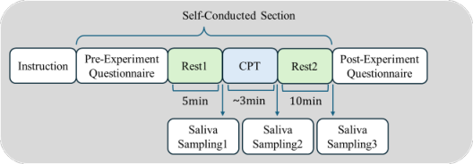
Procedure The overall experimental procedure is illustrated in Figure 1. Upon arrival, participants received an explanation of the study and provided informed consent. The experimenter then confirmed the participants’ understanding of the procedure and assisted them in wearing noise-canceling headphones.
Figure 1: Overview of the experimental procedure.
After preparation, the experiment proceeded in a self-conducted manner following on-screen instructions. Participants first completed the QIDS questionnaire for group assignment, followed by a 5-minute resting phase (Rest 1), during which they sat quietly, breathed naturally, and fixated on a central point displayed on the screen. Immediately after Rest 1, the first saliva sample (S1) was collected by placing a swab under the tongue for 1 minute (Salimetrics LLC, 2011). Participants then underwent the CPT, in which they immersed their hand up to the wrist in ice water (0–4°C) and were instructed to withdraw it at their own discretion when the discomfort became intolerable, with a maximum duration of 180 seconds. Immediately after the CPT, a second saliva sample (S2) was collected using the same procedure as S1.
Subsequently, participants entered a 10-minute guided recovery phase (Rest 2), during which they remained seated and fixated on the central point while following a visual breathing cue displayed on the screen. The cue expanded during inhalation and contracted during exhalation, corresponding to a fixed breathing rhythm of 1.5 seconds for inhalation and 1.5 seconds for exhalation. This procedure was not intended to induce slow breathing or relaxation but to standardize breathing patterns during the recovery period. After Rest 2, a third saliva sample (S3) was collected using the same method. Finally, participants completed a subjective questionnaire, providing retrospective ratings of their experiences across the different experimental phases (see Measures for details).
MEASURES AND ANALYSIS Measures
Behavioral tolerance was assessed using CPT tolerance time, defined as the duration (in seconds) from the onset of hand immersion in ice water until voluntary withdrawal. A maximum duration of 180 seconds was imposed, and participants who reached this limit were recorded as 180 seconds.
Neuroendocrine responses were assessed using salivary cortisol. Saliva samples were collected at three time points (S1, S2, and S3). After collection, samples were stored at −70°C (Garde and Hansen, 2005) and subsequently shipped on dry ice to MACROPHI Inc. (Kagawa, Japan), where cortisol concentrations were determined using an enzyme-linked immunosorbent assay (ELISA) (Lequin, 2005).
Subjective perception was assessed using a retrospective questionnaire consisting of eight items rated on a 7-point scale. The questionnaire covered relaxation and stress during Rest 1, stress and task difficulty during the CPT, and relaxation and stress during Rest 2. Because Rest 2 lasted 10 minutes, participants’ experiences may have differed between earlier and later periods. Therefore, this phase was further divided into an early period (approximately 0–5 minutes) and a late period (approximately 5–10 minutes), which were rated separately to capture dynamic changes during recovery.
Acute Physical Stress Responses in Individuals With Chronic Psychological Stress 631 Statistical Analysis
For CPT tolerance time, survival analysis was conducted to appropriately account for censored observations (participants who sustained immersion until the 180-second limit). A log-rank test was used to assess group differences in survival functions. A Cox proportional hazards model was additionally fitted to quantify the effect size, with the hazard ratio (HR) and 95% confidence interval (CI) reported. For cortisol analyses, to control for individual baseline differences, cortisol values at S2 and S3 were normalized relative to S1 using the formula (S − S1)/S1. Group comparisons of baseline cortisol levels (S1) were performed using the Mann–Whitney U test. In addition, Spearman’s rank correlation analysis was conducted to examine the association between CPT tolerance time and normalized cortisol changes. Fisher’s z test was used to assess whether the correlation coefficients differed significantly between the two groups.
For subjective ratings, group comparisons for each questionnaire item were conducted using the Mann–Whitney U test. Furthermore, to examine within-group changes during Rest 2, ratings from the early and late phases were compared using the Wilcoxon signed-rank test.
For comparisons yielding non-significant results, Bayesian analyses were additionally conducted to quantify the evidence in favor of the null hypothesis. Bayes factors were reported as BF01, interpreted as follows: BF01 > 3 indicates moderate evidence supporting the absence of a group difference; BF01 between 1 and 3 indicates insufficient evidence to draw conclusions; and BF01 < 1 indicates that the data provide more support for the presence of a difference than for its absence.
The significance level for all analyses was set at α = 0.05. Descriptive statistics are presented as median (Mdn) and interquartile range (IQR).
RESULTS
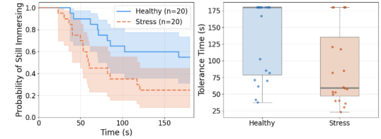
Behavioral and Neuroendocrine Responses to Acute Physical Stress In terms of behavioral responses, the chronic stress group showed a significantly higher risk of withdrawing from the CPT than the healthy control group (log-rank p = 0.023; Cox HR = 2.538, 95% CI [1.105, 5.832], p = 0.028; Figure 2a). The median tolerance time was 180.0 s (IQR = [78.9, 180.0]) in the healthy group and 59.4 s (IQR = [47.1, 135.4]) in the chronic stress group (Figure 2b). Among healthy participants, 11 out of 20 (55%) sustained immersion until the 180-second limit, compared to 5 out of 20 (25%) in the chronic stress group.
Figure 2: Visualization of CPT tolerance time across groups. (a) The Kaplan-Meier curve shows the probability of continued hand immersion over time. (b) The boxplot presents individual data points and group-level distributions of tolerance time.
No significant difference was observed between the two groups in baseline cortisol levels (S1) (U = 214.5, p = 0.500, r = 0.108; BF01 = 2.85). Although the results suggested no clear group difference prior to the experiment, the Bayes factor indicated only limited evidence in favor of the null hypothesis.
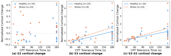
The relationships between normalized cortisol changes and CPT tolerance time are shown in Figure 3. No significant correlations were observed for S2 normalized cortisol in either group (healthy: r = 0.221, p = 0.350; chronic stress: r = 0.323, p = 0.178; Figure 3a). In contrast, S3 normalized cortisol showed significant positive correlations with tolerance time in both groups (healthy: r = 0.464, p = 0.040; chronic stress: r = 0.633, p = 0.004; Figure 3b). Similar results were observed for S3–S2 normalized cortisol (healthy: r = 0.446, p = 0.049; chronic stress: r = 0.604, p = 0.006; Figure 3c). Fisher’s z test revealed no significant differences in correlation coefficients between the two groups (S2: z = −0.318, p = 0.751; S3: z = −0.700, p = 0.484; S3–S2: z = −0.628, p = 0.530).
Bayesian analysis provided limited-to-moderate evidence supporting the absence of group differences in the correlation patterns (S2: BF01 = 3.08; S3: BF01 = 2.64; S3–S2: BF01 = 2.74), suggesting that the relationship between cortisol reactivity and CPT tolerance time showed similar trends across groups.
Figure 3: Relationship between normalized cortisol change and CPT tolerance time at three indices: (a) S2, (b) S3, and (c) S3–S2. Linear regression lines are plotted only for significant correlations in (b) and (c).
Acute Physical Stress Responses in Individuals With Chronic Psychological Stress 633 Subjective Stress Responses Across Experimental Phases
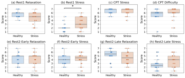
Subjective ratings across experimental phases are shown in Figure 4.
Figure 4: Visualization of subjective ratings across experimental phases. Boxplots show individual ratings and group-level distributions for relaxation and stress during (a-b) Rest 1, (c-d) CPT, (e-f) the early phase of Rest 2, and (g-h) the late phase of Rest 2. (healthy group: n = 20; chronic stress group: n = 20).
During the Rest 1 phase, the chronic stress group reported significantly higher perceived stress than the healthy control group (U = 116.0, p = 0.017, r = 0.377), whereas no significant group difference was observed in relaxation ratings (U = 261.0, p = 0.088, r = 0.270; BF01 = 0.39), though the Bayes factor provided weak evidence favoring the presence of a group difference. During the CPT phase, no significant group differences were found in perceived stress (U = 196.5, p = 0.927, r = 0.015; BF01 = 3.19) or task difficulty ratings (U = 158.5, p = 0.242, r = 0.185; BF01 = 3.00), with Bayesian analysis providing moderate evidence supporting the absence of group differences.
In the early phase of Rest 2, no significant differences were observed between groups in either relaxation (U = 201.5, p = 0.978, r = 0.004; BF01 = 3.22) or perceived stress (U = 176.0, p = 0.514, r = 0.103; BF01 = 3.01), with Bayesian analysis providing moderate evidence supporting the absence of group differences. In the late phase of Rest 2, the chronic stress group showed a trend toward lower relaxation (U = 269.0, p = 0.058, r = 0.300; BF01 = 0.69) and higher perceived stress (U = 130.0, p = 0.056, r = 0.302; BF01 = 0.63), although these differences did not reach statistical significance. The Bayes factors provided weak evidence favoring the presence of group differences.
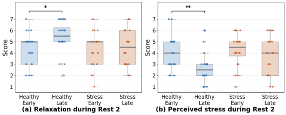
Figure 5: Within-group changes in subjective ratings during Rest 2. Boxplots with individual data points show early- and late-phase ratings during Rest 2 for (a) relaxation and (b) perceived stress in the healthy and chronic stress groups. Brackets indicate significant within-group comparisons between the early and late phases (*p < 0.05, **p < 0.01).
Changes in subjective ratings within the Rest 2 phase are shown in Figure 5. In the healthy group, relaxation ratings increased significantly from the early to the late phase (W = 19.5, p = 0.011; Figure 5a), while perceived stress decreased significantly (W = 14.0, p = 0.003; Figure 5b). In contrast, the chronic stress group showed no significant changes in either relaxation (W = 51.0, p = 0.924; Figure 5a) or perceived stress (W = 54.0, p = 0.278; Figure 5b), with ratings remaining relatively stable over time during Rest 2.
DISCUSSION
The present findings reveal a dissociation across behavioral, neuroendocrine, and subjective levels of stress response. The chronic stress group showed significantly reduced behavioral tolerance to acute physical stress, whereas cortisol reactivity patterns were largely preserved and showed similar associations with CPT tolerance time across groups. In contrast, subjective responses differed between groups, with the chronic stress group reporting higher baseline stress levels and showing reduced subjective recovery during Rest 2. These findings suggest that, at the non-clinical stage, chronic stress may preferentially affect behavioral tolerance and psychological recovery processes before substantial alterations emerge in neuroendocrine responses to acute stress.
At the behavioral level, the chronic stress group exhibited a significantly higher risk of withdrawing from the CPT than the healthy control group. Notably, despite this behavioral difference, no clear group differences were observed in perceived stress or task difficulty during CPT, and Bayesian analyses provided moderate evidence supporting the absence of group differences for these measures. Both groups also reported values close to the upper limit of the scale, suggesting that the subjective experience of distress during CPT was broadly comparable between groups. This dissociation suggests that the increased withdrawal risk observed in the chronic stress group is unlikely to be driven by greater subjective pain during CPT, but may instead be related to their pre-task state. Specifically, the chronic stress group reported significantly higher perceived stress during Rest 1, indicating an
Acute Physical Stress Responses in Individuals With Chronic Psychological Stress 635 elevated baseline stress level prior to exposure to the acute stressor. Entering the task in an already stressed state may reduce available psychological resources for coping with new challenges, leading to earlier withdrawal rather than reflecting heightened pain perception. This interpretation is consistent with the framework of conservation of resources theory (Hobfoll, 1989), which posits that prolonged stress depletes psychological resources, thereby reducing individuals’ capacity to sustain effort when facing additional demands. At the neuroendocrine level, no significant group differences were observed in baseline cortisol levels. Furthermore, normalized cortisol changes (S3 and S3–S2) were significantly positively correlated with CPT tolerance time in both groups, and Fisher’s z-test revealed no significant difference between the correlation coefficients. This indicates that the HPA axis response to acute physical stress appeared largely preserved across groups, with longer tolerance times associated with stronger cortisol responses in both the chronic stress and healthy groups. This pattern contrasts with findings in clinical populations, where disorders such as depression (Lara-Cinisomo et al., 2017) have been associated with dysregulated cortisol responses, including hypo- or hyper-reactivity. In contrast, the non-clinical chronic stress group in the present study showed no clear evidence of impaired neuroendocrine responsiveness to acute stress. These findings suggest that non-clinical chronic stress may represent an early stage of stress-related dysfunction, in which HPA axis reactivity is still relatively preserved and capable of adapting to stress intensity. This further implies that the preserved responsiveness of the HPA axis may help differentiate non-clinical from clinical stress conditions, and highlights a potential window for early intervention. Intervening at this stage may help reduce the risk of progression toward HPA axis dysregulation and more severe stress-related disorders.
At the level of subjective experience, the chronic stress group showed numerically lower relaxation and higher perceived stress in the late phase of Rest 2, although these differences did not reach statistical significance. This pattern mirrors the baseline difference observed in Rest 1, suggesting that individuals with chronic stress may have difficulty achieving subjective relaxation throughout the experimental session, even after exposure to acute stress and a 10-minute guided breathing recovery period. Within- group analyses further revealed the dynamic nature of this difference: the healthy group demonstrated significant subjective recovery during Rest 2, characterized by increased relaxation and decreased perceived stress, whereas the chronic stress group showed no significant changes in either measure. This suggests that the group difference lies not only in the final level of recovery, but also in the recovery process itself. Such a pattern may reflect a characteristic of chronic stress, in which individuals remain trapped in a cycle of incomplete recovery, where elevated baseline stress and impaired recovery mutually reinforce each other. Importantly, this sustained subjective stress was not accompanied by elevated resting cortisol levels, further suggesting that, at the non-clinical stage, dysregulation at the psychological level may precede detectable changes in neuroendocrine function.
Several limitations of this study should be noted. First, subjective ratings were collected retrospectively after the completion of the entire protocol, which may introduce recall bias. Second, only male participants were recruited to control for hormonal influences, resulting in a relatively limited sample size and limiting the generalizability of the findings to female and broader non- clinical populations. Third, although the CPT was employed as a relatively standardized physical stressor, the actual duration of stress exposure differed across participants because immersion could be voluntarily terminated at any time. The present study partially addressed this issue by employing survival analysis for CPT tolerance time and by examining the relationship between cortisol responses and tolerance duration. Nevertheless, unlike paradigms with fixed exposure durations, the total amount of stress exposure remained partially dependent on individual tolerance. Future studies should consider incorporating real-time subjective assessments to enhance ecological validity, as well as including autonomic indices related to the SAM axis to provide a more comprehensive characterization of physiological stress responses. In addition, larger sample sizes and inclusion of female participants are needed to improve external validity.
CONCLUSION
This study systematically examined multi-level responses to acute physical stress in non-clinical adults with and without chronic stress. The findings revealed a consistent dissociation across levels, with impaired behavioral tolerance and difficulties in subjective stress regulation, while neuroendocrine responsiveness showed no clear evidence of impairment.
This pattern has important theoretical implications. It suggests that the impact of chronic stress on the stress system in non-clinical populations is level-specific and may represent an early stage of stress-related dysfunction, in which behavioral and psychological processes are already affected, whereas the core mobilization capacity of the neuroendocrine system remains relatively preserved. In contrast to the global dysfunction often observed in clinical populations, these findings provide a new perspective on the transition from health to disorder and offer empirical support for early identification and intervention in non-clinical chronic stress populations.
REFERENCES
Al Abdi, R.M., et al. (2018). Objective detection of chronic stress using physiological parameters. Medical & Biological Engineering & Computing, 56(12), pp. 2273–2286. Chrousos, G.P. (2009). Stress and disorders of the stress system. Nature Reviews
Endocrinology, 5(7), pp. 374–381. Chu, B., et al. (2024). Physiology, stress reaction. StatPearls [Internet]. Cohen, S., Janicki-Deverts, D. and Miller, G.E. (2007). Psychological stress and disease. JAMA, 298(14), pp. 1685–1687. Garde, A.H. and Hansen, Å.M. (2005). Long-term stability of salivary cortisol.
Scandinavian Journal of Clinical and Laboratory Investigation, 65(5), pp. 433–436.
Acute Physical Stress Responses in Individuals With Chronic Psychological Stress 637 Hellhammer, D.H., Wüst, S. and Kudielka, B.M. (2009). Salivary cortisol as a
biomarker in stress research. Psychoneuroendocrinology, 34(2), pp. 163–171. Hobfoll, S.E. (1989). Conservation of resources: A new attempt at conceptualizing stress. American Psychologist, 44(3), p. 513. Kalmbach, D.A., Anderson, J.R. and Drake, C.L. (2018). The impact of stress on
sleep: pathogenic sleep reactivity as a vulnerability to insomnia and circadian disorders. Journal of Sleep Research, 27(6), e12710. Kaushik, R.V., et al. (2019). Depressive symptoms contribute to increased response during cold pressor test in young adult persons. National Journal of Physiology, Pharmacy and Pharmacology, 9(5), pp. 387–391. Knauft, K., et al. (2021). Perceived chronic stress influences the effect of acute stress
on cognitive flexibility. Scientific Reports, 11(1), p. 23629. Lara-Cinisomo, S., et al. (2017). Perinatal depression, adverse life events, and hypothalamic–pituitary–adrenal axis response to cold pressor stress in Latinas: An exploratory study. Women’s Health Issues, 27(6), pp. 673–682. Lequin, R.M. (2005). Enzyme immunoassay (EIA)/enzyme-linked immunosorbent
assay (ELISA). Clinical Chemistry, 51(12), pp. 2415–2418. Lovallo, W. (1975). The cold pressor test and autonomic function: A review and integration. Psychophysiology, 12(3), pp. 268–282. McEwen, B.S. (2007). Physiology and neurobiology of stress and adaptation: Central
role of the brain. Physiological Reviews, 87(3), pp. 873–904. Mostoufi, S., et al. (2014). Pain sensitivity in posttraumatic stress disorder and other anxiety disorders: A preliminary case control study. Annals of General Psychiatry, 13(1), p. 31. Nouwen, A., et al. (2006). Effects of focusing and distraction on cold pressor–
induced pain in chronic back pain patients and control subjects. The Journal of Pain, 7(1), pp. 62–71. Nugroho, T., Yoshimura, N. and Tejada, L.A.M. (2024). Detection of chronic stress based on electroencephalography responses induced by the Stroop Color Word Test. In: 2024 16th Biomedical Engineering International Conference (BMEiCON). IEEE. Pieritz, K., et al. (2017). Chronic stress moderates the impact of social exclusion
on pain tolerance: An experimental investigation. Journal of Pain Research, pp. 1155–1162. Renaud, P. and Blondin, J.-P. (1997). The stress of Stroop performance: Physiological and emotional responses. International Journal of Psychophysiology, 27(2), pp. 87–97. Riquelme-Aguado, V., et al. (2024). Comparison between conditioned pain
modulation paradigms using cold pressor conditioning stimulus versus ischemic pressure stimulus in fibromyalgia. Biomedicines, 12(10), p. 2222. Rush, A.J., et al. (2003). The 16-item Quick Inventory of Depressive Symptomatology (QIDS). Biological Psychiatry, 54(5), pp. 573–583. Salimetrics LLC (2011). Saliva collection and handling advice. State College, PA:
Salimetrics LLC. World Health Organization (2022). World mental health report: Transforming mental health for all. Geneva: WHO. Yoo, J.-K., et al. (2020). Abnormal sympathetic neural recruitment patterns and
hemodynamic responses to cold pressor test in PTSD. American Journal of Physiology, 318(5), pp. H1198–H1207.
FIND THE COMPLETE VOLUME IN THE AHFE DIGITAL BOOKSTORE
Explore the complete digital volume
Human Systems Engineering and Design (IHSED2026): Future Trends and Applications
Read this individual research paper free through the original publisher, or explore the full digital proceedings book.