Daniel Park, Matthew Reed (2022). Three-dimensional Scalp Shape Prediction from Face and Neck Shapes. In: Digital Human Modeling and Applied Optimization. AHFE Open Access, vol. 46, pp. 1–7. https://doi.org/10.54941/ahfe1001893
The scalp shape is one of the critical factors determining the proper fit for personalized helmets and other head-borne products. Three-dimensional (3D) surface scanning technology enables accurate capture of the 3D shape of an individual’s face, but most head scans do not capture the scalp shape due to hair artifacts because the typical scanning systems do not penetrate through hair. Most head shape studies use an elastic cap that compresses hair, but this does not entirely remove the effects of hair on the head surface shape. Consequently, a method to estimate scalp shape with conventional scanning would be valuable.Objective: This paper presents a model-based approach to predict the 3D scalp geometry from the rest of the head shape, i.e., face and neck, and validates the proposed method.Method: A statistical head shape model (http://humanshape.org/head/) based on 180 ethnically diverse female and male bald head scans was used in this study. A method was developed to predict scalp shapes by fitting the face and neck part of the model to target scans in the model’s shape space defined by 100 principal components. Results: New data from 81 male and female bald head scans not used in the original statistical model development were tested to validate the proposed approach. The prediction results without any information about the scalp shape showed that the mean absolute error at node locations was 2.3 mm on average, and the 95th-percentile absolute error was 6.2 mm across the test scans. When five landmark points digitized on the scalp surface were added in the fitting process, the mean absolute error was reduced to 1.7 mm with the average 95th-percentile of 4.6 mm.Discussion: Given that bald head scans are not generally available, the new method provides a useful and practical solution for obtaining scalp surface information from generic head scans for designing headgear products. The results showed that the scalp shape can be effectively predicted from face and neck shape, and the predictions can be improved through minimal information of the scalp, e.g., a few scalp landmarks or head dimensions. The major limitation of the proposed method is that errors will be larger for the scans with unusual face and neck poses. Another limitation is that although the model we used is based on an anthropometrically diverse population, more work is needed to assess the quality of the predictions for markedly different populations.
The scalp shape is one of the critical factors determining the proper fit for helmets and other head-borne gear (Corner et al. 1997, Liu et al. 2008, Wil- linger et al. 2002, Friess and Bradtmiller, 2003, Lacko et al. 2015, Danckaers et al. 2017). Although three-dimensional (3D) surface scanning technology enables accurate capture of the 3D shape of an individual’s head, the scalp is not accurately measured because optical scanning systems do not penetrate through hair. Most head shape studies use an elastic cap that compresses hair (Xi and Shu, 2009), but this does not entirely remove the effects of hair on the head surface shape. A few studies have utilized CT or MRI image data- bases to overcome the hair artifact issue (Lacko et al. 2015; Yang et al. 2014; Li et al. 2015; Danckaers et al. 2017), but such databases are limited and available only from narrow populations (Shah and Luximon 2018).
This paper presents a new statistical technique to obtain realistic 3D scalp shapes when only the face and neck shape data are available. A statistical
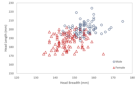
Figure 1: Distribution of head length versus head breadth of the model population.
head shape model with an accurate scalp developed in a previous study (Park et al. 2021, http://humanshape.org/head/) was used for scalp shape predi- ction. This model was developed by analyzing 180 ethnically diverse female and male bald head scans using statistical procedures such as principal com- ponent analysis and multivariate regression. The scalp shape is predicted in this study by fitting the model to the face and neck geometry in the model’s shape space using a rapid fitting method (Park et al., 2014). We also inve- stigated the extent to which additional scalp information such as digitized landmarks or head dimensions can improve the prediction accuracy. The results are then validated by comparing the predicted scalp shapes to data from 81 male and female subjects not included in the development of the original model.
METHODS
Statistical Head Shape Space The statistical head shape model we used in the current study is based on the analysis of high-resolution head scans from a total of 180 adults (100 female and 80 male) with an ethnic diversity of 18 and 59 years old. The male subjects were bald. The scalp contours of the female subjects were obtained using a coordinate measurement technique (Hudson and Mullenger 2020). Figure 1 shows the distribution of the head breadth and length data of the source population. This model accounts for head shape variance without hair artifacts through the principal components (PCs) retained from a PC analysis. This PC analysis linearly transforms the head scan data (Euclidean vertex coordinates) to a new coordinate system with the retained PCs defining a head shape space of the source population. We selected the first 100 PCs to represent the head shape space, which means an entire 3D head shape can be expressed through 100 PC scores/coordinates.
Three-Dimensional Scalp Shape Prediction from Face and Neck Shapes 3
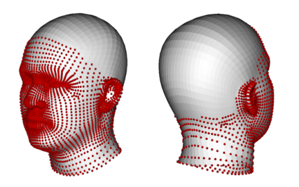
Figure 2: Fitting source vertices on the face and neck area of head shape model (red points).
Model-Based Fitting Method We employed a PC-fitting method (Park et al. 2014, Park et al. 2020) that quickly fits a statistical shape model to target data. The method uses nume- rical optimization to find a set of PC scores that yields the closest shape to the target shape. In each iteration, each fitting vertex of the model is paired to the closest point in the target data, and a set of PC scores are compu- ted using a pseudo-inverse to minimize these local discrepancies between the paired model vertices and the target points. In this study, we paired only the vertices on the face and neck area of the model to the target data for the fit- ting. Figure 2 shows the fitting vertices on the face and neck area to predict the scalp shape. The main benefits of this method are that the fitting perfor- mance is good due to the low dimensionality of the head shape space, and also this method always results in realistic fits even though the target shape is incomplete or noisy since the resulting fits are defined within the shape space.
We also improved this fitting method to allow additional scalp informa- tion for better prediction accuracy. Two types of additional information were considered - (1) digitized scalp landmarks and (2) subject head dimensions such as head breadth, length, and circumference. Scalp landmarks can be considered in the fitting process by finding the closest model vertices to the landmarks and adding these vertices to the fitting vertex list. We applied higher weighting factors for these landmark-vertex pairs, considering the dif- ference in densities between face vertices and scalp landmarks. For the head dimension inputs, a regression model built in the original head model was used. The regression model explains the relationship between the PCs and anthropometric values, such as head length, breadth, and chin arc length. To constrain input head dimensions in the fitting process, dimensions of interest are computed from the PC scores dimensions using the regression model in each iteration to fit the face and neck vertices, and the scores are adjusted to minimize the differences in the dimensions.
RESULTS
Head models with accurate scalp data from 81 adult subjects (ages 18 to 50 years old) that were not used in the original head shape model development
4 Park and Reed
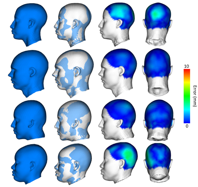
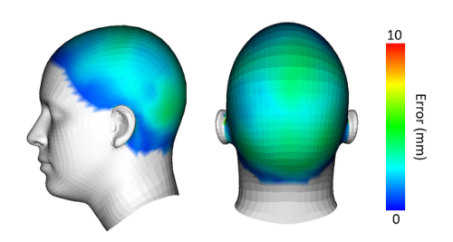
Figure 3: Comparisons between actual head shapes (blue) and prediction results fitted purely based on the face and neck geometry (white) of randomly sampled four sub- jects. The right two columns show the prediction errors colorized using a heatmap (0 mm in blue and 10 mm in red).Figure 4: Colorized mean discrepancies between actual scalp shape and the predicti- ons.
were tested to validate the proposed approach. The scans were aligned to the model’s coordinate system prior to the fitting based on tragion and infraorbitale landmarks.
Three-Dimensional Scalp Shape Prediction from Face and Neck Shapes 5
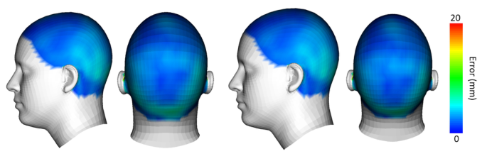
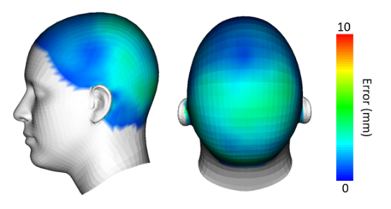
Figure 5: Mean discrepancies of scalp shapes predicted with five scalp landmark points (left two) and ten scalp landmark points (right two).Figure 6: Mean discrepancies between actual scalp shape and scalp predictions with two head dimension constraints – head breadth and length.
Figure 3 compares the actual head shapes of four subjects with the fitting results predicted from the face and neck geometry. The discrepancies between the scan and the fits were computed at every scalp vertex of the model and coded in color using a heatmap with 0 mm in blue and 10 mm in red. The mean absolute error (MAE) in this condition was 2.3 mm across vertices and subjects, and the mean 95th-percentile MAE across all subjects was 6.2 mm. Figure 4 shows the MAE computed at each scalp vertex.
The prediction accuracy increased with additional scalp landmark inputs. We tested two conditions - fitting with five and ten additional scalp landmark points. The landmark positions were selected where the prediction errors are dominant in the scalp area, and we used weighting factors of 10 and 5 for five and ten scalp landmark conditions, respectively. The results showed that adding scalp landmark points improved the prediction accuracy substantially, even with a small number of points. With the five additional scalp landmarks, the mean MAE was reduced to 1.7 mm with 4.6 mm for the mean 95th- percentile MAE. The effect of increasing the number of scalp points from 5 to 10 was modest. The prediction errors with ten scalp landmark points had the mean of 1.5 mm and the 95th percentile error of 4.2 mm. Figure 5 shows
6 Park and Reed
the mean errors at the scalp vertices of these two conditions with additional scalp landmarks.
The improvement from adding head dimensions was similar to the impro- vement from adding scalp points. When head breadth and head length were constrained to the manual measurement values, the mean MAE was 1.9 mm, and the mean 95th-percentile MAE was 5.2 mm. Figure 6 shows the averaged error at each scalp vertex.
DISCUSSIONS AND CONCLUSIONS Despite the importance of scalp shape consideration in the design of head- borne equipment, scalp data are not generally available since optical scanners cannot measure the scalp shape through hair. Many anthropometric data- bases, e.g., the CAESAR database, include whole-body scan data with substantial hair artifacts, particularly female scans. Given the fact that face and neck shape data are generally available in these databases, the method presented in this paper will be a useful and practical solution for obtaining scalp surface information from generic head scans with and without a few additional scalp measures.
The results indicate that the scalp shape is well correlated with the face and neck shape. This is well-aligned with previous anthropometric studies. For example, it has been reported that scalp dimensions are significantly cor- related with age, gender, and ethnicity, as well as face and neck dimensions (Zhuang and Bradtmiller 2005, Sparks and Jantz 2002, Vasavada et al. 2008).
Because the method is based on a statistical model, errors will be larger for the scans with unusual face and neck poses that are not included in the origi- nal head shape population, e.g., open mouth or twisted neck scans, and might be larger for ethnicities not well represented in the training data. Although the model we used is based on an anthropometrically diverse population, more work is needed to assess the quality of the predictions for different populations.
REFERENCES
Corner, B. D., Beecher, R. M., & Paquette, S. (1997, March). Computer-aided fit testing: an approach for examining the user/equipment interface. In Three- Dimensional Image Capture (Vol. 3023, pp. 37–47). International Society for Optics and Photonics. Danckaers, F., Lacko, D., Verwulgen, S., Bruyne, G. D., Huysmans, T., & Sijbers,
J. (2017, July). A combined statistical shape model of the scalp and skull of the human head. In International Conference on Applied Human Factors and Ergonomics (pp. 538–548). Springer, Cham. Hudson, J.A. and Mullenger, C.R. (2020). Female Calvaria Shape Digitization: A Method to Create a Database of Scalp Shapes to be Used in Equipment Design. DTIC Report AD1109544. Lacko, D., Huysmans, T., Parizel, P. M., De Bruyne, G., Verwulgen, S., Van Hulle,
M. M., & Sijbers, J. (2015). Evaluation of an anthropometric shape model of the human scalp. Applied Ergonomics, 48, 70–85.
Three-Dimensional Scalp Shape Prediction from Face and Neck Shapes 7
Li, Z., Park, B. K., Liu, W., Zhang, J., Reed, M. P., Rupp, J. D., ... & Hu, J. (2015).
A statistical skull geometry model for children 0-3 years old. PloS one, 10(5), e0127322. Liu, H., Li, Z., & Zheng, L. (2008). Rapid preliminary helmet shell design based on three-dimensional anthropometric head data. Journal of Engineering Design, 19(1), 45–54. Park, B. K. D., Corner, B. D., Hudson, J. A., Whitestone, J., Mullenger, C. R.,
& Reed, M. P. (2021). A three-dimensional parametric adult head model with representation of scalp shape variability under hair. Applied Ergonomics, 90, 103239. Park, B-K, Lumeng, J.C., Lumeng, C.N., Ebert, S.M., and Reed, M.P. (2014). Child body shape measurement using depth cameras and a statistical body shape model. Ergonomics, 58(2):301-309. Park, B. K. D., Wan, J., Kozak, K., & Reed, M. P. (2020). Model-based characteri-
sation of vehicle occupants using a depth camera. International journal of vehicle design, 83(1), 23–37. Sparks, C. S., & Jantz, R. L. (2002). A reassessment of human cranial plasticity: Boas revisited. Proceedings of the National Academy of Sciences, 99(23), 14636–14639. Vasavada, A. N., Danaraj, J., & Siegmund, G. P. (2008). Head and neck anthropo-
metry, vertebral geometry and neck strength in height-matched men and women. Journal of biomechanics, 41(1), 114–121. Willinger, R., Diaw, B. M., Baumgartner, D., & Chinn, B. (2002). Full face protective helmet modelling and coupling with a human head model. International journal of crashworthiness, 7(2), 167–178. Xi, P., & Shu, C. (2009). Consistent parameterization and statistical analysis of
human head scans. The visual computer, 25(9), 863–871. Yang, B., Tse, K. M., Chen, N., Tan, L. B., Zheng, Q. Q., Yang, H. M., ... & Lee, H. P.
(2014). Development of a finite element head model for the study of impact head injury. BioMed research international, 2014. Zhuang, Z., & Bradtmiller, B. (2005). Head-and-face anthropometric survey of US respirator users. Journal of occupational and environmental hygiene, 2(11), 567–576.
LISTED IN THE AHFE DIGITAL BOOKSTORE CATALOGUE
Explore the complete digital volume
Digital Human Modeling and Applied Optimization
Read this individual research paper free through the original publisher, or explore the full digital proceedings book.
ISBN 978-1-958651-22-3
Explore this ISBN in the AHFE Bookstore ↗Listed in the July 2026 bookstore catalogue; search by ISBN to confirm current availability · Digital book purchase optional