Frank Claes, Jan Berger, Gaetane Stassijns (2021). Arm and Neck Pain in Ultrasonographists. In: Advances in Human Aspects of Healthcare. AHFE Open Access, vol. 13. https://doi.org/10.54941/ahfe100518
The aim of this study was to evaluate the prevalence of upper body quadrant pain among ultrasonographists and to evaluate the association between individual ergonomics, musculoskeletal disorders, and occurrence of neck pain. A hundred and ten Belgian and Dutch male and female hospital ultrasonographists were consecutively enrolled in the study. Data on work-related ergonomic and musculoskeletal disorders were collected with an electronic inquiry, including questions about ergonomics, symptoms and work related factors. Subjects with the screen on their left had significantly more neck pain. Depending on the work space, high-low tables increased the chance on developing neck pain. A screen on eye level caused less neck. Employees with a fixed working space were less susceptible to arm pain. The prevalence of arm pain was significantly higher on the vascular department compared to the radiology, urology and gynecology departments. In the prevention of upper limb pain in ultrasonographists, attention should be paid to the work environment in general, and to the more specific aspects of the ultrasound workstation layout. Primary ergonomic prevention could help the ultrasonographist to work painless during his medical tasks.
In modern medicine, ultrasonography is an important medical diagnostic tool thanks to its non-invasive and radiation-free character. Different medical subdisciplines, such as radiology, abdominal surgery, vascular surgery, cardiology, and gynecology make use of this technique very frequently.
As we know, the prevalence of (low back pain and) neck pain is causing serious public health problems in Western industrialized countries [1-2]. In the Netherlands, the 12-month prevalence of low back pain is estimated to be 44.4% and 28% for neck pain [3] and it is common among the working population as well. Neck pain has unfavorable consequences for the individual worker in terms of pain and disability [1-4], but is also a burden for society and companies in terms of costs due to medical healthcare consumption, work absenteeism, and loss of productivity at work [5-6]. Considering this impact, there is an obvious need for effective preventive strategies.
Personal experience teaches us that ultrasonographists, the people who work with ultrasound machines, are more predisposed to develop neck complaints, potentially in combination with (radiating) pain in the upper limb. However, epidemiologic data about this medical problem are not yet available.
The objective of the present study is to investigate the prevalence, severity, and impact of neck pain caused by working with the ultrasound machine in different medical subdisciplines by means of an electronic inquiry. This study can be the base for further scientific investigation.
Based on our data, we expect to be able to develop custom ergonomic recommendations, so the prevalence of neck pain in the population of ultrasonographists will decrease in the future.
METHODS
The data for this study were obtained through an electronic inquiry about the ergonomic work environment and about the health among Belgian and Dutch ultrasonographists. The self-administered electronic inquiry incorporated 3 parts:
Demographic data: self-reported age (years), gender, institution, medical department, years working as an ultrasonographist.
Ergonomic data: average number of hours per week working with an ultrasound machine, consecutive working hours a day, permanent workplace, high-low table, ergonomic chair, position of the machine compared to their own sitting position, position of the screen compared to their own sitting position, height of the screen, and maneuverability of the screen.
Musculoskeletal data: neck and arm pain (present-past), location of arm pain, evolution of the pain throughout the workday, relation between work and pain.
A hundred and ten male and female ultrasonographists working at a hospital were randomly recruited by e-mail to take part in the study, regardless of their musculoskeletal complaints. An electronic link was placed on the website of the Dutch/Belgian Association for non-invasive vascular diagnosis (http://www.vnivd.nl).
DESCRIPTION OF THE ULTRASONOGRAPHIST TASK Vascular Department: Examination Various Veins
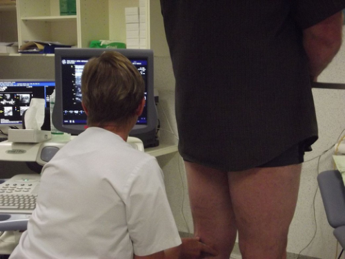
In a standard examination of the various veins, the patient is standing on an adjustable high-low table. The investigator sits on the back side of the patient and holds with his right hand the duplex probe. He uses his left hand for a) manipulating or typing on the duplex machine b) taking ultrasound gel and c) compressing the calf of the lower extremity. The examiner always turns his head left, towards the duplex machine meanwhile his right arm is in the opposite direction, giving compression with the probe on the lower limb. The examiner scans the whole limb, from hip to toe (see Figure 1).
Cardiac Department A standard echocardiogram is also known as a transthoracic echocardiogram (TTE). In this case, the echocardiography transducer (or probe) is placed on the chest wall (or thorax) of the subject, and images are taken through the chest wall. Different views are made: parasternal, apical, subcostal and suprasternal. This is a non- invasive, highly accurate, and quick assessment of the overall health of the heart. The technician can quickly assess a patient's heart valves and degree of heart muscle contraction (an indicator of the ejection fraction). The images are displayed on a monitor, and are recorded by digital techniques.
An echocardiogram can be used to evaluate all four chambers of the heart. It can determine strength of the heart (an indicator of the ejection fraction), the condition of the heart valves (morphological and functional) and the aorta. It can be used to detect a heart attack, hypertrophy of the heart and infiltration of the heart with an abnormal substance . With advanced measurements of the movement of the tissue with time (tissue doppler), it can measure diastolic function (=filling).
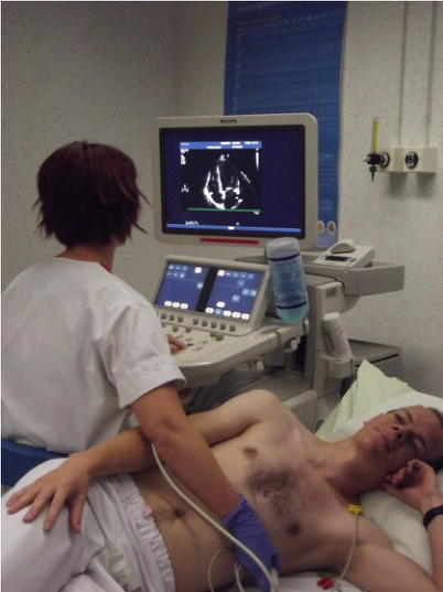
To obtain the best image quality the room needs to be darkened. The subject has to undress his upper body and lie down on his left side with his left hand under his head and the right hand on his side. Electrodes will be connected on the chest of the subject to record the heart rate of the patient. To have better image quality on the screen, we put gel on the probe. This is needed because otherwise there is a small space between the skin and the probe. Air is bad for image quality.
When all images are gathered, we give the subject some tissue so he can clean of the gel. He may put on his clothes again and wait for his cardiologist to receive the results (see Figure 2).
Figure 1. Examination of the various veins
Vascular department, standard examination of the various veins, the patient is standing, the investigator sits on the back side of the patient and holds with his right hand the duplex probe. The examiner always turns his head left, towards the duplex machine meanwhile his right arm is in the opposite direction, giving compression with the probe on the lower limb.
Figure 2. Echocardiography transducer.
Cardiac department, the echocardiography transducer (or probe) is placed on the chest wall of the subject. The images are displayed on a monitor on the left side of the technician.
DISCUSSION
This study was conducted to determine the prevalence of neck and arm pain in ultrasonographists. This resulted in different findings.
Subjects with the screen on their left had significantly more neck pain.
When performing work with the hands and fingers, the muscles in the neck/shoulder region must act as stabilizers.
Static contraction of the trapezius and other shoulder muscles is needed to keep the arms at right angles, a necessary posture when using the keyboard. This contraction is accentuated when there is also rotation or bending of the neck when the computer screen is placed to the side of the worker. The recommended position is in front of the worker.
Continuous rotation of the neck throughout the working day causes neck pain. It should be analyzed what the impact is on different structures located in the neck area and which anatomic structures are the root cause of the pain development. In addition, the effect of head rotation, combined with the position of the arm on the development of a specific complaint pattern in the upper quadrant should be monitored.
In our population, only two people (1.82%) worked with the screen on their right side. This sub-population was too small to consider their test results in our statistical analysis.
A screen on eye-level caused less neck pain.
In the scientific literature there is consensus that poor ergonomic conditions at workstations contribute to musculoskeletal symptoms. Studies have shown that holding the neck in a bent posture and working in the same posture for prolonged periods of time were significantly associated with neck pain Ranasinghe suggest that modification of incorrect postures at work and improvements in the ergonomic designs of workstations could be important, not only as primary preventive strategy, but also as a secondary preventive strategy in those with symptoms. Ariens found a trend for a positive relation between neck flexion and neck pain, although not significant, suggesting an increased risk of neck pain for those who spent a high percentage of the working time with their neck at a minimum of 20° of flexion.
Depending on the work space, height adjustable tables increase the chance on developing neck pain.
Left-handed people suffered significantly less arm pain than right-handed people. In our study population we found 15 left-handed people (13.76%), of which only four (3.70%) held the ultrasound probe with their left hand.
Employees with a fixed working space were less susceptible to arm pain.
When people can use ergonomic furniture, we expect that they adjust their table to the right height so the chance on arm or/and neck pain will decrease. Poor ergonomic knowledge is a significant predictor of complaints in the neck, forearm and hand regions. Implementation of a worksite ergonomics program is known to be effective in reducing work-related complaints in the workforce. The height of standard tables has been designed with respect to the average length of humans. Abuse or non-use of a height-adaptable table possibly has a negative impact on various structures in the upper quadrant. Further research is needed to determine which structures might be hurt by this and how we could assess the ideal individual working height for health care staff. Based on these findings, every staff member performing ultrasound scans should complete a thorough training.
Ultrasonographists with less than 10 years of working experience had less arm pain than those who have more than 10 years of experience. However, this effect was found to be marginally significant, Cagnie also found a trend for a positive relation between years of working experience and arm pain. The increase with age can be explained by the increasing degeneration of the cervical spine with age.
The prevalence of arm pain was significantly higher at the vascular department compared to the radiology, urology and gynecology departments Further research will have to clarify to which extent the working conditions, such as position of the screen or subject, of the vascular department staff is different compared to those of the ultrasound staff of the other departments mentioned above. There might be great differences in working methods between the departments, which might explain why the vascular department staff seems to be more susceptible to complaints than others.
CONCLUSIONS
This study shows there is an important problem of arm and neck pain in ultrasonographists. Worldwide, a large number of people is working as an ultrasonographist. Hence, we think there is a need for a larger and more profound study concerning this problem.
Based on the results of our study, further research should consider multiple aspect of the workspace of ultrasonographists.
First, there is the ultrasound machine itself. A more ergonomic approach to the machine configuration is appropriate. Since most ultrasonographists have their own workplace, separating the screen and the keyboard from the machine is advisable. This will result in more individualized configurations.
Second, we should reconsider the position of the ultrasonographist. It is possible to identify some individual parameters by which we can set up an appropriate and individualized ultrasound configuration. The sitting height of each person is an individual parameter. It is possible to calculate this for every ultrasonographist. Depending on the sitting height, we can search for the ideal table height. We should also encourage examiners to shift their position more often. The current technology allows us to remember the position of our car seats. Why not do the same for the ultrasonographist’s seat?
Finally, we should also reconsider the position of the patient. It is possible to redesign the examination table in a more ergonomic way so the ultrasonographist can work in more ergonomic conditions. This redesign varies with the different medical subdisciplines. The working conditions in the cardiology department are not the same as in the radiology or vascular department.
Further research on all aspects of the work situation mentioned above is necessary and implementing these necessary changes will result in an improvement of the health status of our ultrasonographists.
REFERENCES
Ariens GA, Bongers PM, Hoogendoorn WE, van der Wal G, van Mechelen W: High physical and psychosocial load at work and sickness absence due to neck pain. Scand J Work Environ Health 2002, 28:222-31 Ariens G, Bongers P, Douwes M, Miedema M, HoogendoornW, Van der Wal G, Bouter L, van Mechelen W (2001) Are neck
flexion, neck rotation and sitting at work risk factors for neck pain? Results of a prospective cohort study. Occup Environ Med 58:200–207 Cagnie, B. Æ L. Danneels Æ D. Van Tiggelen Æ V. De Loose Æ D. Cambier Individual and work related risk factors for neck pain among office workers: a cross sectional study Eur Spine J (2007) 16:679–686 DOI 10.1007/s00586-006-0269-7 Cole DC, Hogg-Johnson S, Manno M, Ibrahim S, Wells RP, Ferrier SE, Worksite Upper Extremity Research Group: Reducing
musculoskeletal burden through ergonomic program implementation in a large newspaper. Int Arch Occup Environ Health 2006, 80:98-108. Côté P, van der Velde G, Cassidy JD, Carroll LJ, Hogg-Johnson S, Holm LW, et al. The burden and determinants of neck pain in workers: results of the Bone and Joint Decade 2000-2010 Task Force on Neck Pain and Its Associated Disorders. Spine (Phila Pa 1976). 2008;33(4 Suppl):S60–S74. Dagenais S, Caro J, Haldeman S. A systematic review of low back pain cost of illness studies in the United States and
internationally. Spine J. 2008;8(1):8–20. doi:10.1016/j. spinee.2007.10.005 Freburger JK, Holmes GM, Agans RP, Jackman AM, Darter JD, Wallace AS, Castel LD, Kalsbeek WD, Carey TS. The rising prevalence of chronic low back pain. Arch Intern Med. 2009;169(3):251–8. doi:10.1001/archinternmed.2008.543. Gerr F, Monteilh CP, Marcus M: Keyboard use and musculoskeletal outcomes among computer users. J Occup Rehabil 2006,
16:265-277. Henschke N, Maher CG, Refshauge KM, Herbert RD, Cumming RG, Bleasel J, York J, Das A, McAuley JH. Prognosis in patients with recent onset low back pain in Australian primary care: inception cohort Ijmker S, Huysmans MA, Blatter BM, van der Beek AJ, van Mechelen W, Bongers PM: Should office workers spend fewer
hours at their computer? A systematic review of the literature. Occup Environ Med 2007, 64:211-222. Lambeek LC, van Tulder MW, Anema JR, Swinkels ICM, van Mechelen W. Trend in societal costs of low back pain in The Netherlands in the period 2002 - 2007. Spine (Phila Pa 1976.). 2010. Picavet HS, Schouten JS. Musculoskeletal pain in the Netherlands: prevalences, consequences and risk groups, the DMC(3)-
study. Pain. 2003;102(1–2):167–78. doi:10.1016/s0304-3959(02)00372-x. Priyanga Ranasinghe, Yashasvi S Perera, Dilusha A Lamabadusuriya, Supun Kulatunga, Naveen Jayawardana, Senaka Rajapakse and Prasad Katulanda, Work related complaints of neck, shoulder and arm among computer office workers: a crosssectional evaluation of prevalence and risk factors in a developing country. Environmental Health 2011, 10:70
LISTED IN THE AHFE DIGITAL BOOKSTORE CATALOGUE
Explore the complete digital volume
Advances in Human Aspects of Healthcare
Read this individual research paper free through the original publisher, or explore the full digital proceedings book.
ISBN 978-1-4951-2093-0
Explore this ISBN in the AHFE Bookstore ↗Listed in the July 2026 bookstore catalogue; search by ISBN to confirm current availability · Digital book purchase optional