M. Mazzolaa, L. Forzonib, S. D’Onofriob, C.E. Standolia, G. Andreonia (2020). Using Santos DHM to Design the Working Environment for Sonographers in Order to Minimize the Risks of Musculoskeletal Disorders and to Satisfy the Clinical Recommendations. In: Advances in Applied Digital Human Modeling. AHFE Open Access, vol. 11. https://doi.org/10.54941/ahfe100406
Publisher statistics will load when you open Metrics.
AHFE OPEN ACCESS OPEN ACCESS CONFERENCE PROCEEDINGS PAPER
Using Santos DHM to Design the Working Environment for Sonographers in Order to Minimize the Risks of Musculoskeletal Disorders and to Satisfy the Clinical Recommendations
The percentage of sonographers reporting consequences of pain and discomfort is close to 80% and the Society of Diagnostic Medical Sonography demonstrates that sonographers, on average, experience pain or Musculoskeletal disorders within 5 years of entering the profession. Digital Human Models (DHM) can be an essential tools, supporting the definition of a correct medical environment to perform Sonography in response to the regulatory aspects that standardize the Health Care design and in setting up the ergonomics requirements. The methodology proposed to perform an optimal setting of the workspace considers the different aspects of a diagnostic Ultrasound (US) examination room in a clinical setting: sonographer's seating and examination bed, US system and probe. Vascular and Abdominal applications were considered. The aim of this study is to present an example of how an Advanced DHM can support the design of the working environment for sonographers in order to minimize the risks of muscle-skeletal disorders and to satisfy the clinical recommendations. Results were compared with data presented in previous studies about ergonomics in professional sonography and they demonstrate to be coherent with the plan for an ideal set-up.
The Ergonomic evaluation and assessment of sonographers’ workspace are very important. The percentage of sonographers reporting consequences of pain and discomfort is close to 80% and the Society of Diagnostic Medical Sonography (SDMS) demonstrates that sonographers, on average, experience pain or Musculoskeletal disorders (MSDs) within 5 years of entering the profession. Higher anatomical sites experienced discomfort reported by sonographers are in the shoulder, neck, low back, wrist and hand/fingers. (Society of Diagnostic Medical Sonography, 2003, Village and Trask, 2007).
Sonographers are exposed to a variety of ergonomics-related risk factors when they perform specific tasks, dealing with the transportation of equipment, the positioning of patients and equipment and the daily use of Ultrasound (US) systems.
Several studies in this research area identified nine major factors of interest (Andreoni et al., 2013, Murpy and Russo, 2000). Among these factors, the transducer design, the US system user interface and control panel design and the sonographer’s body posture seem to be relevant for injury and risk prevention. Indeed, a comfortable chair and correct body position protected the sonographer from the onset of neck and back discomfort. These studies contributed to define the best practices for a correct positioning of US equipment, in order to minimize the MSDs risks for professional sonographers.
Nonetheless, the correct observance of these recommendations depends on the sonographer’s diligence and this is due to the lack of tools that quantify the distances between the sonographer, the patients and the equipment, but also that can support the design of the clinical environment, in order to verify the correct positioning.
Digital Human Models (DHM) can be important tools that support the definition of a correct medical environment to perform diagnostic US examinations in response to the regulatory aspects that standardize the Health Care design and in setting up the ergonomics requirements.
The aim of this study is to present how an Advanced DHM, i.e. Santos Digital Human, can support the sonographers’ working environment design, in order to minimize the risks of muscle-skeletal disorders and to satisfy the clinical recommendations.
Sonography can be performed in different applications. Moreover, thanks to the availability of portable US systems with high level of diagnostic performance , US examinations can be performed in many different settings, ranging from the typical hospital setting examination room, to emergency Departments, patient bedside, patient home, emergency vehicles and so on.
The presented study is mainly focused on Abdominal and Vascular applications to meet differing conditions to simulate a scan and it considers a classic hospital setting environment. In addition, various probes (Linear probe used for Vascular application and Convex probe used for Abdominal application) are used with different consoles and dissimilar grasps are applied for diverse applications.
MATERIAL AND METHODS Preparation of the Simulation Environment
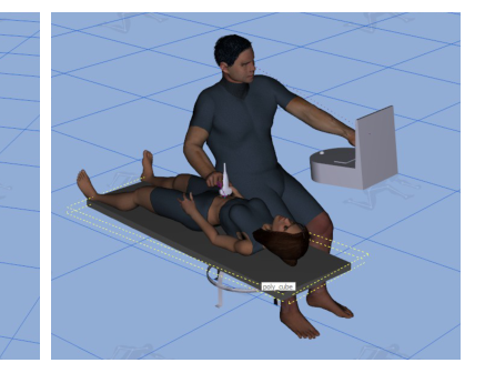
In order to design the optimal sonographer’s workplace, it is necessary to consider the mutual position of the sonographer and the objects that compose the simulation environment; in particular these objects include: the US console, the patient’s bed and the sonographer’s sitting chair. In this work, Santos DHM was used to prepare the simulation environment, in accordance with the literature description of the most preferable working conditions (Murphy and Russo, 2000). The workspace was designed considering the possibility to adjust the objects height and their distances from the sonographer’s body.
As a consequence, the evaluation of the joint angles were compared, assuming both sitting work posture chair height and bed height as adjustable.
The methodology proposed to perform an optimal setting of the workspace consists of the following steps:
1) To design the most comfortable set-up, the seating and bed height are assumed to be adjustable, as well as the console position.
2) The Avatar was anthropometrically scaled, representing the 5th, 50th and 95th percentile. In this work, the evaluation was conducted only for the male gender.
3) Preparation of the simulation environment. The Height Regulation of the seat was determined to guarantee that the avatar, in neutral sitting position, had the feet resting on the floor.
4) The position of the bed, both for Abdominal and Vascular application and the US console position were determined using the Santos Zone Differentiation (ZD) analysis tool.
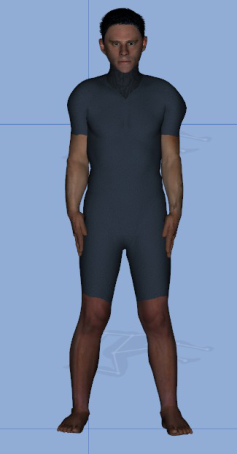
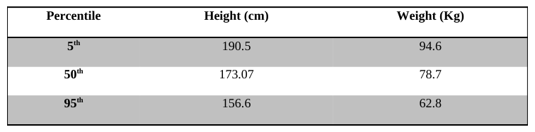
Anthropometrical Characterization of the Avatar The avatars were anthropometrically differentiated by somatotypes and by percentile: 5th, 50th and 95th male percentiles were used to scale the Avatars body sizes. The 5th percentile corresponds to the somatotype defined as “Short, Lean and V-shaped”, the 50th percentile corresponds to the normal Santos somatotype, and the 95th percentile corresponds to “Tall, Heavy, H-shaped” somatotype. Avatar’s body weight and limb lengths are based on the data of the Standard ISO 3411. Table 1 present body height and weight of the different percentiles.
Figure 1: From left to right: frontal picture of Avatar corresponding to the 5th, 50th and 95th percentiles.Table 1: Anthropometrical measures of the different percentiles, according to the ISO-3411 Percentile Height (cm) Weight (Kg)
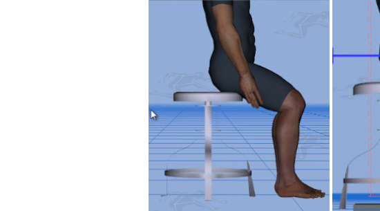
Seat Height Regulation To assess the correct height regulation of the sitting work posture on the chair, the Avatar is firstly positioned in the Neutral Sitting posture. Therefore, the seat height has been dimensioned to guarantee that the Avatar’s feet rest on the floor.
Figure 2: On the left: the avatar is positioned in the neutral sitting posture. In the middle, the seat height is oversized, and the avatar is not able to touch the floor with his feet. On the right, the correct dimension of the seat height, adopted for the simulations.
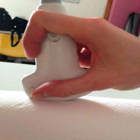
Grasps The orientation of the transducer within the hand, which must be rotated to obtain different images, determined the type of grip used. There are 3 different types of grasp, depending on the analyzed probe application: Longitudinal Pincer Grip, Transversal Pincer Grip and Palmar Grip. An example of each grasp, real and DHM simulated, is presented in Figure 3.
Figure 3: On the top left, the Longitudinal Pincher Grip, DHM simulated and observed. On the top right, the Transversal Pincher Grip and in the second line the Palmar grip.
The aim of the present work is the general assessment of the workspace environment and it considers the posture as the most relevant parameter to be evaluated. As a consequence, each workspace condition can be assumed independently from the specific grasp adopted.
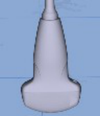
Probes 4 different US probes (Convex array probes CA631 and AC2541; Linear array probes LA523 and SL1543; Esaote S.p.A., Firenze, Italy; see Figure 4) were used for ergonomic analysis; each probe was used with a specific console and with three different grasps, as a consequence of the specific application (Abdominal/Vascular). AC2541 and
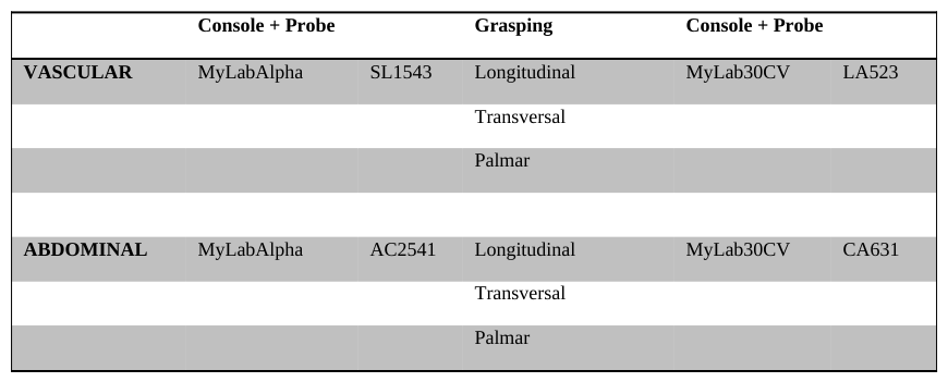
SL1543 are ergonomically designed probes, appleprobe Design transducers. Each probe can be grabbed with the three different grasps described above, while each of them is associated with a single application and a specific console. In table 2, for each probe, the related US console and the possible grasps are listed.
Figure 4. From left to right: probe CA631, AC2541, LA523, SL1543Table 2: Correspondence of US application, Consoles, Probes and Grasps Console + Probe Grasping Console + Probe
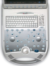
Consoles Two different Esaote portable US systems were used for the investigation: MyLab Alpha and MyLab30CV (Esaote S.p.A., Firenze, Italy). They’re presented in figure 5 together with the simplified model imported in the DHM and the reaching regions of interest, that correspond to the most used areas of the user interface.
Figure 5: On the left: MyLab30CV Console presented as a simplified .obj model imported in the Santos environment, and a picture of the real interface is presented. In red are showed the different areas of investigations for the simulation conditions of the Left Hand. On the Right: The MyLabAlpha console presented as a simplified .obj model imported in the Santos environment, and a picture of the real interface. In this case, the zones objects of the simulations are showed in blue on the left
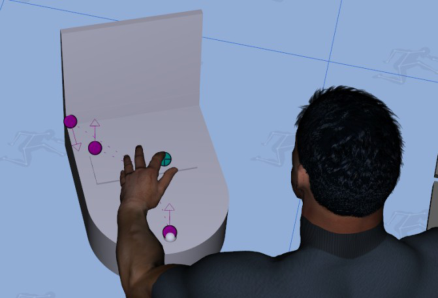
In figure 6, the specific target points objects of investigations are showed. In particular, there are 3 markers points (Top Left of the Qwerty Keyboards, Top Left of the Soft Key Menu on the screen and the Trackball) for the
MyLab30CV model and two marker points (Top Left of the Touch screen and the Trackball) for the MyLabAlpha model.
Figure 6: On the Left, the 3 target points of the MyLab30CV US system; on the right, the 2 target points of the MyLabAlpha US system
Zone Differentiation for US Probe Reaching Zone Santos ZD tool was used to optimize the US probe and the console reaching zones. The ZD system allows the user to analyze information according to posture-based performance measures (Yang et al., 2004, 2006, 2008). In this study, the performance measure adopted to determine the optimal reaching zones was the Effort Performance Measure, in a percentage of 80%, and the Joint Displacement for 20%.
The effort performance measure models the tendency to gravitate towards one’s initial position. In sonography, the movement of both left hand (controls adjustment on US system console) and right hand (probes holding, pressing and moving for the exploration of the patient’s body) can be described as consecutive small changes respect to the last posture adopted by the avatar. As a consequence, the effort performance measure represents the most convenient performance measure for the ergonomics evaluation of the working environment in sonography. A 20% of Joint Displacement performance measure was set, in order to avoid the possible increase of the distance from the neutral sitting posture of the avatar, that could generate unacceptable postures.
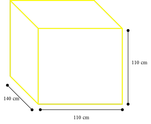
New ZD were generated for both the right and the left hand of the avatar, for each percentile. The computed zone were defined with a volume of 110 x 140 x 110 cm, with a resolution of 64x64x64.
Figure 7. volume of the computed Zone Data
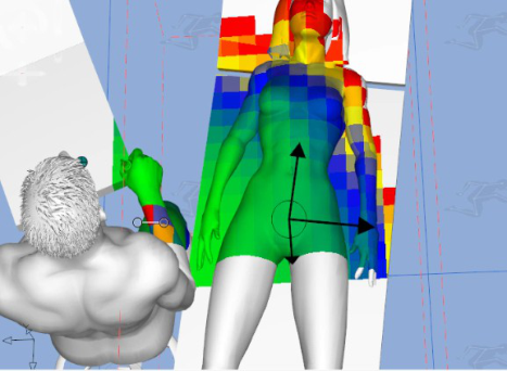
Figure 8 presented the procedure adopted for the optimal positioning of the different workspace objects for the 5 th percentile avatar somatotype. The optimal position is determined selecting the position that presents the most green colored area for the relative object, that represents the maximum comfort according to the performance measured used for computation (i.e., effort).
Figure 8. On the top left, the Volume of the computed data for the left end the effector (left hand) is presented in yellow, for the 5th percentile avatar somatotype. On the top right, a sphere has been used to graphically represent the result of the ZD data computed. On the bottom line, the optimal position obtained using the ZD application is presented for the abdominal application.
Posture Evaluation The posture evaluation was conducted considering the Maximum Holding Time (MHT) Index for the Shoulder, and the Right Shoulder Abduction Angle, considering all the subjects right handed, grasping the probe with the right hand and reaching the US console with the left hand.
RESULTS AND DISCUSSION
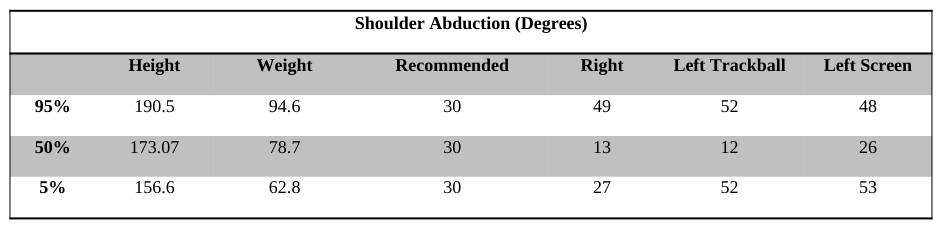
Table 3 presents results of the simulations. In addition to the anthropometrical data of the three avatars, the recommended value of Shoulder Abduction angles, as defined in the Report of Murphy and Russo, is reported. This value represents the reference value and it has been compared with: the Abduction angle of the Right Shoulder, the Left Shoulder in reaching the closest target point (US system trackball) and the Left Shoulder while reaching the touchscreen of the monitor.
Results demonstrate that the 50th percentile avatar somatotype presents optimal values, always lower than the reference, for both the right and left shoulder, in all the conditions. Results for the 5th percentile avatar are opposite; the abduction angle of the right shoulder is lower than the reference, while the angle of the left shoulder is higher. The 95th percentile presents the worst results, being always over the recommended threshold. Results are reported without the differentiation between vascular and abdominal application, due to the fact that, once the operating volume has been defined and the postural comfort zone determined, the patient should be located there with the specific body part, without modifying the postural asset.
Table 3. Results of the Simulation. Shoulder joints angles are presented and compared with the recommended reference value.Figure 9. Results of the simulation for 5th, 50th (top line) and 95th percentile avatar somatotypes.
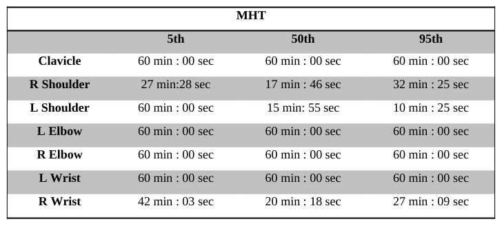
Results of the MHT Analysis are presented in Table 4. Results for the 5th percentile avatar somatotypes demonstrates that potential problems and risk of fatigue could arise at the right shoulder level, while the other joints do not present any critical situation, except for a small fatigue for the right wrist. On the contrary, both 50th and 95th percentiles present possible fatigue at the right and left shoulder level and at the right wrist level. These results could be determined by the mass property of the different percentile and the corresponding force property. Also the limb length can influence the postural evaluation and increasing the level of fatigue of the bigger percentiles. Fatigue at the wrist level is due to the probe grasp. Possible future development could investigate the relation between the MHT, the probe model and the grasp adopted.
Table 4. Maximum Holding Time Index results MHT
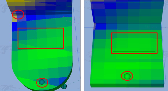
Figure 10 presents the comfort area determines for the two different US consoles. This result is very promising and interesting. On the left it is possible to visualize the MyLab30CV console model, with the three different target points considered and what it can be noticed is that the position of the US console trackball and the touch screen on the monitor are not ergonomically in optimal position and are outside of the preferred comfort zone (green). Also the keyboard presents a decrease in the comfort level, moving the hand from the right to the left.
On the contrary, the position of the controls of the MyLabAlpha US model seems to be optimal, for both the US console trackball and the touchscreen.
Figure 10. Results of the comfort zone evaluation for MyLab30CV (Left) and MyLabAlpha (Right).
CONCLUSIONS
In this study, a methodology to design the optimal workspace for a diagnostic US examination was suggested. The ergonomic analysis proposed is based on the use of the Santos DHM software, applying its capability in providing a coherent ergonomics design tool, in accordance to referred practical recommendations.
The simulation process studied the optimal positioning of the sonographer’s seat, the patient’s bed and the US system console, according to the biomechanical characteristic of three different somatotype scaled avatars.
Two US systems were ergonomically evaluated. Results demonstrates that the procedure adopted is coherent with the experimental recommendation for the 50th percentile and the 5th percentile, considering the Shoulder Abduction angles and the MHT Index.
The 95th percentile demonstrates to assume, in the optimal position, a posture that can determine fatigue hazard, but its comparison with the reference value could be refined, considering the anthropometrical differences among the different somatotype.
Results were compared with data presented in previous studies about ergonomics in sonography and they demonstrates to be coherent with the plan for an ideal set-up.
In conclusion, this work demonstrates that DHM can be a promising tool in the early design of the sonographer’s working environment, respecting the recommendations and the best practice suggested in literature to minimize the risk of MSDs.
Further developments could propose this DHM approach as an effective tool to design medical workplaces.
REFERENCES
Andreoni G., Costa F., Mazzola M., Fusca M., Romero M., Carniglia E., Zambarbieri D., Santambrogio G.C. (2013), “A multifactorial approach and Method for Assessing Ergonomic Characteristics in Biomedical Technologies”, Advances in Human Aspects of Healthcare, edited by Duffy V., Chapter 1, 1- 12. Gregory, V. (1998), “Musculoskeletal Injuries: Occupational Health and Safety Issues in Sonography”, Sound
Effects 30. International Standard ISO 3411 (2007), “Earth-moving machinery – Physical dimensions of operators and minimum operator space envelope”, Fourth edition. Magnavita, N., Bevilacqua, L., Mirk, P., Fileni, A, and Castellino, N. (1999), “Work-related Musculoskeletal Complaints in
Sonologists”, J Occup Environ Med, Nov; 41(11):981-8. Mirk, P., Magnavita, N., Masini., L., Bazzocchi., M ., and Fileni., A. (1999), “Frequency of Musculoskeletal Symptoms in Diagnostic Medical Sonographers. Results of a Pilot Survey”, Radiol Med, Oct; 98 (4):236-41. Murphy, C. and Russo, A. (2000), “Report: An Update on Ergonomic Issues in Sonography”, Healthcare Benefit Trust, EHS -
Employee Health and Safety Services. NIOSH Technical Report, HETA 99-0093-2749 Smith, A.C., Wolf, J. G., Xie, G.Y, and Smith, M.D. (1997), “Musculoskeletal Pain in Cardiac Ultrasonographers: Results of a Random Survey”, J. Am. Soc Echocardiogr, May; 10(4):357-62. Smith and Sainfort (1989) in Kuorinka et al. (1995), “Work-Related Musculoskeletal Disorders: A Reference for Prevention”,
Philadelphia: Taylor & Francis. Society of Diagnostic Medical Sonography (2003), “Industry Standards for the Prevention of Work-Related Musculoskeletal Disorders in Sonography”.Village J., Trask C. (2007), “Ergonomic analysis of postural and muscular loads to diagnostic sonographers”, Int. Jour. Ind. Ergonomics 37, 781-789. Yang, J., Marler, T., Beck, S., Abdel-Malek, K., and Kim, H.-J. (2006), “Real-Time Optimal-Reach Posture Prediction in a New
Interactive Virtual Environment”, Journal of Computer Science and Technology, 21(2), 189-198. Yang, J., Marler, R. T., Kim, H., Arora, J. S., and Abdel-Malek, K. (2004), “Multi-Objective Optimization for Upper Body Posture Prediction”, 10th AIAA/ISSMO Multidisciplinary Analysis and Optimization Conference, Albany, NY. Yang, J., Verma, U., Penmatsa, R., Marler, T., Beck, S., Rahmatalla, S., Abdel-Malek, K., and Harrison, C. (2008),
“Development of a Zone Differentiation Tool for Visualization of Postural Comfort”, SAE 2008 World Congress, Detroit, MI.
LISTED IN THE AHFE DIGITAL BOOKSTORE CATALOGUE
Explore the complete digital volume
Advances in Applied Digital Human Modeling
Read this individual research paper free through the original publisher, or explore the full digital proceedings book.
ISBN 978-1-4951-2094-7
Explore this ISBN in the AHFE Bookstore ↗Listed in the July 2026 bookstore catalogue; search by ISBN to confirm current availability · Digital book purchase optional