Ricarda Lehmanna, Kerstin Thurowa, Regina Stollb, Matthias Weippertab (2022). Human vs. Machine in Life Science Automation: Comparing Effectiveness of Manual and Automated 3-D Cell Culturing Processes. In: Advances in Ergonomics In Design, Usability & Special Populations: Part II. AHFE Open Access, vol. 19. https://doi.org/10.54941/ahfe1001219
There is an ongoing shift from 2-D cell culturing to 3-D approaches, particularly because three dimensional cell cultures better simulate in vivo conditions. Thus, automation of 3-D culturing processes is a current issue in life science automation, as it is supposed to provide standard qualities and the potential of large-scaled production of 3-D tissues. In the traditional production process, human sensory-motor and evaluation skills are essential for successful 3-D cultivation. This study aimed to evaluate the effectiveness and efficiency of an semi-automated HeLa-cell culturing process compared to traditional manual processing. HeLa cells were manually and automatically encapsulated in alginate matrices and cultivated in media with (+P/S) and without antibiotic (-P/S). The manual steps were carried out by skilled laboratory staff. The automated procedures were performed by the Biomek® Cell Workstation. The proliferation rates and toxicities were evaluated on day 1, 14 and 35. Further, measurements of the duration of the processes and an expert interview as well as process observation of novice and expert staff have been carried out. The proliferation rates of manual produced alginate beads +P/S were significantly higher compared to the rates of the semi-automated produced beads. Average proliferation rates of the manually and semi-automatically produced alginate beads –P/S were similar on all post production days. Further, our results showed a significant increase of the cytotoxicity +P/S and –P/S from day 1 to day 35 for both types of production. On day 1, the cytotoxicity of manual produced beads was significantly higher compared to semi-automated produced beads. Particularly on day 35 toxicity of alginate beads +P/S was significantly increased compared to beads –P/S. Concluding, single process duration of the automated alginate bead production is higher and effectiveness and efficiency of the manual bead production is slightly worse compared to manual processing. However, the main benefits of automated processes are a stable quality, high reproducibility and increased absolute sample production (24/7-operation).
Cell culturing is now moving from monolayer (2-D) to three dimensional cell culturing (3-D). This shift is motivated by the need of optimally mimicking the functions and environmental conditions of living tissue and the general recognition that monolayer ‘petri dish’ based approaches are not representative for physiological tissues (Pampaloni et al. 2007; Jonitz et al. 2011b; Kumachev et al. 2011; Ghidoni et al. 2008). Automation of 3-D culturing processes might be fundamental to provide standard qualities as well as large-scaled production of 3-D tissues (Tebenkova 2011). Typical 3-D culture systems are alginate beads: adherent cells are encapsulated and arrested in an alginate matrix (Jonitz et al. 2011b; Ghidoni et al. 2008). Many field in the biotech domain, e g. cancer research, regenerative medicine and compound screening (Cassidy et al. 1996) use alginate beads to simulate in-vivo conditions. However, 3-D culturing is complex and some sub-processes require human sensory-motor skills and evaluation tasks as well. However, to improve qualities and to upscale quantities the automation of these processes is a current issue in life science automation. Provided sufficient quality and efficiency automation of the complex 3-D culturing will replace traditional manual 3-D cell production. Generally, the automated process is supposed to reduces costs, human errors and ensure a stable quality of alginate beads (Blow 2008; Carere et al. 2002; Chapman 2003; Markin, Whalen 2000) This study compared the quality and quantity of an automated HeLa-alginate bead manufacturing process against the traditional manual method (effectiveness). Furthermore, performance/workshift ratios of the processes were evaluated (efficiency) by task analysis and 24 –hour projection. Results will provide a basis for the identification of potential limitations of automation in the field of 3-D cell culturing and might help to improve automation performance and productivity.
MATERIALS AND METHODS
Task Analysis Task analysis was carried out by an ergonomic investigator who accompanied biotech experts and novices for several hours during routine manual and semi-automated alginate bead production process. Commercially available software (TaskArchitect 3.1.7) was used to document the observed process steps for quantitative analysis and subsequent efficiency projection.
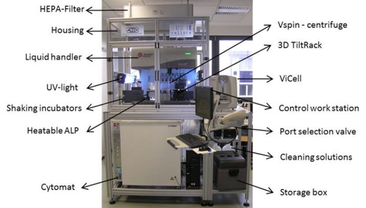
Instrumentation - Automated cell culture system The automated cell culture processes were performed by the proprietary Biomek® Cell Workstation (Center for Life Science Automation, Rostock, Germany) (Figure 1). This automated system enables the cultivation of adherent and suspension cell cultures as well as the production of 3–D cell cultures. The central Span 8 liquid handler (Beckman Coulter, Brea, CA) pipettes solutions and the integrated gripper provides transport of well plates and flasks. Different automated laboratory positioners (ALP) are integrated on the liquid handler deck. The 3-D tilt racks simulates manual handling (proviting, knocking) of the cell culture flasks. The heatable ALP (Beckman Coulter) is used for warming solutions. The positive positioning ALP (Beckman Coulter) enables the exact positioning of well plates. Furthermore, short incubations at 37°C are realized by two shaking incubators. The V-spin™ centrifuge (Velocity 11, Palo Alto, CA), ViCell cell counter (Beckman Coulter) and Cytomat incubator (37°C, 5% CO 2) (Thermo Fisher Scientific, Waltham, MA) are located close to the liquid handler deck. Additionally, the port selection valve enables the change of six different media. Sterile working conditions are ensured by UV-lights (Vilber, Eberhardzell, Germany) and housings equipped with a HEPA-Filter system (Camfil, Stockholm, Sweden).
Edited by T. Ahram, W. Karwowski and T. Marek
Figure 1. automated cell workstation (Lehmann R., Vetter L., Person L., Thurow K. 2013)
Manual cell cultivation HeLa cells were cultivated in 75 cm² cell culturing flasks at 37 °C and 5 % CO 2. Dulbecco's modified eagle medium (DMEM) was supplemented with (1 %) (+P/S) and without penicillin/streptomycin (-P/S) (SIGMA-ALDRICH) and 10 % fetal bovine serum (FBS). For the splitting process, cells were washed three times in phosphate-buffered saline (PBS) (PAA, Cölbe, Germany) and treated with Trypsin/EDTA (SIGMA-ALDRICH, Seelze, Germany) for 4 minutes at 37 °C. Trypsin/EDTA was then neutralized with DMEM and the cell suspension was split into new culture flasks.
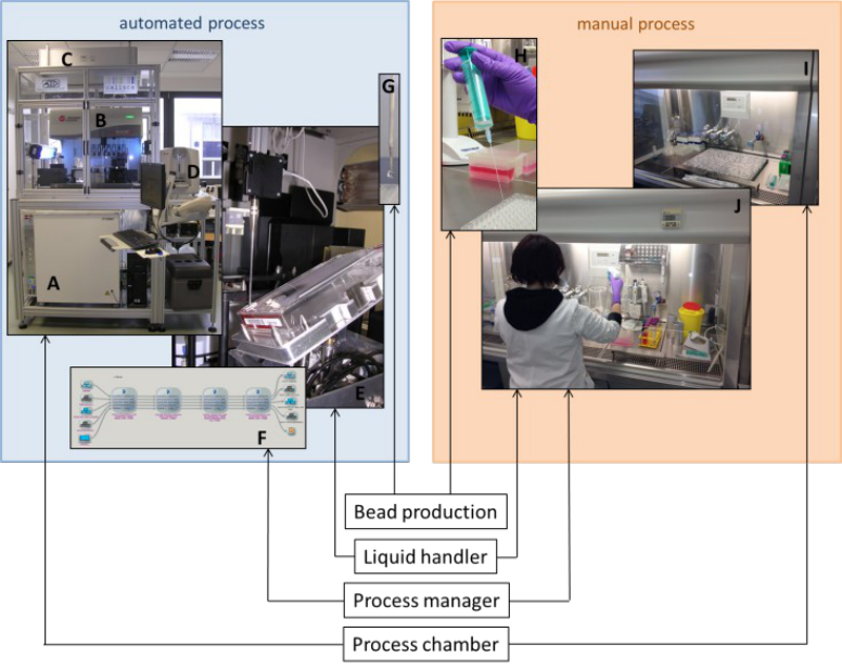
Automated cell cultivation The automated cell culturing was carried out by the Biomek® Cell Workstation (Figure 1,2). Greiner CELLSTAR® AutoFlasks™ were transferred by a liquid handling robot to different positions of the workstation. HeLa cells were washed three times in PBS. The 3D Tilting-ALP enables the homogenous distribution of PBS. Cells were detached by incubation with Trypsin/EDTA for 4 minutes using an on deck incubator. After neutralization of Trypsin/EDTA with DMEM the cell suspension was transferred into a modular reservoir.
Manual Production of Alginate Beads For HeLa cell detachment, the conditioned media was removed and cells were washed three times in PBS. Afterwards, the detachment of cells was realized by adding Trypsin/EDTA and an incubation time of 4 minutes at 37 °C and 5 % CO2. After stopping the detachment process with DMEM, the cell suspension was collected in a 50 mL centrifuge tube. After cell count measurement and centrifugation, 2 x 10⁶ cells/ml were encapsulated in alginate and the cell/alginate suspension was added dropwise to CaCl2 with a disposable syringe. Alginate beads were washed three times in NaCl and fresh media was added (Figure 2).
Automated Production of Alginate Beads
Edited by T. Ahram, W. Karwowski and T. Marek Also the automated production of alginate beads was performed using the Biomek® Cell Workstation (Figure 1 and 2). Quality of the cell suspension of the automated cell culturing of HeLa cells was evaluated by cell count assessments by the Vi-CELLTM XR system. After cell suspension was transferred into a deep well and centrifuged, cells were embedded in alginate (2 x 10⁶ cells/mL) and dropped into CaCl2 in a 96 well microtiter plate. Alginate beads were washed three times in NaCl. Finally, fresh media was added.
Figure 2. Comparison of key features of the automated (left panel) and manual (right panel) cell culture systems. Major processing components of the Biomek Cell Workstation are labelled: Cytomat (A), Biomek Span-8 liquid handler (B), ViCell cell counter (C), Housing with HEPA filter (D), 3D-Tilt Rack with AutoFlask™ and penetrating steal cannula (E); Sami Software – program (F); automated alginate beads dropping (G); manual alginate beads dropping by a syringe with a needle (H), work bench for manual handling under sterile conditions (I), work bench with human operator (J)
EZ4U - Cell Proliferation- and Cytotoxicity-Assay The proliferation rate was detected by the EZ4U - Cell Proliferation Assay Kit (Biomedica Medizinprodukte GmbH & Co KG, Wien, Austria). EZ4U is a non-radioactive proliferation and toxicity assay based on the reduction of tetrazolium salt to colored formazan in living cells. The reagent (1/10 volume) was added to the fresh media of the 3D cell cultures, followed by an 3-hour incubation at 37 °C and 5 % CO2. The absorbance (OD 450 nm) of the supernatant was detected by the PHERAstar reader (BMG Labtech, Ortenburg, Germany).
Adenylate Kinase - Cytotoxicity-Assay Direct toxicity was evaluated by the Adenylate Kinase-Assay Kit (PromoCell, Heidelberg, Germany). The AK- toxicity assay is based on the detection of ubiquitous adenylate kinase from cells with damaged membrane. The Ergonomics In Design, Usability & Special Populations II
Edited by T. Ahram, W. Karwowski and T. Marek toxicity was distinguished by 1/1 dilution of 100 µl supernatant with the reagent in a white well plate. After 5 minutes of incubation at room temperature the luminescence was detected by the PHERAstar reader (BMG Labtech).
Statistical Analysis Statistical analysis was performed by calculating the mean values, the standard deviations and standard errors. Significances were calculated by the student's t-test. Differences were considered to be significant when p<0.05 (*), p<0.01 (**) and p<0.001 (***).
RESULTS
Task Analysis
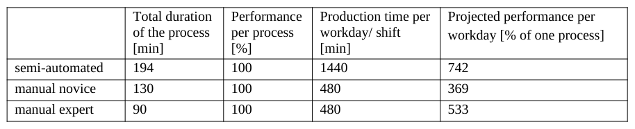
Figure 3 visualizes the processing steps and duration of the semi-automated and manual alginate bead production. If the alginate bead production is carried out manually process duration is approximately 130 minutes (when carried out by novice staff), while the semi-automated process takes about 194 minutes. However, total operator time during the semi-automated processing is 59 minutes, which corresponds to > 50 % time savings. At this, the operator work steps are sample preparation, quality control of the beads and cleaning of the workstation. Even when the manual bead production is carried out by expert staff (approximately 90 minutes process duration) there is still a time saving of 31 operator minutes. As preparation steps at the beginning of the semi-automated production are not time critical, semi-automated production has the potential of 24-hour operation without the need of operators doing shift work. When performance is projected for 24 h operation, efficiency – measured as a workshift-to-performance index – of the semi-automated process is much higher. Provided similar qualities during the different processes, efficiency is 40% higher compared to manual production by expert staff and doubled compared to novice staff (Table 1). This calculation bases on a projection of an 8-hour operator shift (manual process) and a 24-hour workshift for the semi- automated process.
Edited by T. Ahram, W. Karwowski and T. Marek
Figure 3. Timeline of work steps durations during the manual (left) and the semi-automated alginate bead production (right)
Figure available in the original PDF paper.
Table 1. Projected efficiency for manual and semi-automated alginate bead production
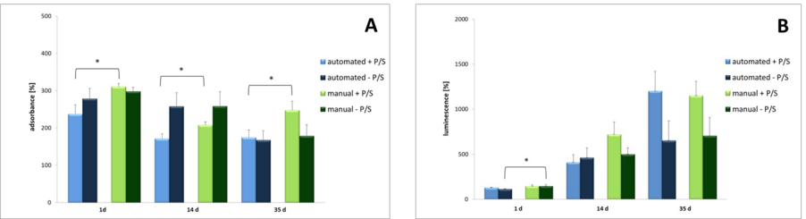
Performance measures Performance of the cell culturing was evaluated by the proliferation rate of the processed samples, detected by the EZ4U - Cell Proliferation Assay Kit (Biomedica Medizinprodukte GmbH & Co KG, Wien, Austria) and their direct toxicity, using an Adenylate Kinase-Assay Kit (PromoCell, Heidelberg, Germany). Both measures mirror the quality and quantity of the cultured cells. Effectiveness in terms of proliferation and toxicity was assessed on day 1, 14 and 35 post manufacturing. The proliferation rates of manual and automated produced alginate beads –P/S decrease from day 1 to day 35. The proliferation rates of both types of production +P/S also decrease from day 1 to day 14. Average proliferation rate increased from day 14 to day 35; however, this increase was not significant. The proliferation rates of manual produced alginate beads cultivated in media with antibiotic were significantly higher compared to the rates of the semi-automated produced beads. Average proliferation rates of the manually and semi-automatically produced alginate beads –P/S were similar on all post production days (Figure 4 A). Further, our results showed a significant increase of the cytotoxicity with and without antibiotic from day 1 to day 35 for both types of production. On day 1, the cytotoxicity of manual produced beads was significantly higher compared to semi-automated produced beads. Particularly, the toxicity of alginate beads +P/S was significantly increased on day 35 compared to beads –P/S (Figure 4 B, no p-values shown).
Figure 4. Manually and automatically produced alginate beads with and without antibiotic on day 1, 14 and 35. A) Proliferation rates and B) toxicity of cells embedded in alginate matrix + standard error. Significances (Comparison of automated and manual produced alginate beads): * p < 0,05; ** p < 0,01; *** p < 0,001, n ≥ 6.
Edited by T. Ahram, W. Karwowski and T. Marek DISCUSSION At present, there has been a trend from 2 dimensional monolayer cell cultures to cell cultivation in a 3 dimensional construct. Background of this shift is the improved mimicking of in vivo conditions and the simulation of human tissue (Pampaloni et al. 2007; Jonitz et al. 2011b). We translated a traditional manual alginate bead production into a semi-automated process. Effectiveness of the two types of alginate bead production was evaluated by proliferation rates and cytotoxicities, efficiency by performance/workshift ratios. The semi-automated alginate bead production consists of manual preparation and postprocessing steps and the automated bead production. The manual bead production is accomplished completely manually. The automated beads production takes longer because of disinfection steps and incubation periods. Furthermore, the span-8 pipetting head requires a longer period for the automated washing steps. If projected for 24 hours the automated solution generates a higher amount of alginate beads (24-hour process) compared to an 8-hour manual shift. In general, automated processes are thought to guarantee stable qualities across complete series. Manual processing can be influenced the skill-level of the operators, their motivation, health status and well being and other factors. This can have varying effects on process qualities and quantities. In this special case, efficiency in terms of performance- to-work shift is almost doubled for the semi-automated production (provided a 24-hour shift as standard for the semi-automated production and an 8-hour shift as standard for the traditional manual production), whereas effectiveness in terms of quality is almost similar for the two methods. There is one exception: Proliferation rate of the alginate beads + P/S, was significantly higher for the traditional manual production (day 1 about 73.8%, day 14 about 36.8% and day 35 about 73%). This is due to the more sensitive handling of the cell suspensions during the manual process, especially during supernatant-removing after centrifugation, which finally results in a higher number of encapsulated cells. Furthermore, automatically produced alginate beads are exposed to higher mechanical stress during liquid handling. The manual and automated produced alginate beads cultivated in media without antibiotic showed a decrease of proliferation rate combined with an increase in cytotoxicity from day 1 to day 35. On the contrary, alginate beads cultivated in media with antibiotics showed an increased proliferation rate on day 35 combined with an increased cytotoxicity on all measurement days. This might be a result of a higher cell aggregate formation and clogging of the pore space with metabolic end products (Malda et al. 2007; Jonitz et al. 2011a). Furthermore, the effect of penicillin/streptomycin might have been diminished. This finding is in contrast to Duewelkhenke et al., who found penicillin and streptomycin having no effect on toxicity and metabolic activity at HeLa cells (Duewelhenke et al. 2006). Our results support the notion of Cohen et al. who found a decreased proliferation rate of cells cultivated in media with P/S (Cohen et al. 2006). To summarize, human skills seem to contribute significantly to an increased performance during 3-D cell production. It can be further concluded from this prototype-study, that a semi-automated 3-D cell culturing process is able to provided acceptable qualities, savings of operator times as well as larger absolute quantities when projected for 24-hour operation.
Acknowledgment: We thank the Federal Ministry of Education and Research (BMBF Germany) for the financial support (FKZ: 03Z1KN11). Furthermore we thank Mrs. Grit Koch (University of Rostock) and Mrs. Carolin Gallert (Center for Life Science Automation) for bead productions and screenings.
REFERENCES
Blow, N. (2008), "Lab automation: tales along the road to automation", in Nat Meth 5 (1), pp. 109–112 Carere, A. Stammati, A. Zucco, F. (2002)," In vitro toxicology methods: impact on regulation from technical and scientific advancements", in Toxicology Letters 127 (1-3), pp. 153–160
Edited by T. Ahram, W. Karwowski and T. Marek Cassidy, M. B. Lee, H. Trevors, J. T. (1996), "Environmental applications of immobilized microbial cells: A review", in Journal
of Industrial Microbiology 16 (2), pp. 79–101 Chapman, T. (2003), "Lab automation and robotics: Automation on the move", in Nature 421 (6923), pp. 661, 663, 665-6
Cohen, S. Samadikuchaksaraei, A. Polak, J.M. Bishop, A.E. (2006), "Antibiotics Reduce the Growth Rate and Differentiation of Embryonic Stem Cell Cultures", in Tissue Engineering 12 (7), pp. 2025–2030 Duewelhenke, N. Krut, O. Eysel, P. (2006), "Influence on Mitochondria and Cytotoxicity of Different Antibiotics Administered in
High Concentrations on Primary Human Osteoblasts and Cell Lines", in Antimicrobial Agents and Chemotherapy 51 (1), pp. 54–63 Ghidoni, I. Chlapanidas, T. Bucco, M. Crovato, F. Marazzi, M. Vigo, D. et al. (2008), "Alginate cell encapsulation: new advances in reproduction and cartilage regenerative medicine", in Cytotechnology 58 (1), pp. 49–56
Jonitz, A. Lochner, K. Lindner, T. Hansmann, D. Marrot, A.Bader, R.(2011a), "Oxygen consumption, acidification and migration capacity of human primary osteoblasts within a three-dimensional tantalum scaffold", in Journal of Materials Science: Materials in Medicine 22 (9), pp. 2089–2095
Jonitz, A. Lochner, K. Peters, K.Salamon, A. Pasold, J. Mueller-Hilke, B. et al. (2011b), "Differentiation Capacity of Human Chondrocytes Embedded in Alginate Matrix", in Connective Tissue Research 52 (6), pp. 503–511 Kumachev, A. Greener, J. Tumarkin, E. Eiser, E. Zandstra, P.W. Kumacheva, E. (2011), "High-throughput generation of
hydrogel microbeads with varying elasticity for cell encapsulation", in Biomaterials 32 (6), pp. 1477–1483 Lehmann, R. Vetter, L. Person, L. Thurow, K. (2013), "Evaluation of Antibiotic-Free Cell Cultures in Manual and Automated Cell Handling", in American Laboratory, available online at http://www.americanlaboratory.com/914-Application- Notes/148434-Evaluation-of-Antibiotic-Free-Cell-Cultures-in-Manual-and-Automated-Cell-Handling/
Malda, J. Klein, T.J. Upton, Z. (2007); "The Roles of Hypoxia in the In Vitro Engineering of Tissues", in Tissue Engineering 13 (9), pp. 2153–2162 Markin, R. S. Whalen, S. A. (2000), "Laboratory automation: trajectory, technology, and tactics", in Clinical chemistry 46 (5),
pp. 764–771 Pampaloni, F. Reynaud, E.G. Stelzer, E.H.K. (2007), "The third dimension bridges the gap between cell culture and live tissue", in Nature Reviews Molecular Cell Biology 8 (10), pp. 839–845
Tebenkova, I. (2011), "Vergleich zweier trägerfreier Kultursysteme bei der chondrogenen Differenzierung humaner mesenchymaler Stammzellen", in Ludwig-Maximilians-Universität München, available online at http://edoc.ub.uni- muenchen.de/13240/1/Tebenkova_Irina.pdf
LISTED IN THE AHFE DIGITAL BOOKSTORE CATALOGUE
Explore the complete digital volume
Advances in Ergonomics In Design, Usability & Special Populations: Part II
Read this individual research paper free through the original publisher, or explore the full digital proceedings book.
ISBN 978-1-4951-2107-4
Explore this ISBN in the AHFE Bookstore ↗Listed in the July 2026 bookstore catalogue; search by ISBN to confirm current availability · Digital book purchase optional