Marcos Gadoy, Filipa Carvalho (2022). Development of a new foot device for Hallux Valgus correction: preliminary results. In: Ergonomics In Design. AHFE Open Access, vol. 47, pp. 539–548. https://doi.org/10.54941/ahfe1001981
CIAUD, Centro de Investigação em Arquitetura, Urbanismo e Design, Faculdade de Arquitetura, Universidade de Lisboa, Lisboa, 1349-055, Portugal; Laboratório de Ergonomia, Faculdade de Motricidade Humana, Universidade de Lisboa, Cruz Quebrada, 1499-002, Portugal ↗
Hallux Valgus is a progressive subluxation of the first metatarsal-phalangeal joint, represented by a lateral deviation of the hallux in relation to the first metatarsal bone in more than 15°, which it affects. 20% of the population aged between 18 and 65 years and over 35% of patients aged ≥65 years with a higher prevalence in women (30%) than in men (13%). The main objective of this study is to develop a new device (foot piece) to attach to the inner surface of the shoe insole (anterior region of the first ray of the foot) promoting a small elevation and, consequently, the hypothesis of correction in alignment of this region and relief of symptoms caused by alterations resulting from Hallux Valgus. Some inclusion criteria were defined that will be verified through a physical evaluation and brief anamnesis. To verify the functional alterations and possible signs and symptoms of the pathology, the Brazilian version of Foot Function Index (FFI) were applied, in the initial and final phase of the study. To assess the alignment of the hallux, computerized biophogrammetry were performed using specific software to record and compare the evolution after a defined period of using the foot piece. The main objective of this manuscript will be presenting the preliminary results of the study. The results obtained suggest that there may be improvements both in terms of pain symptoms and in the correction of hallux deviation.
Hallux Valgus (HV) is a progressive subluxation of the first metatarsal- phalangeal joint (Piqué-Vidal, Solé and Antich, 2007), represented by a lateral deviation of the hallux in relation to the first metatarsal bone by more than fifteen degrees (> 15°) (McCluney and Tinley, 2006). This three- dimensional first ray deformity is estimated to affect more than 20% of patients aged between 18 and 65 years, and more than 35% of patients aged ≥65 years with a higher prevalence in women (30%) than in men (13%) (Nix, Smith and Vicenzino, 2010).
This orthopedic dysfunction presents in some cases a painful and disa- bling condition that can negatively impact the quality of life and physical function of affected individuals (Menz et al., 2011). HV significantly affects balance and represents an important risk factor for falls in the elderly (Koski et al., 1996; Menz and Lord, 2005). In addition, it can also modify gait in this population, especially when walking on uneven ground (Menz and Lord, 2005). In the literature, there are no reports of devices or static orthoses that
improve HV deformity in a simple way, without mechanical traction to favor alignment. The dynamic orthoses used are difficult to adapt to shoes due to the space they occupy when positioned on the foot, reducing the prospect of adherence to use. The complexity of this dynamic device limits footwear options due to the increase in internal volume or because it causes discomfort during its use (Guimarães et al., 2006).
Considering the above, the present research aims to develop a device to fix the inner surface of the insole of the shoe, named a foot piece, which will be positioned in the anterior region of the first ray of the foot, promoting a small elevation. Foot pieces are reliefs used in insoles, placed at specific points in order to correct the positioning of the foot and body (Przysezny, 2015). The use of this foot device aims to improve the distribution of plantar pressure in the anterior region of the foot and, consequently, the hypothesis of correction in the alignment of the first metatarsophalangeal joint, since individuals with HV present different loads in the metatarsal regions, being greater when compared to others with no orthopedic pathology (Eshraghi, Esat and Mohagheghi, 2018).
Another issue is related to the reduction of the pronation movement during gait, since the foot piece promotes an elevation in the anterior region of the foot responsible for impulsion in the final phase of human gait. Thus, the development of a foot piece, adjusted to the needs of each individual, that promotes the biomechanical efficiency of the functions of the foot promoting an alignment of the hallux and probable relief of the signs and symptoms caused by the HV and easiness use justify the investigation proposal of this study.
METHOD AND MATERIAL Participants, Inclusion Criteria and Procedures
Participants were invited to participate in this study voluntarily, after an explanation of the procedures, inclusion criteria and associated risks. An informed written consent was previously obtained.
To participate in the study the following inclusion criteria should be ensured: a) subjects must have a lateral deviation of the first toe with a meta- tarsophalangeal angle ≥15 degrees; b) subjects did not have other orthopedic disorders or surgical procedures that influenced the lower limb, especially at the foot; c) subjects were not undergoing physical therapy treatment in the lower limbs; d) subjects were not using plantar orthoses or other devices for the treatment of HV or discontinuing use during the study period; e) subjects with age ≥18 years f) subjects reported an absence of pain or immediate discomfort when experimented the foot pieces.
The study was submitted and approved by the Research Ethics Committee (CEP) of UniRV – University of Rio Verde, Rio Verde – GO - Brazil and by the Ethics Council of the Faculdade de Motricidade Humana - Universidade de Lisboa, Lisbon – Portugal.
This study comprised 3 fundamental stages: 1st – Screening stage; 2nd - Evaluation of the hallux misalignment (in degrees) and the dysfunction index
Development of a New Foot Device for Hallux Valgus Correction: Preliminary Results 541 caused by HV; 3rd – reevaluation of the hallux misalignment and the dysfu- nction index (reapplication of the FFI questionnaire), after using of the foot piece.
The Screening Stage was performed to confirm the orthopedic dysfunction of HV through the use of a goniometer positioned in the upper region of the foot, extending from the base of the first metatarsal to the first toe, with the metatarsophalangeal joint as the axis, with the individual in the standing and barefoot position, verifying alignment in both feet, performing the interven- tion only in the segment that presented an angle greater than 15 degrees. All measures were carried out by the principal researcher.
To quantitatively measure the index of dysfunctions caused by HV, each participant fulfilled the Brazilian version of FFI questionnaire – (Foot Functi- onal Index). A complete description of the instrument can be found in articles written by Martinez et al. (2016). This procedure was made before and after the use of the foot piece for further results comparation. With this instrument we analyzed the level of Pain (using a Numerical Pain Rating Scale - NRS), the Index of Disability (in %], the Index of Difficulty (in %) and the Index of Pain (in %).
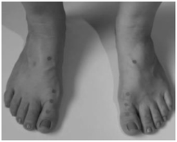
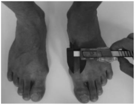
To standardize and to determine the angle formed by the misalignment of the segment of the first toe and the I metatarsal, 4 points were marked on the feet of the participants, in anatomical regions with 3 mm circular stickers in the anterosuperior region of the first ray (head and base of the proximal phalanx of the hallux and the head and base of the first metatarsal) follow- ing the procedures adopted by Yamaguchi et al (2017) (Figure 1). To locate the determined regions, palpation techniques and methods were standardized using a digital caliper with a level in the horizontal plane, to centralize the circular patches proportionally to the anatomical structures of reference in the transverse plane, recording the average values of the three evaluations in each participant (Dissaneewate et al., 2022) (Figure 2).
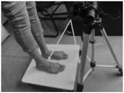
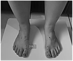
The participant was positioned on a wooden platform with an area of 40X40 cm at 10 cm from the floor, barefoot, in an orthostatic position and with parallel feet, maintaining a usual space. A Webcam was placed on a tripod to record images of the upper region of the foot, maintaining an angle perpendicular to the evaluated structures, at a distance of 45 cm (Figure 3). The recording of the images of the determined region was analyzed using a specific software called Kinovea® (Figure 4).
All measures were performed by the same researcher. To know the margin of error in the evaluation and reevaluation of the angle formed in the misa- lignment of the hallux, the fixation of the adhesives and the analysis of the images in the software were performed twice in a subsample of 5 participa- ting individuals, with an interval of 5 days. The margin of error was fixed in ±0.9 degrees (mean = 0.12; SD = 0.45; range: -0.60; 0.9).
After the initial assessments, the participant received guidance from the researcher on the use of the device such as the placement and adaptation of the foot piece in the shoe and for a few minutes took short walks in the research data collection environment to report any discomfort or incidence of immediate pain and only after this information, the experimental protocol was continued. The use of the foot piece had a duration of 30 days, with
542 de Godoy and Carvalho
Figure 1: Placement and position of marking stickers.Figure 2: Placing the standardized stickers with the digital caliper.Figure 3: Capture of the image of the feet through the camera.Figure 4: Image after analysis of Kinovea® software.
a minimum use of 5 days a week, for a period of at least 7 hours a day, regardless of the footwear used. The use of the foot piece in the period of 30 days takes into account other studies using plantar orthoses to correct the pathology under study, where plantar pressures and comfort related to HV were analyzed (Farzadi et al., 2014; Dissaneewate et al., 2022). The follow-up
Development of a New Foot Device for Hallux Valgus Correction: Preliminary Results 543
during this period was via telephone contact, or messages for questions, or early return of evaluation with the researcher for possible adjustments or suspension of the intervention in the research. After completing 30 days using the foot piece all procedures were repeated to further results comparation and evaluation if improvement in both pain symptoms and correction of hallux deviation were obtained.
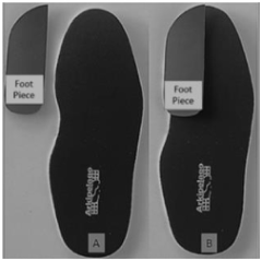
Manufacture of the Foot Piece The foot pieces were made of EVA (ethylene vinyl acetate) specific for the manufacture of postural or proprioceptive insoles with a thickness of 4 mm, being manually cut in a personalized way for each participant, the measure- ments being determined by the longitudinal filling of the entire anterior region of the first ray of the foot (from the base of the first metatarsal extending to the end of the hallux) and the width was determined by measuring 1/3 of the distance between the heads of the I and V metatarsals, verified with a digital caliper. The foot pieces were adapted to the shoes used by the participant, in the insole or support surface of the foot in the shoes in models without insole (Figure 5), fixed with an adhesive tape, and can be used in other shoes used by the participant, removed and placed by themselves.
Figure 5: Ready foot piece next to the insole (A) and the foot piece adapted to the insole (B).
Descriptive analyses were performed using measures of location (freque- ncy, means, medians or percentages) and dispersion (amplitude and standard deviation) to summarize the results of both the sample characterization, the FFI questionnaire and the misalignment of the segment.
To compare the results before and after using the foot piece, instead of only analyzing the angle formed in the misalignment of the hallux before (MisalBefore) and after (MisalAfter) of using the foot piece, difference between both values obtained were computed according to equations (1).
Equation reproduced from the original PDF.
544 de Godoy and Carvalho
The same procedure was used for the other variables, obtained with FFI questionnaire: the Numerical Pain Rating Scale (NRS), Disability index (Dis) Difficulty index (Dif) and Pain index (Pain), where the Difference between both values (Before and After) were obtained by computing the equations (2 to 5), respectively.
Equation reproduced from the original PDF.
Considering the margin of error, the obtained results in variable - DifMsial - can be interpreted as follow: DifMsial < −0.9, means that there was a wor- sening of the hallux misalignment; when the DDifMsialD results are between [−0.9 and 0.9] it means that there are no changes and when DDifMsiD are > 0.9 means that there has been improvement in misalignment of the hallux.
Additionally, for the remaining variables analyzed, positive/negative values mean that there were improvements or worsening for the variable under study, respectively. Values equal to Zero mean that there was no change in the analyzed variables.
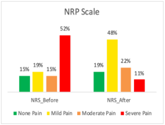
For a better interpretation of the NRS, a four level scale was used as purposed by (McCaffery et al., 1989), where: (1) No-Pain; (2) Mild-Pain; (3) Moderate-Pain and (4) Severe-Pain. The same procedures were performed for the other 3 variables used in FFI, where: (1) No-Disability/Difficulty/Pain; (2) Mild-Disability/Difficulty/Pain; (3) Moderate-Disability/Difficulty/Pain and (4) Severe-Disability/Difficulty/Pain Finally, for continuous variables, the normality was assessed with Shapiro- Wilk test and a Paired Samples T-Test or the Wilcoxon tests were performed to assess the difference in deviation values associated to each variable. The significance level for all tests was set at 0.05.
RESULTS AND DISCUSSIONS At this time, a total of 43 subjects volunteered to participate in the study, but only 27 (63%) were readable to participate. Nine out of these were males and 18 were females; participants aged between 19 and 72 years (mean = 43.59
Table 1. Comparison of mean, SD, minimum and maximum values of the hallux misalignment (in Degrees) between Right (R) and Left (L) foot, Before and After using the foot piece.
Table available in the original PDF paper.
R-Foot (N = 27) L-Foot (N = 22) Before After Before After Mean 24.73 23.69 25.14 24.24 Std. Deviation 5.68 6.04 5.71 5.44 Minimum 15.80 13.90 17.20 17.10 Maximum 35.40 35.10 35.90 33.50
Development of a New Foot Device for Hallux Valgus Correction: Preliminary Results 545
years; SD = 14.89 years) and had an average weight of 61.63 Kg (SD = 12.18 Kg; range: 43–85 Kg), an average height of 155 cm (SD = 11 cm; range: 146–179 cm) and an average BMI of 24.11 kg/m2 (SD = 3.27 kg/m2, range: 19.15–30.96 kg/m2) where, at least 37% of the individuals were overwei- ghted. The use of the foot piece was carried out for an average duration of 8 hours (SD = 1.07 h, range: 6–10 h). Table 1 shows the comparations of the hallux misalignment (in Degrees) between Right and Left foot, before and after using the foot piece. The
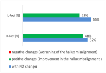
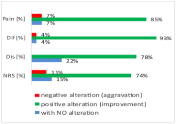
Figure 6: Changes in Hallux misalignment, after using the foot piece.Figure 7: Changes at NRS and in Pain, Difficulty, Disability and Pain Indexes, after using the foot piece.Figure 8: Comparation between NRS, before and after using the foot piece.
546 de Godoy and Carvalho
Wilcoxon test and the Paired Samples T-Test revealed that there were signi- ficant statistical differences between the hallux misalignment after using the foot piece (Z = −4.366; p = .00) and (t(21) = 5.148; p = .00), for R-foot and L-Foot, respectively. Positive changes (which means improvement in the hal- lux misalignment) were present in 48% and 45% of the hallux misalignment for Right and Left foot, respectively (Figure 6). Similar results were obtained
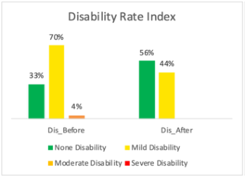
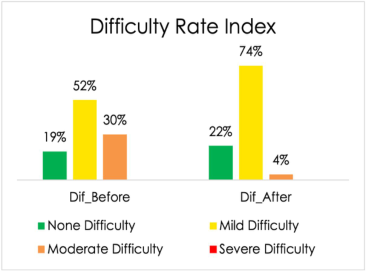
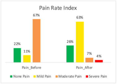
Figure 9: Comparation between Disability Index, before and after using the foot piece.Figure 10: Comparation between Difficulty Index, before and after using the foot piece.Figure 11: Comparation between Pain Index, before and after using the foot piece.
Development of a New Foot Device for Hallux Valgus Correction: Preliminary Results 547
for all the other variables assessed by FFI, where there were statistically dif- ferences in NRS, Disability Index; Difficulty Index, and Pain Index (p < .05), with predominantly positive changes (Figure 7 to Figure 11).
CONCLUSION
Concerning the results obtained, it can be concluded that the use of the foot piece for a 30-days period showed an improvement in the alignment of the hallux, at least, in 45% of the participants, and a higher improvement in the other variables assessed by FFI questionnaire with values ≥75%. The results seem to indicate that the foot piece has the potential to be used as a resource in conservative treatment and rehabilitation, indicating a possible solution for individuals who have disorders caused by orthopedic HV dysfu- nction. As future perspectives, it will be important to increase the size of the sample to have a greater representation of cases with different degrees of misalignment and thus analyze the impact that this may have on the various dimensions addressed throughout this manuscript Another important issue to be analyzed is the dynamic use of the foot piece to verify the biomechanical and functional behavior of the foot during gait.
ACKNOWLEDGMENT
This work is financed by national funds through FCT - Fundação para a Ciência e a Tecnologia, I.P., under the Strategic Project with the references UIDB/04008/2020 and UIDP/04008/2020.
REFERENCES
Dissaneewate, T. et al. (2022) ‘Comparison between the plantar pressure effects of toe separators and insoles in patients with hallux valgus at a one-month follow- up’, Foot and Ankle Surgery. European Foot and Ankle Society, 28(1), pp. 93–99. doi: 10.1016/j.fas.2021.02.005. Eshraghi, S., Esat, I. and Mohagheghi, A. A. (2018) ‘Characterization of gait using plantar force transfer trajectory in individuals with hallux valgus deformity’, Gait and Posture. Elsevier, 62(August 2017), pp. 186–190. doi: 10.1016/j.gaitpost.2018.03.023. Farzadi, M. et al. (2014) ‘Effect of medial arch support foot orthosis on plantar
pressure distribution in females with mild-to-moderate hallux valgus after one month of follow-up’, Prosthetics and Orthotics International, 39(2), pp. 134–139. doi: 10.1177/0309364613518229. Guimarães, C. et al. (2006) ‘Fatores associados à adesão ao uso de palmilhas biomecânicas’, Revista Brasileira de Fisioterapia, 10(3), pp. 271–277. doi: 10.1590/s1413-35552006000300004. Koski, K. et al. (1996) ‘Physiological factors and medications as predictors of inju-
rious falls by elderly people: A prospective population-based study’, Age and Ageing, 25(1), pp. 29–38. doi: 10.1093/ageing/25.1.29. Martinez, B. R. et al. (2016) ‘Validity and reliability of the Foot Function Index (FFI) questionnaire Brazilian-Portuguese version’, SpringerPlus. Springer International Publishing, 5(1). doi: 10.1186/s40064-016-3507-4. McCaffery, M. et al. (1989) Pain: Clinical manual for nursing practice. St. Louis,
MO: Mosby.
548 de Godoy and Carvalho
McCluney, J. G. and Tinley, P. (2006) ‘Radiographic Measurements of Patients With Juvenile Hallux Valgus Compared With Age-Matched Controls: A Coh- ort Investigation’, Journal of Foot and Ankle Surgery, 45(3), pp. 161–167. doi: 10.1053/j.jfas.2006.02.002. Menz, H. B. et al. (2011) ‘Impact of hallux valgus severity on general and foot-specific
health-related quality of life’, Arthritis Care and Research, 63(3), pp. 396–404. doi: 10.1002/acr.20396. Menz, H. B. and Lord, S. R. (2005) ‘Gait instability in older people with hallux valgus’, Foot and Ankle International, 26(6), pp. 483–489. doi: 10.1177/107110070502600610. Nix, S., Smith, M. and Vicenzino, B. (2010) ‘Prevalence of hallux valgus in the general
population: A systematic review and meta-analysis’, Journal of Foot and Ankle Research, 3(1), pp. 1–9. doi: 10.1186/1757-1146-3-21. Piqué-Vidal, C., Solé, M. T. and Antich, J. (2007) ‘Hallux Valgus Inheritance: Pedigree Research in 350 Patients With Bunion Deformity’, Journal of Foot and Ankle Surgery, 46(3), pp. 149–154. doi: 10.1053/j.jfas.2006.10.011. Przysezny, W. L. (2015) Manual de Podoposturologia: Reorganização Neuro
Músculo Articular Através da Es-timulação dos Neurossensores Podais. Brusque: Centro de Pesquisa em Podoposturologia da Podaly do Brasil. Yamaguchi, S. et al. (2017) ‘Non-Radiographic Measurement of Hallux Valgus Angle Using Self-Photography’, Foot and Ankle Surgery, 23, pp. 1–8.
LISTED IN THE AHFE DIGITAL BOOKSTORE CATALOGUE
Explore the complete digital volume
Ergonomics In Design
Read this individual research paper free through the original publisher, or explore the full digital proceedings book.
ISBN 978-1-958651-23-0
Explore this ISBN in the AHFE Bookstore ↗Listed in the July 2026 bookstore catalogue; search by ISBN to confirm current availability · Digital book purchase optional