Yuqian Wang, Yue Wei, Yan Luximon (2024). A pilot fMRI study for understanding the facial perception of 3D human faces. In: Creativity, Innovation and Entrepreneurship. AHFE Open Access, vol. 125, pp. 61–67. https://doi.org/10.54941/ahfe1004721
1 Laboratory for Artificial Intelligence in Design, Hong Kong Science Park, Hong Kong SAR, China; School of Design, The Hong Kong Polytechnic University, Hong Kong SAR, China ↗
2 School of Psychology, Shenzhen University, Shenzhen, China ↗
The human face is one of the most powerful communication tools, especially when people meet for the first time. Facial perception can be influenced by many factors. Although fMRI has been used to study the neural mechanisms when people look at pictures, how people perceive facial stimuli under different movements has not been studied in detail. In this pilot study, adult participants were scanned with a block design of static faces, dynamic faces, and corresponding scrambled face stimuli with a one-back task for attention. The activation of the right fusiform face area (FFA) validates the setting of this facial perception experiment. The dynamic nature of facial movements engages primary visual cortex V1 and other visual areas, resulting in heightened neural activity compared to static faces, as they analyze and extract information from the changing visual inputs. This result provides new insights for further research work in the neural activities related to dynamic face processing.
Human face is one of the most powerful communication tools (Jack & Schyns, 2015; Lieberman, 2013), and it is also valid for online communi- cation, even when using virtual faces (Balas, Tupa, & Pacella, 2018). How people assess a human face is always an essential problem in social cognition and it affects many social outcomes.
In the research area, the majority of facial related studies focus on static facial pictures which only provide static 2D information. In facial perception experiments, the majority of studies typically employ 2D front faces (Balas et al., 2018; Oosterhof & Todorov, 2008). However, some studies utilize both front and angled faces (Zhan et al., 2021; Zhang, Luximon, Shah, & Li, 2023) for their investigations. However, in daily life, people always see dynamic faces when face-to-face interaction.
According to the previous fMRI studies, the face area (FFA) in the inferior temporal lobe of the brain is specialized for faces (Grill-Spector, Knouf, & Kanwisher, 2004; Kanwisher, McDermott, & Chun, 1997) and also synthetic faces with lower activation (Loffler, Yourganov, Wilkinson, & Wilson, 2005). Compared to other brain area, face-sensitivity emerges first in the right FFA (Rossion, Hanseeuw, & Dricot, 2012).
Different regions that work for 3D effect. Additionally, when comparing dynamic landscapes to static ones, researchers discovered heightened activity in the middle temporal gyrus (MT/V5) and hippocampus during aesthetic evaluations of the dynamic landscapes (Zhao et al., 2020).
This pilot fMRI study aims to compare the activated cortical regions when individuals view a dynamic facial video versus static ones. By examining these differences, the study seeks to enhance our understanding of how people eval- uate human faces in everyday life. Additionally, the research aims to uncover the underlying mechanisms involved in human interaction with 3D virtual faces.
METHOD
Participants Four paid young adult participants (with an average age of 28.9 years) joined this pilot study. All of them did not have clinical neurological or psychiatric disorders. Before the scanning, all of them had normal vision or they were asked to wear a pair of MRI compatible glasses to achieve the corrected normal vision.
The pilot study was approved by the Hong Kong Polytechnic University Institutional Review Board, set up under the Research Committee. All par- ticipants were introduced to all considerations and signed the consent form before their participation.
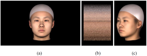
Stimuli The static and dynamic stimuli were generated from method called 3D Com- prehensive Morphable Models 3DCMM (Zhang, Luximon, Zhu, & Li, 2022). This model extract 3D shape principal components from a set of 733 healthy Chinese adults between the ages of 18 and 30, taken from the 3DCMM dataset, were used to develop the Chinese 3D model face for this study. 3D head shape mesh was randomly generated. For all stimuli, the same Asian-average texture were used (Zhang, Luximon, Wan, & Li, 2023) with a gray swimming cap on the head (see Fig. 1a). The scrambled stimuli were used to provide a base line for facial stimuli (see Fig. 1b).
The 3D dynamic stimuli are turning face from left 45
to right 45
video in 2s. The left 45 degree is shown in Fig. 1c.
Figure 1: (a) An illustration of a full-screen front head stimulus; (b) the center part of corresponding scrambled stimulus (with left and right blank cropped); (c) the center part of a left 45-degree stimulus.
A Pilot fMRI Study for Understanding the Facial Perception of 3D Human Faces 63
Experimental Setting A research-dedicated whole-body 3.0T Siemens Prisma scanner system equipped with 32 -channel head coil and a mirror-screen visual stimulation system for the pilot experiment. The screen is 2.44 m far away from par- ticipants’ eye position (consider the reflection of mirror) and the size of the screen is 55 inch (890x500 mm) with 4K resolution and 60 fps resulted in a visual angle of 21.3*11.3 degree.
For the scan setting, the participants will be able to undergo a T1 scan- ning (TR = 1900ms; TE = 2.15s; TI = 904ms; Flip Angle = 9 deg; FoV = 256x256x176mm; voxel size = 1x1x1mm; total time 4:26min) first and then two repeated EPI scans (TR = 2s; TE = 30ms; Flip Angle = 90 deg; voxel size = 2x2x2mm; FoV = 208x208x144mm; 72 slice in total).
An E-prime3 program was developed to synchronize the MRI system and the stimuli display system. A block design of different types of stimuli was adopted. There are 5 blocks in total, three static blocks (left 45-degree view, front view, right 45-degree view), one dynamic block (turning from left 45-degree view to right 45 degree) and a scrambled stimuli block. The inter-stimuli interval is 500ms. Each stimulus was shown for 2000 ms.
To ensure participants’ attention, a one-back task was utilized. During the scan, They were asked to click the button on their left hand when they found this stimulus was the same as the last one; otherwise, they were required to click with their right hand. In order to make sure participants were able to perform the task within the allowed time interval, they were asked to have training of the one-back task with same experimental design but using different stimuli items.
Data Analysis The fMRI data (EPI scans) will be firstly pre-processed using the SPM 12 software on Matlab. The packaged software DPABI (Yan, Wang, Zuo, & Zang, 2016) is utilized for data preprocessing. For pre-processing step1 is slice timing correction based on the slice order. Step2 head motion correction was calculated only when the participants’ maximum head motion is within 1.5mm, the data were not eliminated (Power, Barnes, Snyder, Schlaggar, & Petersen, 2012). There are 3 participants’ data remaining.
Then the neck part of T1 image is cropped and reoriented to synchronize to the fMRI data. T1 images were co-registered into functional images, and then the images were segmented into gray matter, white matter, and cere- brospinal fluid. Regression on interfering covariates was performed, then functional images were normalized to the MNI space DARTEL method (Ash- burner, 2007) and resliced to 2.0 × 2.0 × 2.0 mm3. Normalized images were subjected to spatial smoothing with a full width at half maximum (FWHM) of 4.0 × 4.0 × 4.0 mm3.
Then statistical analysis with the framework of the general liner model will be applied firstly on individual level and then group level to characterize the different brain activation from face stimuli to scrambled faces stimuli and dynamic to static face stimuli. Paired T-tests were used to obtain statistical significance for task-related BOLD signals, corrected for FWE using random field theory (PFWE < 0.05).
RESULTS
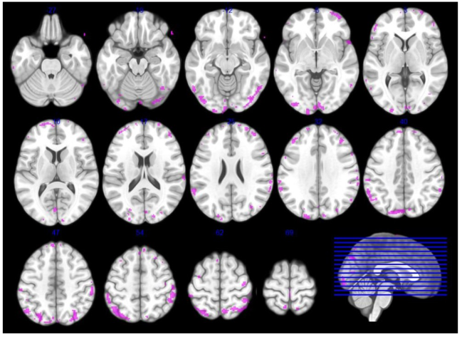
The second level contrast between the face and scrambled face stimuli result is shown in Fig. 2. The result turned out that generally a various cortex area is activated.
Figure 2: The contrast of face stimuli and scrambled face stimuli.
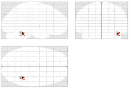
However, when the FWE adjustment is adopted, only the activated area is larger or equal to 30 voxels (Fig. 3) is verified to be significant.
Figure 3: The contrast of dynamic face stimuli and static face stimuli. The red marker pointed at [40, -46, -26] of the standard brain space in SPM 12.
As a result, only a cluster at right FFA is still significant, which leads to the result that all face stimuli activated the right FFA for all participants.
A Pilot fMRI Study for Understanding the Facial Perception of 3D Human Faces 65
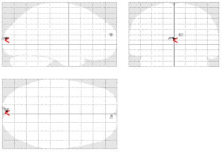
Then the contrast between the statistic face stimuli and dynamic stimuli turned out that the mental processing of dynamic face stimuli involving more cerebral cortex related to primary visual cortex (V1). The significant regions are shown in Fig. 4 with a height threshold T = 10.21 (p< 0.001 unc.) and extent threshold k = 9 voxels.
Figure 4: The contrast of dynamic face stimuli and static face stimuli. The red marker pointed at [-2, -96, 10] of the standard brain space in SPM 12.
DISCUSSIONS The processing of dynamic visual face stimuli is a complex and distributed process involving interactions among multiple brain regions. The V1 and right FFA areas are some of the key regions implicated in this process, but they do not represent an exhaustive list due to the limit of participants sample size.
Fusiform Face Area (FFA) located in the fusiform gyrus of the ventral tem- poral lobe, is particularly associated with face perception. It is involved in processing both static and dynamic facial information, such as facial identity, emotional expressions, facial movements and social face dimensions (Said, Dotsch, & Todorov, 2010). Hence the activation of FFA validated both static and dynamic face stimuli were passively perceived and processed as faces in the brain area, while the scrambled stimuli were not.
The reason the primary visual cortex (V1) is significantly more activated by dynamic face than static face stimuli can be attributed to some factors. Firstly, V1 is involved in the processing of visual motion (Beckers & Zeki, 1995). The motion signals conveyed by dynamic faces engage V1 in analyzing and representing the spatiotemporal aspects of facial movements. Besides, dynamic faces tend to capture more attention compared to static faces (Stoesz & Jakobson, 2014). The presence of face motion draws more attention from and results in increased neural activity in V1.
CONCLUSION
In summary, the processing of dynamic visual face stimuli involves multi- ple brain regions, including the fusiform face area (FFA) and the primary visual cortex (V1). The FFA is associated with face perception and processes
both static and dynamic facial information. V1 is more activated by dynamic faces due to its involvement in visual motion processing and its response to the attention-capturing nature of dynamic faces. However, the list of impli- cated brain regions may not be exhaustive due to the limited sample size of participants.
ACKNOWLEDGMENT
Thanks to University Research Facility in Behavioral and Systems Neuro- science in the Hong Kong Polytechnic University providing the guidance and facilities of the Human MRI scanning.
The work described in this paper was fully supported by a grant from the Research Grants Council of the Hong Kong Special Administrative Region, China (Project No. GRF/PolyU 15609523).
REFERENCES
Ashburner, J. (2007). A fast diffeomorphic image registration algorithm. Neuroimage, 38(1), 95–113. Balas, B., Tupa, L., & Pacella, J. (2018). Measuring social variables in real and
artificial faces. Computers in Human Behavior, 88, 236–243. Beckers, G., & Zeki, S. (1995). The consequences of inactivating areas V1 and V5 on visual motion perception. Brain, 118(1), 49–60. Grill-Spector, K., Knouf, N., & Kanwisher, N. (2004). The fusiform face area
subserves face perception, not generic within-category identification. Nature neuroscience, 7(5), 555–562. Jack, R. E., & Schyns, P. G. (2015). The human face as a dynamic tool for social communication. Current Biology, 25(14), R621–R634. Kanwisher, N., McDermott, J., & Chun, M. M. (1997). The fusiform face area: a
module in human extrastriate cortex specialized for face perception. Journal of Neuroscience, 17(11), 4302–4311. Lieberman, M. D. (2013). Social: Why our brains are wired to connect: OUP Oxford. Loffler, G., Yourganov, G., Wilkinson, F., & Wilson, H. R. (2005). fMRI evidence for the neural representation of faces. Nature neuroscience, 8(10), 1386–1391. Oosterhof, N. N., & Todorov, A. (2008). The functional basis of face evaluation.
Proceedings of the National Academy of Sciences, 105(32), 11087–11092. Power, J. D., Barnes, K. A., Snyder, A. Z., Schlaggar, B. L., & Petersen, S. E. (2012).
Spurious but systematic correlations in functional connectivity MRI networks arise from subject motion. Neuroimage, 59(3), 2142–2154. Rossion, B., Hanseeuw, B., & Dricot, L. (2012). Defining face perception areas in the human brain: A large-scale factorial fMRI face localizer analysis. Brain and cognition, 79(2), 138–157. Said, C. P., Dotsch, R., & Todorov, A. (2010). The amygdala and FFA track both
social and non-social face dimensions. Neuropsychologia, 48(12), 3596–3605. Stoesz, B. M., & Jakobson, L. S. (2014). Developmental changes in attention to faces and bodies in static and dynamic scenes. Frontiers in Psychology, 5, 193. Yan, C.-G., Wang, X.-D., Zuo, X.-N., & Zang, Y.-F. (2016). DPABI: data processing
& analysis for (resting-state) brain imaging. Neuroinformatics, 14, 339–351. Zhan, J., Liu, M., Garrod, O. G., Daube, C., Ince, R. A., Jack, R. E., & Schyns, P. G.
(2021). Modeling individual preferences reveals that face beauty is not universally perceived across cultures. Current Biology, 31(10), 2243–2252. e2246.
A Pilot fMRI Study for Understanding the Facial Perception of 3D Human Faces 67
Zhang, J., Luximon, Y., Shah, P., & Li, P. (2023). 3D Statistical Head Modeling for Face/head-Related Product Design: A State-of-the-Art Review. Computer-Aided Design, 103483. Zhang, J., Luximon, Y., Wan, J., & Li, P. (2023). Capture My Head: A Convenient
and Accessible Approach Combining 3D Shape Reconstruction and Size Mea- surement from 2D Images for Headwear Design. Computer-Aided Design, 159, 103487. Zhang, J., Luximon, Y., Zhu, L., & Li, P. (2022). 3DCMM: 3D Comprehensive Morphable Models for Accurate Head Completion. Paper presented at the Proceed- ings of the 18th ACM SIGGRAPH International Conference on Virtual-Reality Continuum and its Applications in Industry. Zhao, X., Wang, J., Li, J., Luo, G., Li, T., Chatterjee, A., He, X. (2020). The neu-
ral mechanism of aesthetic judgments of dynamic landscapes: An fMRI study. Scientific Reports, 10(1), 1–11.
LISTED IN THE AHFE DIGITAL BOOKSTORE CATALOGUE
Explore the complete digital volume
Creativity, Innovation and Entrepreneurship
Read this individual research paper free through the original publisher, or explore the full digital proceedings book.
ISBN 978-1-964867-01-4
Explore this ISBN in the AHFE Bookstore ↗Listed in the July 2026 bookstore catalogue; search by ISBN to confirm current availability · Digital book purchase optional