Usa Karukunchitab, Manida Swangnetrac, Rungthip Puntumetakulab, Wichai Eungpinichpongab, Alongkot Emasithib (2018). Prevalence of Lower Extremity Malaignment in Rice Farmers. In: Advances in Physical Ergonomics and Human Factors: Part II. AHFE Open Access, vol. 2. https://doi.org/10.54941/ahfe100057
Rice farming activities in Thailand are still based on manual work and human-machine interaction. Most tasks are involved awkward postures, repetitive motion, high force and prolonged work. Long-term exposure to ergonomics risks and lower extremity disorders can produce lower extremity malalignment, which has been reported to increase risk of lower extremity injury and disabilities. The objective of this study was to investigate the prevalence of lower extremity malalignment in rice farmers. A cross-sectional survey was conducted with 292 rice farmers in Khon- Kaen province, Thailand. Participants were required to have experience at least one year in rice cultivation process and have no current injury on back and lower extremity or any previous history that affected the lower extremity alignment. The measurement included pelvic angle, femoral antetorsion, quadriceps (Q) angle, tibiofemoral angle, genu recurvatum, tibial torsion, rearfoot angle and medial longitudinal arch angle. Results revealed the prevalence of lower extremity characteristics to be highest in foot pronation (20.89%), followed by knee valgus (18.49%), respectively. Other malalignment included external tibial torsion (11.64%), anterior pelvic tilt (10.96%), femoral antetorsion (6.85%), knee hyperextension (5.82%), and knee varus (3.43%). The findings indicated lower extremity malalignment to be common among Thai rice farmers, especially foot and knee malalignment. The knowledge gained from this study will be beneficial for healthcare providers in order to advise farmers on lower extremity malalignment prevention and self-management.
Rice farming is one of the most important agricultural industries in Thailand. Rice is a top ten principal export value of Thailand (The customs department, Thailand, 2012). In 2012, the export production quantity was more than 6 billion tons, which was approximately 143 billion bahts (about 4.4 billion dollars) (Thai rice exporters association, 2012). Typical rice cultivation processes include plowing, seeding, planting, nursing and fertilizing and harvesting (Mokkamul, 2006). In Thailand and other Asian countries, these activities are still based on manual work and human-machine interaction. Most tasks are performed with bare feet and involve awkward postures, repetitive motion, high force and prolonged standing and walking in extreme work environment conditions (Pengseesang,
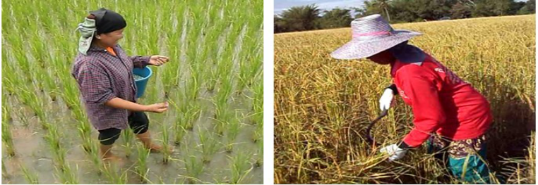
2009; Swangnetr et al., 2012). Specifically, the rice field plowing is performed in slippery tilted walking surface and involves a use of heavy vibrating machinery (see Figure 1a). The seeding (Figure 1b), nursing and fertilization processes (Figure 1d) involve the heavy lifting, carrying and prolonged walking on muddy surface filled with water. The planting task requires high repetitive trunk forward bending and laterally twisting, as well as prolonged standing on muddy surface filled with water (see Figure 1c). The harvesting process involves prolonged stoop, trunk twisting and walking on dry and rough soil (see Figure 1e). These risk exposures can result in repetitive strain injuries and may lead to legs and feet musculoskeletal disorders (MSDs) (Reid et al., 2010; Messing et al., 2008; Walker-Bone and Palmer, 2002; Jaffer et al., 2011; da Costa and Vieira, 2010; Petrass LA and Twomey DM, 2013). Rice farmers were reported to have a high prevalence lower extremity MSDs ranging from 10-41% (Osborne et al., 2012). Long- term exposure to lower extremity disorders and ergonomics risks, including overuse of muscles, abnormal compensatory adaptation, microtraumatic damage and imbalanced weight distribution, can produce lower extremity malalignment (Reid et al.,2010;Jaffer et al.,2011; Daneshmandi et al., 2009; Shultz et al., 2009; Solberg,2008; Oatis, 2009).
(a) (b) (c)
(d) (e)
Figure 1. Typical rice cultivation processes including: a) plowing; b) seeding; c) planting; d) nursing and fertilizing and; e) harvesting
Basic interstructural support of joints includes articular surfaces, muscle and ligaments. These structures control anatomical alignment and normal biomechanical function. Structural injury may cause compensation of segmental alignment (Murphy et al, 2003). Lower extremity malalignment may influence abnormal biomechanical change and neuromuscular dysfunction to control body alignment; and therefore, result in abnormal joint loading, muscle imbalance and proprioceptive deficit (Daneshmandi and Saki, 2009; Nguyen and Shultz, 2009). In additional, alteration in one alignment may cause changes in an adjacent body segment (Nguyen and Shultz, 2009; Daneshmandi et al, 2011). Many studies revealed the relationship between lower extremity malalignment and lower limb injury. Lower extremity malalignment has been reported as risk factors of lower extremity injuries, including hip and knee osteoarthritis (Suri et al, 2012), patellofemoral pain symptom (Power et al,2002), anterior cruciate ligament injury (Daneshmandi and Saki, 2009; Myer et al,2008; Griffin, 2006; Hertel et al, 2004) and medial tibial stress syndrome (Raissi et al, 2009).Moreover, lower extremity malalignment can cause lower limb pain which limits activity living and decreases work performance (Suri et al, 2012; Power et al,2002; Daneshmandi and Saki, 2009; Myer et al,2008; Griffin, 2006; Hertel et al, 2004; Raissi et al, 2009). Lower extremity pain report in Thai rice farmers included 41% hip pain, 35.4% knee pain, and 10.3% ankle and foot pain (Puntumetakul et al., 2011). However, there have not yet been reported the prevalence of lower extremity in rice farmers in Thailand. The objective of this study was to examine the prevalence of lower extremity malalignment among rice farmers. The study also identifies the problematic body parts and risk of disability that might be resulted from rice cultivation activities. High prevalence malalignment body parts should be emphasized in order to further develop guideline to prevent such malalignment incidence.
METHODS
Study Design and Participants A cross-sectional survey was conducted with rice farmers in Khon Kaen province, the northeastern region of Thailand. The participants (age between 20-59 years) were selected using cluster sampling technique in order to represent the total population with economical sample. Two hundred and ninety two (292) rice farmers completed the questionnaire and the clinical measures of lower extremity alignment. Participants were required to have at least one year of experience in rice cultivation process. Participants were excluded if they had current injury to lower limbs or any previous history that would relate to lower limbs alignment, such as fracture and/or surgery, gouty arthritis, rheumatoid arthritis, or ankylosing spondylitis. Participants were asked to read and sign a consent form that had been approved by the human ethics committee of Khon Kaen University before participation. Subsequently, demographic data, including gender, age, height, weight, average working hours per day, and years of work experience, were recorded and lower extremity anatomical alignment characteristics were measured for each participant.
Measurement of Lower Extremity Alignment Lower extremity alignment characteristics examined in this study included pelvic angle, femoral antetorsion, quadriceps (Q) angle, tibiofemoral angle, genu recuvatum, tibial torsion, rearfoot angle, and medial longitudinal arch angle. All measurements were repeated 3 times by a single examiner who had excellent test-retest reliability on all lower extremity measures (ICC range of 0.89-0.98). Participants were asked to be clothed in shorts that exposed lower limb parts needed for testing. For lower extremity measured in a standardized standing position, participants were asked to stand barefoot with the feet positioned biacromial width apart and the toes taking forward. The details of lower extremity alignment measurement were as follows:
- Pelvic angle was measured in standardized standing position. Participants were measured the angle between a line from the anterior superior iliac spines (ASIS) and posterior superior iliac spines (PSIS) and horizontal plane (see Figure 2a), using a PALM inclinometer (Performance Attainment Associates, St Paul, MN) (Leard et al., 2009). For normal alignment, pelvic angle ranges between 70 and 150 (Magee, 2002). Anterior pelvic tilt (i.e., positive angle) was presented when the ASIS position was lower than PSIS. A posterior pelvic tilt (i.e., negative angle) was presented when the ASIS position was higher than the PSIS (Shultz et al., 2006; Herrington, 2011).
- Femoral antetorsion angle was measured using the Craig’s test (Nguyen and Shultz, 2009) in prone position with knee flex at 900 (see Figure 2b). Participants’ femur was moved in passive internal rotation motion in order to bring the greater trochanter to the maximum lateral position. A standard goniometer was used to measure the angle formed by a true vertical line and the shaft of the tibia. Normal femoral antetorsion value ranges from 80 to 300. Excessive femoral antetorsion was classified as the femoral antetorsion greater than 150 (Magee, 2006; Feller et al., 2007; Solberg, 2008; Oatis, 2009).
- Q angle was measured in standardized standing position. It represented the angle between a line from the ASIS to the center of patella and a line from the center of patella to the tibial tuberosity (see Figure 2c), measured by the standard goniometer (Nguyen and Shultz, 2009). The normal angle for male and female was approximately 100-130 and 150-180, respectively (Green, 2005; Oatis, 2009). The Q angle greater than 180 was found to be associated with genu valgum (knock-knee) (Magee, 2006).
- Tibiofemoral angle was measured in standardized standing position with the standard goniometer. It was identified as the angle (see Figure 2d) formed by a line from the anatomical axis the femur (midpoint between ASIS and greater trochanter) to center of knee and a line from the center of knee to the ankle center (midpoint between medial and lateral malleolar distance) (Nguyen and Shultz, 2009). The normal tibiofemoral angle was between 173-1800. Participant knees were classified as the genu valgum condition when tibiofemoral angle value was less than 1730. Genu varus condition (bow-leg) was identified when more than 1800 of tibiofemoral angle value was observed (Sogabe et al., 2009).
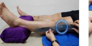
- Genu recuvatum was measured in supine position. A bolster was placed under participants’ distal tibia. Participants were asked to extend knee passively until firm resistance was felt in supine position. The angle was measured between a line connected the femur and the tibia and the saggital plane (see Figure 2e) using a standard goniometer (Nguyen and Shultz, 2009). Genu recuvatum angle value greater than 10 0 was classified as knee hyperextension (Medina, 2009; Devan, 2004).
- Tibial torsion was measured in supine position with knees extended. Participants’ femur was passively rotated to move the femoral epicondyles parallel to the horizontal plane. The angle was represented by a line between the true vertical and a line bisecting medial a lateral malleolar (see Figure 2f) (Nguyen and Shultz, 2009). The normal tibia rotation angle is approximately 200. The angle more than 400 represented tibial torsion condition (Fouilleron et al., 2010; Drexler et al., 2012).
- Rearfoot and medial longitudinal arch angles were measured based on Jonson and Gross’s method (Tsai et al., 2006). Rearfoot angle was represented by the angle between the calcaneus bisecting line and the distal one third leg bisecting line (see Figure 2g). Medial longitudinal arch angle was measured by the angle between a line from medial malleolus to navicular tuberosity and a line from navicular tuberosity to medial aspect of the first metatarsal head (see Figure 2h). Standing foot alignment was classified as pronate, neutral or supinate foot. Rearfoot angle between 30 and 90 and medial longitudinal arch angle between 1340 and 1500 were considered as the neutral foot. The pronated foot was identified if rearfoot angle was greater than 90 and the medial longitudinal arch angle was less than 1340. The supinated foot was identified if rearfoot angle was less than 30 and medial longitudinal arch angle was greater than 1500 (Tsai et al., 2006).
ANALYSIS AND RESULTS Descriptive statistics were used to describe characteristics of participants and lower extremity alignment variables. Continuous variables, including age, height, weight, average working hours per day, and years of work experience, were represented by mean and standard deviation (SD). Categorical variables, including gender and lower extremity malaligment characteristics, were presented in terms of frequency and percentage.
Demographic characteristics of study population were presented in Table 1. There was similar number of male and female participants in this study. Participant age ranged between 20 to 59 years with average age of 45 years. Their rice farming experience ranged from 1 year to as high as 51 years.
Table 1: Characteristics of study population (N=292) Characteristics N (%) Mean ± SD
Daily working hours (hours per day) 7.86 ± 1.35 Experience (years) 25.53 ± 11.50
For the prevalence of lower extremity malalignment, the average of three lower extremity alignment measurements was used for analyses. Table 2 presented the prevalence of lower extremity malalignment observed in this study. Foot pronation and knee valgus were found to be the first and second highest prevalence among rice farmer, respectively. Other lower extremity malalignment included external tibial torsion, anterior pelvic tilt, femoral antetorsion, knee hyperextension, and knee varus. No posterior pelvic tilt and supinate foot conditions were observed in this study.
Table 2: Prevalence of lower extremity malalignment (N=292) Characteristics Malalignment
Table available in the original PDF paper.
N % Anterior pelvic tilt 32 10.96
Excessive femoral antetorsion 20 6.85 Knee valgus 54 18.49
The previous studies have not yet reported the prevalence of lower extremity in agricultural workers involved in rice cultivation process. This is the first study describing the prevalence of lower extremity among rice farmers. The findings indicated that foot and knee malalignment to be common among Thai rice farmers. The highest prevalence of lower extremity malalignment among rice farmers were found in foot pronation (20.89%) and knee valgus (18.49%). Other lower extremity malalignment included external tibial torsion, anterior pelvic tilt, femoral antetorsion, knee hyperextension, and knee varus. The findings of this study were partial in line with the prevalence of lower extremity MSDs among rice farmers in Thailand reported by Puntumetakul et al. (2011), indicating high prevalence was found for both knee pain and malalignment. However, Puntumetakul et al. (2011) study found hip to be the highest prevalence of all lower limb regions (41%); while the current study indicated foot area to have the highest prevalence of malalignment. This might be due to the difference of sample characteristics. Excessive foot pronation may also be resulted from excessive internal femoral rotation, external tibial rotation or anterior pelvic tilt position (Khamis and Yizhar, 2007; Barwick et al, 2012). These malalignment conditions might contribute to hip muscle strain and pain (Chuter and Janse de Jonge X, 2012).
This study found the highest prevalence of lower extremity was foot pronation. Although the association of workrelated factors and excessive foot pronation remains poorly understood, it might be explained that abnormal physical ergonomic factors were associated with ankle and foot MSDs. The abnormal biomechanics of ankle and foot might be due to adverse effects between ground reaction force and abnormal rotational alignment of lower extremity. Such effects usually occurred on the weight-bearing surface during prolonged walking in the stance phase of gait (Letafatkar et al, 2013; Donatelli and Wooden, 2010; Murphy et al., 2003). Fertilization process, which is performed on hard surface, may increase friction force or ground reaction force to farmer feet and lead to musculoskeletal function overload and injury (Murphy et al., 2003). Other rice cultivation tasks, specifically field plowing and planting tasks, were performed with bare feet in slippery tiled walking surface. This environment condition would increase instability of ankle and foot and challenge for controlling body alignment (Donatelli and Wooden, 2010); and therefore, may lead to risk of ankle and foot injury (Messing and Stock, 2006; Yu et al., 2012).
Individual with pronate foot has shorter single stance duration, as compared with person with neutral foot. The shorter stance duration induced poor postural control and increased risk of fall injury (Cobb et al, 2004). Excessive pronated foot has been found to increase lower extremity strain, abnormal compressive force on knee joint and overuse injury resulting in lower extremity pain and MSDs (Chuter and Janse de Jonge, 2012). Excessive foot pronation can also lead to neuromuscular disorders, including trauma, ligament laxity, gastrosoleus tightness, lower limb rotational deformities (Donatelli and Wooden, 2010). Several studies revealed pronated foot to be risk factors of lower extremity injuries, including plantar fasciitis, stress fractures of the foot and tibia, medial tibial stress syndrome and patellofemoral pain symptom (e.g., Chuter and Janse de Jonge X, 2012; Barnes et al, 2008; Yates and White, 2004; Levinger and Gilleard, 2007).
According to knee alignment, distribution loading at knee joint may result from control alignment of hip, knee and ankle (Daneshmandi and Saki, 2009; Nguyen and Shultz, 2009). Knee valgus may contribute to excess abnormal knee loading and lead to lower extremity injury (Tanamas et al., 2009). Most tasks in rice cultivation processes were performed prolonged standing or walking, stooping, bending that might result in increasing risk factors for knee injury (Jone et al, 2007, Allen et al,2010, D’Souza et al,2008; Yu et al, 2012; Baker et al, 2003; McWilliams et al, 2011). These exposures were risk factor of knee joint degenerative change, osteoarthritis, patellofemoral pain syndrome and might indicate abnormal knee alignment including knee valgus (Daneshmandi et al, 2011; Nguyen and Shultz, 2009). Knee valgus has been found to contribute to patella maltracking or stress on patella and increase in compression loading on lateral tibiofemoral and patellofemoral joint. These excessive loads on knee joints may result in osteoarthritis, patellofemoral pain syndrome (McWilliams et al., 2010; Petersen et al., 2013). Moreover, excessive knee valgus may increase Q angle. Greater Q angle had associated with excessive femoral internal rotation, external tibial rotation, or tibiofemoral angle (Daneshmandi et al., 2011; Nguyen and Shultz, 2009).Other lower extremity malalignments found in this study included external tibial torsion, anterior pelvic tilt, femoral antetorsion, knee hyperextension, and knee varus. Long-term exposure to ergonomic risk factors can lead to progressive abnormal biomechanical functions resulting in such malalignments (Daneshmandi et al., 2009; Shultz et al., 2009; Solberg, 2008; Oatis, 2009). Repetitive work with awkward posture, especially repetitive trunk forward bending and laterally twisting, in planting and harvesting tasks can lead to hip muscular discomfort and risk of injury (Jaffar et al., 2011). Plowing, seeding and nursing and fertilization processes required high force exertion in lifting or carrying may overload lower extremity muscles and tendons; and therefore, lead to risk of lower extremity injury (Jaffar et al., 2011; Messing et al, 2008). Prolonged standing in planting and harvesting tasks causes static contraction and mechanical compression on legs and feet tissue. This would lead to muscle fatigue and damage; and therefore, increase lower limb discomfort and MSDs (Messing et al., 2008; Reid et al., 2010). Related to associated lower extremity injuries, anterior pelvic tilt might lead to increase in lumbar hyperlordosis resulting in low back pain and hamstring muscle strain (Panayi, 2010). Excessive femoral antetorsion and external tibial torsion can cause abnormal shear force loading on patellofemoral and tibiofemoral joint; and therefore, lead to patellofemoral pain syndrome and knee osteoarthritis (Kessler et al., 2008; Piva et al., 2009; Krackow et al., 2011; Schwartz and Lakin, 2003; Vincent et al., 2012). Knee hyperextension or genu recurvatum, commonly resulted from knee ligament laxity, can increase compression force loading on knee, anterior tibia translation and anterior cruciate ligament injury (Terauchi et al, 2011).
Although, some relationships between ergonomics risk factors and lower extremity malalignment have been found in literature, specific rice cultivation tasks and activities contributed to malalignment are still unclear. Therefore, systematic ergonomic risk factors evaluation of each task in rice cultivation process should be conducted to identify exact causes of these lower extremity malalignments. Besides work-related factors, prior research suggested that the body alignments to be resulted from other factors, for example the individual characteristics. Gender was found to attribute to anatomical and functional differences (Nguyen and Shultz, 2007; McKeon and Hertel, 2009; Zyroul et al., 2013; Nguyen et al., 2009; Tamari et al., 2006). Degenerative joint associated with aging was found to lead to repetitive injury and develop lower extremity anatomical adaptation (Zyroul et al., 2013; Tamari et al., 2006). Individual with more weight was related to increase of joint loading and could develop lower extremity anatomical alignment (Nicolella et al., 2012; Viester et al., 2013). Psychosocial factors, including high mental stress, low social support, were found to be associated with knee MSDs (Jensen et al., 2005) and increase knee pain and disability (Piva et al., 2009). Future prospective epidemiological studies on effects of individual factors on lower extremity malalignment should be examined to develop occupational health and safety program for specific individual characters in order to reduce and prevent lower extremity malalignment.
CONCLUSIONS
This study revealed high prevalence of lower extremity malalignment among Thai rice farmers. The highest prevalence of lower extremity malalignment was found in abnormal knee and foot alignment. Preventive and corrective actions are needed to prevent lower extremity malalignment as well as potential permanent disability, which will ultimately restrict work performance.
Future research will include identifying work-related risk factors of lower extremity malalignment, focusing on feet and knees. Individual characteristics will also be examined for additional contribution to lower extremity malalignment. The knowledge gained from the study will be beneficial for healthcare providers in order to advise farmers on lower extremity malalignment prevention and self-management. The long-term goal is to develop the preventive and corrective work guideline for rice cultivation and to design ergonomic intervention for reducing and preventing lower extremity malalignment. Such interventions are expected to help maintaining farmer health and safety.
ACKNOWLEDGEMENTS
This study was primary supported by grants from Thailand Research Fund (TRF), Office of the Higher Education Commission (OHEC) and Khon Kaen University (No. MRG5680009). The opinions expressed in this paper are those of the authors and do not necessarily reflect the views of TRF, OHEC or Khon Kaen University. We also thank the Back Neck and Other Joint Pain Research Group for partially support in this study.
REFERENCES
Allen, K.D, Chen, J.C, Callahan, L.F, Golightly, Y.M, Helmick, C.G, Renner, J.B., Jordan, J.M. (2010). Associations of occupational tasks with knee and hip osteoarthritis: the Johnston County Osteoarthritis Project. The Journal of Rheumatology, 37, 842-50. Baker, P., Reading I., Cooper, C., Coggon D. (2003). Knee disorders in the general population and their relation to occupation.
Occup Environ Med, 60,794-797. Barnes, A. Wheat, J. Milner C. (2008). Association between foot type and tibial stress injuries: a systematic review. Br J Sports Med, 42, 93-8. Barwick, A., Smith, J., Chuter, V. (2012). The relationship between foot motion and lumbopelvic-hip function: A review of the
literature. The foot, 12,224-231 Cobb, S.C., Tis,L.L., Johnson, B.F., Higbie, E.J. (2004). The effect of forefoot varus on postural stability. J Orthop Sports Phys Ther, 34,79-85.
Chuter, V.H. and Janse de Jonge, X. (2012). Proximal and distal contributions to lower extremity injury: a review of the literature. Gait and Posture,36,7-15. da Costa, B.R., Vieira, E.R. (2010). Risk factors for work-related musculoskeletal disorders: A systematic review of recent
longitudinal studies. Am J Ind Med, 53, 285–323. Daneshmandi, H., and Saki F. (2009). The study of static lower extremity posture in female athletes with ACL injuries. Harkat Sport Medicine, 1,75-91. Daneshmandi, H., Saki, F., Shahheidari, S., khouri, A. (2011). Lower extremity malalignment and its linear relation with Q angle
in female athletes. Procedia Social and behavioural Sciences, 15, 3349-3354. Devan, M., Pescatello, L., Faghrit, P., Anderson, J. (2004). A Prospective study of overuse knee injuries among females athletes with muscle imbalances and structural abnormalities. Journal of athletic training, 39,263-267. Donatelli, R.A ., and Wooden, M.J.(2010). Orthopaedic Physical therapy (4th ed.). USA: Churill Livingstone. D’Souza, J., Werner, R., Keyserling, W., Gillespies, B., Rabourn, R., Ulin, S., et al. (2008). Analysis of the third national health
and Nutrition examination survey (NHANES III) using expert ratings of job categories. American Journal of Industrial Medicine, 51,37-46. Feller, J.A., Amis, A.A., Andrish, J.T., Arendt, E.A., Erasmus, P.J., Power, C.M. (2007). Surgical biomechanics of the patellofemoral joint. Arthroscopy: The journal of Arthoscopic and related surgery, 23, 542-553 Green,S.T. (2005). Patellfemoral syndrome. Journal of bodywork and movement therapies, 9, 16-26. Griffin, L.Y. (2006). Understanding and prevention noncontact anterior cruciate ligament injuries. Am J Sport Med, 34,1512-
1531. Herrington, L. (2011) Assessment of the degree of pelvic tilt within a normal asymptomatic population. Manual therapy, 16,646-
648. Hertel, J., Dorfman, J., Braham, R. (2004). Lower extremity malalignment and anterior cruciate ligament injury history. J Sci Med Sport, 3,220-225. Jaffer, N., Abdul-Tharim, A.H., Mohd-Kamar, I.F., Lop, N.S. (2011).A Literature Review of Ergonomics Risk Factors in
Construction Industry. Procedia Engineering, 20, 89-97. Jensen, R., Hystad, T., Baerheim, A. (2005). Knee function and pain related to psychological variables in patients with long-term patellofemoral pain syndrome. J Orthop Sports Phys Ther, 35(9), 594-600. Jones, G.T., Harkness, E.F., Nahit, E.S., McBeth, J., Silman, A.J., Macfarlance, G.J. (2007). Predicting the onset of knee pain:
Results from a 2-year prospective study if new workers. Ann Rheum Dis ,66,400-406. Kessler, O., Pati, S., Colwell, C., D’Lima, D. (2008). The effect of femoral component malrotation on patellar biomechanics. Journal of biomechanics, 41, 3332-3339. Khamis, S.and Yizhar, Z. (2007). Effect of feet hyperpronation on pelvic alignment in a standing position. Gait & posture ,25,127-134. Krackow, K., Mandeville, D., Rachala, S., Bayers-Thering, M., Osterning, L. (2011). Torsion deformity and joint loading for
medial knee osteoarthritis. Gait and Posture, 33, 625-629. Leard, J.S., Crane, B.A., Ball, K.A. (2009). Intrarater and interrater reliability of 22 clinical measures associated with lower quarter malalignment. Journal of manipulative and physiological therapeutics, 32,270-276. Letafatkar, A., Zandi, S., Khodayi, M, Vashmesara, J.B. (2013). Flat foot deformity, q angle and knee pain are interrelated in
wrestlers. J Nov Physiother, 3(2), 138. Levinger, P. Gilleard, W.(2007) Tibia and rearfoot motion and ground reaction forces in subjects with patellofemoral pain syndrome during walking. Gait Posture, 25(1), 2-8. Magee, D.J. (2006). Orthodedic Physical Assessment .Philadelphia, PA: Saunder. McKeon, J.M., and Hertel, J. (2009). Sex Differences and Representative Values for 6 Lower Extremity Alignment Measures.
Journal of Athletic Training, 44,249-255. McWilliam, D., Leeb, B., Muthuri, S., Doherty, M., Zhang, W. (2011). Occupational risk factors for osteoarthritis of the knee: a meta-analysis. Osteoarthritis and cartilage, 19, 829-839. McWilliam, D.F., Doherty, S., Maciewicz, R.A., Muir, K.R., Zhand, W., Doherty, M. (2010). Self-reported knee and foot
alignments in early adult life and risk of osteoarthris. Arthis care and research, 62(4), 489-495. Messing, K., Tissot, F., Stock, S. (2006). Lower limb pain, standing, sitting and walking: the importance of freedom to adjust one’s posture. Proceedings of the 16th Congress of the International Ergonomics Association, Maastricht, Netherlands. Amsterdam, the Netherlands: Elsevier. Messing, K., Tissot, F., Stock, S. (2008). Distal lower-extremity pain and work postures in the Quebec population. Am J Public
H, 98, 705-713. Mokkamul, P. (2006) .Ethno botanical Study of Rice Growing Process in Northeastern Thailand.
[http://scholarspace.manoa.hawaii.edu/handle/10125/251]. Murphy, D.F., Connolly, D.A., Beynnon, B.D. (2003). Risk factors for lower extremity injury: a review of the literature. Br J Sports Med, 37,13-29. Myer, G.D., Kevin, R.F, Pareno, M.V., Nick, T.G., Hewett, T.E. (2008). The effect of general joint laxity on risk of anterior
cruciate ligament injury in young female athletes. Am J Sport Med, 36, 1078-1080. Nguyen, A.D., and Shultz, S.J. (2009). Identifying relationships among lower extremity alignment characteristics. Journal of Athletic training, 44,511-518.
Nguyen, A.D., Boling, M.C., Levine, B., Shultz, S.J. (2009). Relationships Between Lower Extremity Alignment and the Quadriceps Angle. Clin J Sport Med, 19,201-206. Nicolella, D.P., O’Connor, M., Enoka, R.M., Boyan, B.D., Hart, D.A., Resnick, E., Berkley, K.J., Sluka, K.A., Kwoh, C.K, Tosi,
L.L., Coutts, R.D., Havill, L.M. and Wendy, M.K. (2012). Mechanical contributors to sex differences in idiopathnic knee osteoarthritis. Biology of sex difference, 3, 28. Oatis, C.A. (2009). Kinesiology: The mechanics and patjomechanics of human movement. (2nd ed). India: Lippincott Williams and Wilkins. Osborne, A., Blake, C., Fullen, B.M., Meredith, D., Phelan, J., McNamara, J., Cunningham, C. (2012). Prevalence of
musculoskeletal disorders among farmers: A systematic review. Am J Ind Med, 55,376–389. Panayai, S. (2010).The need for lumbar-pelvic assessment in the resolution of chronic hamstring strain. Journal of bodywork and movement therapies, 14,294-298. Pengseesang, S. (2009). Ergonomic problems and risk factors of farmers in Sriwichai Subdistrict Wanon Niwat District of Sakon
Nakon Province. Unpublished master’s thesis, Chiang Mai University, Chiang Mai, Thailand. Petrass, L.A., and Two, D.M. (2013). The relationship between ground conditions and injury: What level of evidence do we have?. Journal of Science and medicine in sport, 105-112. Petersen, W., Ellermann, A., Gösele-Koppenburg, A., Best, R., Rembitzki, I.V., Brüggemann, G.P., Liebau, C. (in press, 2013).
Patellofemoral pain syndrome. Knee Sur Sports Traumatol Arthrosc. Piva, S.R., Fitzgerald, G.K., Irrgang, J.J., Fritz, J.M., Wisniewski, S., Mcgenty, G.T., Childs, J.D., Domenech, M.A., Jones, S., Delitto, A. (2009). Assocaited of physical function and pain in patients with patellofemoral pain syndrome. Arch Phys Med Rehabili, 90(2), 282-295.Power, C.M., Chen, P.Y., Reischl, S.F., Perry, J. (2002). Comparison of foot pronation and lower extremity rotation in persons with and without patellofemoral pain. Foot Ankle Int, 23,634-640. Puntumetakul, R., Siritaratiwat, W., Boonprakob, Y., Puntumetakul, M. (2011). Prevalence of Musculoskeletal Disorder in
Farmer: Case study in Sila, Muang Khon Kaen, Khon Kaen Province. J Med Tech Phy Ther, 23, 298-303. Raissi, G.R., Cherati, A.D., Mansoori, K.D., Razi, M.D. (2009). The relationship between lower extremity alignment and Medial Tibial Stress Syndrome among non-professional athletes. Sports Medicine, Arthroscopy, Rehabilitation, Therapy & Technology ,1, 11. Reid, C.R., Bush, P., Karwowski, W., Durrani, S. (2010). Occupational postural activity and lower extremity discomfort: A
review. International Journal of Industrial Ergonomisc, 40,247-256. Schwartz, M. and Lakin, G. (2003). The effect of tibial torsion on the dynamic function of the soleus during gait. Gait and Posture ,17,113-118. Shultz, S.J., Nguyen, A., Windley, T.C., Kulas, A.S., Botic, T.L., Beynnon, B.D. (2006). Intratester and intertester reliability of
clinical measures of lower extremity anatomical characteristics; implications for multi-center studies. Clin J Sport Med ,16,155–161. Shultz, S.J., Nguyen, A., Levine, B.J. (2009) .The relationship between lower extremity alignment characteristics and anterior knee joint laxity. Athletic Training ,54-60. Solberg,G. (200). Postural Disorder and Musculoskeletal Dysfunction: Diagnosis, Prevention and Treatment. (2th ed.). China:
Churchill Livingstone. Suri, P., Morgenroth, D.C., Hunter, D.J. (2014). Epidemiology of osteoarthritis and associated comorbidities. PE R, 4, 10-19. Swangnetr, M., Namkorn, P., Phimphasak, C., Saenlee, K., Kaber, D., Buranruk, O., Puntumetakul, R. (2012). Ergonomic analysis of rice field plowing. The 4th International Conference on Applied Human Factors and Ergonomics. San Francisco, CA. Tanamas, S., Hanna, F., Cicuttini, F., Wluka, A., Berry, P., Urquhart, D. (2009). Does knee malalignment increase the risk of
development and progression of knee osteoarthritis? A systematic review. Arthritis & Rheumatism, 61, 459-467. Terauchi, M., Hatayama, K., Yanagisawa, S., Saito, K., Takagishi, K.. (2011). Sagittal alignment of the knee and its relationship to noncontact anterior cruciate ligament injuries. The American journal of sports medicine, 39,1090-1094.Thai rice exporters association. Retrieved January 28, 2012, from: http://www.thairiceexporters.or.th/Press%20release/2012/Press %20Release%20-%2016%20Jan%202012.pdf The customs department, Thailand. Customs report. Retrieved January 28, 2012, from:
http://www.customs.go.th/wps/wcm/connect/0c69831d-bd84-45d8-a8bd-9da1dafb8beb/CustomsReport_August2013.pdf? MOD=AJPERES&CACHEID=0c69831d-bd84-45d8-a8bd-9da1dafb8beb Tsia, L.C., Yu, B., Mercer, V.S., Gross, M.T. (2006). Comparison of different structural foot types for measures of standing postural control. Journal of Orthopaedic & Sport Physical Therapy, 36,942-953 Vicent, K., Conrad, B.P., Fregly, B., Vincent, H. (2012). The pathophysiology of osteoarthritis: a mechanical perspective on the
knee joint. The American academy of physical medicine and rehabilitation, 4, S3-S9.Viester, L., Verhagen, E.A., Hengel, K.M., Koppes, L., van der Beek, A.J. (2013). The relation between body mass index and musculoskeletal symptoms in the working population. BMC Musculoskeletal Disorders, 14,238. Walker-Bone, K., and Palmer, K.T. (2002). Musculoskeletal Disorders in Farmers and Farm Workers. Occup Med ,52,441–50. Yates, B. White, S. (2004). Incidence and risk factors in the development of medial tibial stress syndrome among naval recruits.
Am J Sports Med, 32(3), 772-80. Yu, S., Lu, M., Gu, G., Zhou, W., He, L., Wang, S. (2012). Musculoskeletal symptoms and associated risk factors in a large sample of Chinese workers in Henan province of China. American journal of industrial medicine, 55,281-293.
LISTED IN THE AHFE DIGITAL BOOKSTORE CATALOGUE
Explore the complete digital volume
Advances in Physical Ergonomics and Human Factors: Part II
Read this individual research paper free through the original publisher, or explore the full digital proceedings book.
ISBN 978-1-4951-2105-0
Explore this ISBN in the AHFE Bookstore ↗Listed in the July 2026 bookstore catalogue; search by ISBN to confirm current availability · Digital book purchase optional