Thibaud De Barnier, Mickael Causse (2022). Can Vigilance be Enhanced by Flashing Visual Stimuli? An EEG Study. In: Human Error, Reliability, Resilience, and Performance. AHFE Open Access, vol. 33, pp. 148–156. https://doi.org/10.54941/ahfe1001579
In aviation, poor vigilance has been cited as an important factor of incidents and accidents. Consequently, many studies are conducted in order to detect the level of vigilance and thus reduce the risk of occurrence of low task engagement or low awareness. Being able to enhance someone’s level of vigilance could be useful in many fields, such as aviation or the navy, in particular when a particularly high level of awareness is required (critical maneuvers, occurrence of warning alarms etc.). With the evolution of the knowledge about brain waves, it seems now possible to influence cortical activity by inducing specific wave patterns, using stimuli such as binaural sounds or visual flashes. In this study, we assessed the possibility of artificially increasing the level of vigilance by stimulating brain activity using flashing visual stimuli. Twelve participants performed a vigilance test (Mackworth clock test) while visual flashing stimuli were displayed around the task on the screen border, and at different flashing frequencies (0 Hz, 4 Hz, 8 Hz, 40 Hz). Brain waves were recorded continuously with a 32-channel electroencephalogram (EEG). Results revealed that reaction times during the vigilance task were shorter with the 40 Hz flashing stimuli. However, subjective mental workload was increased by the presence of each type of visual flash. With the 40 Hz flash, gamma activity (roughly oscillating to the frequency of the 40 Hz flash) in the visual cortex was much higher than with the other flashing frequencies. Interestingly, this increased gamma activity was extended to the frontal regions. In addition, the theta/beta ratio was generally higher on frontal electrodes with lower flashing frequency (4 Hz) than with faster flashing frequencies (8 Hz and 40 Hz). It suggests that the vigilance level was poorer with lower flashing frequency. Indeed, higher theta/beta ratio has been associated with lower vigilance levels and mind wandering episodes. Despite these rather encouraging results, replications of such studies are needed to confirm that visual flashing stimuli can modulate vigilance level and elicit specific brain waves, in particular in regions outside the visual cortex.
Maintaining an appropriate level of vigilance in the flight deck is a key ele- ment of flight safety. Vigilance is a state of concentration that helps people to detect specific stimuli. Thus, low vigilance states can diminish the abi- lity to react effectively, for example after a long period of low workload during the cruise phase. It is thus desirable to be able to ensure that aler- tness remains at sufficient levels, especially to improve responsiveness of the
Can Vigilance be Enhanced by Flashing Visual Stimuli? An EEG Study 149
crew to unexpected events. Since most of the incidents can only be avoided by the human pilots and cannot be predicted, improving vigilance can help reduce the numbers of incidents. This led researchers to seek for a way to increase awareness during critical phases or long phases of inactivity.
It is well known that the brain exhibits different waves frequencies such as: delta (0.5–3 Hz), one of the slowest bandwidths produced by the brain, associated with “deep meditation” or sleep; theta (3–8 Hz), that was asso- ciated with learning processes (Van Someren et al, 2011); alpha (8–12 Hz), dominant during present thoughts and during mental coordination, increa- sed perception, or and acquisition of information; beta (12–38 Hz), related to when a subject is engaged in a task; and gamma (38–42 Hz), related to “extreme brain activity” and “heavy workload”. Also, it has been proposed that synchronized gamma-band oscillations in neurons of the visual cortex constitute the neural correlate of visual awareness (Sewards, 1999). Other authors support the hypothesis that the theta/beta ratio in the prefrontal cor- tex is associated with the level of vigilance. An increased ratio (and thus more theta and less beta activity) would be associated with lower levels of vigila- nce and the occurrence of mind wandering episodes (van Son et al. 2019). On the contrary, decreased theta/beta ratio would be associated with greater executive control over attention to mild and high threat, and thus probably with a more vigilant state (Angelidis et al. 2018). In general, a predominance of faster brain waves (ex. beta) vs slower brain waves (ex. theta) is reflecting a more “vigilant” or “aroused” state (Hima et al, 2020).
Interestingly, the brain waves can be influenced when subjected to binau- ral beats (Garcia Gaés, 2018). Temporal modulation of visual stimuli is a very powerful tool for capturing attention. It has been widely used to attract attention on key information (Pinto et al, 2015). Interestingly, studies led by Herrmann (2001) has shown that stimuli which flicker at gamma frequencies are processed faster by the brain than when they flicker at other freque- ncies. This effect could be due to natural neural oscillators, which preferably oscillate at certain frequencies (so-called resonance frequencies). Moreover, another studies showed the link between conscious awareness of a visual sti- mulus and activity in the gamma frequency range (Summerfield et al, 2002). Other study from Luo et al (2009) also focused on the gamma frequency when studying awareness. One might assume that it could be possible to artificially modulate brain waves, and thus awareness, using flashing visual stimuli. A flash may influence different bandwidths, to switch from one “brain state” to another in order to help the brain rest or be active. Seve- ral applications could be found with this technique: 1) A temporary increase of vigilance in case of an unexpected event that requires concentration and rapid problem solving (failure, necessity to take control back rapidly…), 2) A temporary increase of vigilance during a warning event in order to faci- litate awareness of the alert, 3) A sustained increase/maintenance of vigilance during particular phase of flight (landing) or during long period of inactivity (cruise).
Whereas it is already know that the neural oscillations in the occipital cor- tex can be influenced by flickers (Herrmann et al., 2001), little is known about the possibility to “contaminate” brain frequencies of other regions
150 de Barnier and Causse
with flashing visual stimuli. If possible, influencing prefrontal cortex acti- vity, a region associated with rationality, reasoning and decision-making (Clark et al, 2004), could be interesting to modulate the level of vigilance. The principle would be quite analog to the technique used by the Rhythm company (Dreem headband). Their device records five types of physiological signals via 3 types of sensors embedded in the device: brain cortical activity via 5 EEG dry electrodes, movements, positions, breathing frequency via a 3- D accelerometer located over the head; and heart rate via a red-infrared pulse oximeter (Arnal et al, 2019). Based on these measures, an auditory closed- loop stimulation of sleep, which consists in stereophonic clicks of pink noise with rising and falling slopes (Navarette et al, 2020), is used to reduce time falling asleep and nocturnal awakenings (Debellemaniere et al, 2018), leading to a feeling of being better rested with less hours of sleep.
The current work aimed at examining whether it is possible to modulate brain waves in the visual and prefrontal cortices thanks to a visual flashing stimulus displayed around a focal task. The objective would be to increase the processing efficiency of unexpected stimuli and the level of vigilance. We hypothesized that visual flashing in the gamma band (40 Hz) should shorten reaction times, increased gamma spectral power, and decreased the theta/beta ratio.
METHOD
Participants Twelve participants, 8 males and 4 females, from 22 to 28 years old performed the experiment. Given the potential effects, information about medication and drug use were verified before starting the experiment. The study was performed in agreement with the Declaration of Helsinki. All par- ticipants signed an informed consent and were reminded that they could withdraw from the experiment at any time.
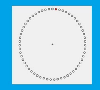
Mackworth Clock Test We selected the Mackworth clock test (Figure 1) because it is long enough to observe vigilance decrement and it requires sustained concentration despite low stimulations. The participant has to focus on a clock in which a red dot moves in short jumps like the second hand of an analog clock. The task requires following the red dot going around the circle at a rate of 1 Hz. At infrequent and irregular intervals, the dot jumps a position, and the partici- pant has to press a button as fast as possible in this situation. The duration of the task was set to 5 minutes. We measured reaction times to indicate the unpredictable double jump and the number of misses. Subjective workload was also evaluated on a 1-100 scale.
Visual Flashing Around the Task A blue square was displayed around the test (Figure 1). This square was still (0 Hz), or flashed (alternating blue and white color) at 4 Hz, 8 Hz, or 40 Hz.
Can Vigilance be Enhanced by Flashing Visual Stimuli? An EEG Study 151
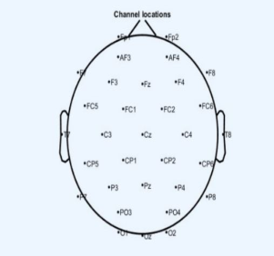
Figure 1: The Mackworth clock test (vigilance task) with the blue square around. This blue square remained still (0 Hz) or was flashed at different frequencies (4 Hz, 8 Hz, or 40 Hz).Figure 2: The 32 channels used in the experiment: the placement was made according to the International 10–20 System.
Mehta and Zhu (2009) showed that blue color is less stress inducing than red. Thus, it was decided to choose this color.
EEG Measurements EEG was amplified and recorded with an ActiveTwo BioSemi system (Bio- Semi, Amsterdam, Netherlands) from 30 Ag/AgCl active electrodes mounted on a cap and placed on the scalp according to the International 10–20 System, see Figure 2. Two additional electrodes placed close to Cz, the Common Mode Sense (CMS) active electrode and the Driven Right Leg (DRL) pas- sive electrode, were used to form a feedback loop that maintains the average potential of the participant as close as possible to the AD-box reference poten- tial. Six external electrodes were also placed, 4 electrodes around the eyes, and 2 on the mastoids, the latter serves as a reference.
Electrode impedance was kept below 5 k for scalp electrodes, and below 10 k for the four eye channels. Skin-electrode contact, obtained using electro-conductive gel, was monitored, keeping voltage offset from the CMS
152 de Barnier and Causse
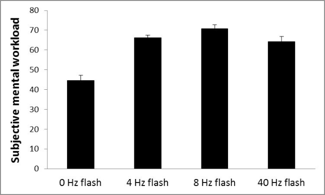
Figure 3: Subjective workload in the Mackworth clock test for each flash frequency.
below 25 mV for each measurement site. All the signals were (DC) amplified and digitized continuously with a sampling rate of 512 Hz with an anti- aliasing filter with 3 dB point at 104 Hz (fifth-order sinc filter). EEG data was then off-line re-referenced to the average activity of the two mastoids and band-pass filtered (0.1–50 Hz, 12 dB/octave). We measured the spectral power in the Gamma (38-42Hz) band at Oz since it is known to reflect awa- reness of visual stimuli (Summerfield 2000). We also explored the Gamma band at Fz to examine possible projection of Gamma waves from the occipi- tal cortex to anterior part of the brain. The Theta/Beta ratio at Fz was also calculated, it is an important marker of vigilance. It was computed by divi- ding the theta by the beta power at the Fz channel (van Son et al. 2019). EEG data from each participant and each condition were averaged to obtain group analysis.
PROCEDURES The experiment took place in a quiet room. The vigilance task was displayed on a 22’ screen. The total experiment duration was approximatively 1 hour, including 20 minutes for the EEG placement and 20 minutes for all 4 task conditions. A 3 minutes break was performed between each task condition.
RESULTS OF THE EXPERIENCE Subjective Workload
We found a main effect of the flash frequency on subjective workload F(3, 33) = 27.42, p < .001, η2 p = .73, with a lower mental workload without flash (i.e., 0 Hz) vs all other flash frequencies (LSD, ps < .001 in all comparisons), see Figure 3. The other comparisons were not significant (LSD, p > .05).
Can Vigilance be Enhanced by Flashing Visual Stimuli? An EEG Study 153
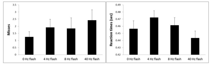
Figure 4: Number of misses (left) and reaction times (right) in the Mackworth clock test for each flash frequency.
Misses We found no significant effect of the flash frequency on the number of misses F(1, 33) = 1.05, p = .379, η2 p = .08. A visual inspection of the Figure 4 shows that the lower number of misses was during the condition without visual flash.
Reaction Times We found a main effect of the flash frequency on reaction times F(3, 33) = 3.46, p = .027, η2 p = .24, with faster reaction times with the 40 Hz flash vs the 4 Hz and the 8 Hz ones (LSD, ps < .05 in all comparisons), see Figure 4. The other comparisons were not significant (LSD, ps > .05).
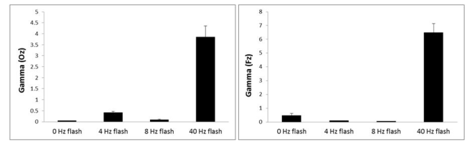
Gamma Bandwidth We found a main effect of the flash frequency on the gamma band on occipital regions (Oz electrode) F(3, 33) = 54.71, p < .001, η2 p = .83, with a much higher spectral power with the 40 Hz flash vs all other flashing frequencies (LSD, ps < .001 in all comparisons), see Figure 5. The other comparisons were not significant (LSD, ps > .05 in all comparisons).
We also found a main effect of the flash frequency on the gamma band on frontal regions (Fz electrode), F(3, 33) = 54.71, p < .001, η2
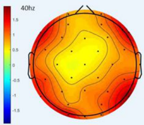
p = .83, with a much higher spectral power with 40 Hz flash vs all other flashing frequencies (LSD, ps < .001 in all comparisons), see Figure 5. The other comparisons were not significant (LSD, ps > .05 in all comparisons). A Scalp map during the 40 Hz condition is displayed Figure 6.
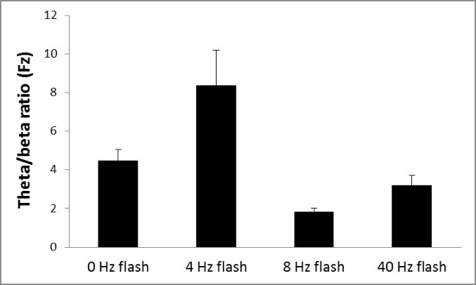
Theta/Beta Ratio We also found a main effect of the flash frequency on the theta/beta ratio on frontal regions (Fz electrode) F(3, 33) = 8.70, p < .001, η2 p = .44, with a higher ratio with 4 Hz vs all other flashing frequencies (LSD, ps < .005 in all comparisons), see Figure 7. The ratio was also marginally higher with 0 Hz vs 8 Hz flashes (LSD, p = .057). The other comparisons were not significant (LSD, ps > .05 in all comparisons).
154 de Barnier and Causse
Figure 5: Spectral EEG power in the gamma band at Oz (left) and Fz (right) electrode in the Mackworth clock test for each flash frequency.Figure 6: Gamma brainwaves power spectrum in the Mackworth clock test for the 40 Hz visual flash (values are pondered by the mean power spectrum).Figure 7: Theta/Beta ratio on each electrode in the Mackworth clock test for each flash frequency.
Can Vigilance be Enhanced by Flashing Visual Stimuli? An EEG Study 155
DISCUSSION
This work examined whether visual stimuli flashing at a rapid pace may help reduce reaction times to a vigilance task and increase specific brain waves, in particular the gamma band. The results showed that the 40 Hz visual flash markedly increased activity in the gamma band in the occipital and frontal regions, reduced the frontal theta/beta ratio, and was associated with shorter reaction times during the vigilance task. On the contrary, slower flash fre- quency (in particular 4 Hz) was associated with an increase in the theta/beta ratio and increased reaction times.
Taken together, our results suggest that a stimulus flashing at a rate analog to the gamma band (i.e., 40 Hz) may have an effect on awareness. Indeed, increased gamma activity has been associated with more efficient awareness of visual stimuli. Interestingly, the increased gamma power was observed in both posterior and anterior part of the brains, suggesting that modula- tion of brain waves may have extended from visual to prefrontal regions. According to Carmel et al. (2006), activity of higher-level cortical areas such as prefrontal region is important for awareness of stimuli such as flickers. The other important result is the decreased theta/beta ratio with the 40 Hz flash. A lower ratio is considered to reflect a higher level of awareness (van Son et al., 2019). Despite these encouraging results, one must notice that subjective mental workload was increased with each flashing frequency in comparison to the condition without flash. This result indicates that even if flash may prove to be useful to promote awareness, high-frequency tempo- ral modulation of visual stimuli should be used during short period of times or preferably during critical events, i.e., for example in contexts that require immediate actions (Teng Cao et al, 2012).
CONCLUSION
We found that a 40 Hz visual flashing displayed around a vigilance task gene- rated an improvement of the reaction times, and influenced brain waves, not only in the visual cortex, but also in higher level regions such as the prefrontal cortex. Gamma band was increased, and theta/beta ratio was decreased, both being markers of better awareness. On the contrary, 4 Hz flashing seemed to be the worst frequency, and was reported as annoying by the participants. If replicated, rapid visual flashes displayed in the field of vision may be a solution to increase vigilance and reactivity of operators during adverse situations.
REFERENCES
Angelidis, A., Hagenaars, M., van Son, D., van der Does, W., Putman, P., 2018. Do not look away! Spontaneous frontal EEG theta/beta ratio as a marker for cognitive control over attention to mild and high threat. Biological Psychology 135, 8–17. Arnal, P. J., Thorey, V., Ballard, M. E., Hernandez, A. B., Guillot, A., Jourde, H., ...
& Sauvet, F., 2019. The Dreem Headband as an Alternative to Polysomnography for EEG Signal Acquisition and Sleep Staging (preprint). Neuroscience. Bowen, H.M., 1964. Vigilance as a Function of Signal Frequency and Flash Rate.
Percept Mot Skills 18, 333–338.
156 de Barnier and Causse
Carmel, D., Lavie, N., & Rees, G. (2006). Conscious awareness of flicker in humans involves frontal and parietal cortex. Current biology, 16(9), 907–911. Clark, L., Cools, R., Robbins, T.W., 2004. The neuropsychology of ventral prefrontal
cortex: Decision-making and reversal learning. Brain and Cognition 55, 41–53. Debellemaniere, E., Chambon, S., Pinaud, C., Thorey, V., Dehaene, D., Léger, D., Chennaoui, M., Arnal, P.J., Galtier, M.N., 2018. Performance of an Ambulatory Dry-EEG Device for Auditory Closed-Loop Stimulation of Sleep Slow Oscillations in the Home Environment. Front. Hum. Neurosci. 12, 88. Garcia Gaes, L., 2018. Binaural beats: Brain wave induction and the use of binaural
beats to induce brain wave patterns. Curr Res Integr Med 03. Herrmann, C.S., 2001. Human EEG responses to 1-100 Hz flicker: resonance phenomena in visual cortex and their potential correlation to cognitive phenomena. Experimental Brain Research 137, 346–353. Hima, C.S., A, A., Nair, C.C., Nair, S.M.J., U, F.B., 2020. A Review on Brainwave
Therapy. World Journal of Pharmaceutical Sciences 59–66. Kim, T., Allen, E.A., Pasley, B.N., Freeman, R.D., 2015. Transcranial Magnetic Stimulation Changes Response Selectivity of Neurons in the Visual Cortex. Brain Stimulation 8, 613–623. Luo, Q., Mitchell, D., Cheng, X., Mondillo, K., Mccaffrey, D., Holroyd, T., Carver,
F., Coppola, R., Blair, J., 2009. Visual Awareness, Emotion, and Gamma Band Synchronization. Cerebral Cortex 19, 1896–1904. Mehta, R., Zhu, R. (Juliet), 2009. Blue or Red? Exploring the Effect of Color on Cognitive Task Performances. Science 323, 1226–1229. Navarrete, M., Schneider, J., Ngo, H.-V.V., Valderrama, M., Casson, A.J., Lewis, P.A.,
2020. Examining the optimal timing for closed-loop auditory stimulation of slow- wave sleep in young and older adults. Sleep 43, zsz315. Pinto, Y., van Gaal, S., de Lange, F.P., Lamme, V.A.F., Seth, A.K., 2015. Expectations accelerate entry of visual stimuli into awareness. Journal of Vision 15, 13. Sewards, T.V., Sewards, M.A., 1999. Alpha-band oscillations in visual cortex: part
of the neural correlate of visual awareness? International Journal of Psychophysi- ology. Summerfield, C., Jack, A.I., Burgess, A.P., 2002. Induced gamma activity is associated with conscious awareness of pattern masked nouns. International Journal of Psychophysiology 44, 93–100. Teng Cao, Feng Wan, Peng Un Mak, Pui-In Mak, Mang I Vai, Yong Hu, 2012. Fla-
shing color on the performance of SSVEP-based brain-computer interfaces, IEEE, San Diego, CA, pp. 1819–1822. Van Someren, E.J.W., Van Der Werf, Y.D., Roelfsema, P.R., Mansvelder, H.D., Lopes da Silva, F.H., 2011. Slow brain oscillations of sleep, resting state, and vigilance, in: Progress in Brain Research. Elsevier, pp. 3–15. van Son, D., De Blasio, F.M., Fogarty, J.S., Angelidis, A., Barry, R.J., Putman, P.,
2019. Frontal EEG theta/beta ratio during mind wandering episodes. Biological Psychology 140, 19–27.
LISTED IN THE AHFE DIGITAL BOOKSTORE CATALOGUE
Explore the complete digital volume
Human Error, Reliability, Resilience, and Performance
Read this individual research paper free through the original publisher, or explore the full digital proceedings book.
ISBN 978-1-958651-09-4
Explore this ISBN in the AHFE Bookstore ↗Listed in the July 2026 bookstore catalogue; search by ISBN to confirm current availability · Digital book purchase optional