Hoi Yan Cheung, Joanne Yip, Kit Lun Yick, Sun Pui Ng (2022). Preliminary wear trial of anisotropic textile brace designed for adolescent idiopathic scoliosis. In: Healthcare and Medical Devices. AHFE Open Access, vol. 51, pp. 117–124. https://doi.org/10.54941/ahfe1002106
1 Institute of Textile and Clothing, The Hong Kong Polytechnic University, Hung Hom, Kowloon, Hong Kong ↗
2 Division of Science, Engineering and Health Studies, Hong Kong Community College, The Hong Kong Polytechnic University, Hung Hom, Kowloon, Hong Kong ↗
Adolescent idiopathic scoliosis (AIS) is a common condition that involves the curvature of the lateral spine and rotation of vertebrae often found in adolescents from age 10 to skeletal maturity. There are various kinds of treatments that prevent the natural progression of the spinal curvature, such as bracing and surgery. However, spinal surgery is mainly reserved for patients with severe scoliosis (spinal curvature that exceeds 45 degrees). For those with moderate scoliosis (spinal curvature larger than 21 but less than 40 degrees), bracing treatment is usually recommended as a non-operative treatment. In this study, the anisotropic textile brace (ATB) is designed to help those with moderate AIS to stop any further progression of their curvature. A case study of a female patient with AIS who has participated a 2-hour wear trial with the ATB is reported. The result of her Cobb angle values shown in the in-brace radiograph is compared without wearing a brace so as to evaluate the immediate effects of this treatment with a soft brace. Besides, the in-brace radiograph is also compared with a supine radiograph to determine the effectiveness of the bracing treatment. A positive result is found in that there is an immediate reduction of the spinal curvature.
Adolescent idiopathic scoliosis (AIS) is a prevalent three-dimensional spinal deformity defined as the lateral curvature of the spine of more than 10°. AIS usually appears during adolescence as either an “S” or a “C”-shape of the spine. There is no known cause for AIS. According to epidemiological stu- dies (Weinstein et al., 2008), around 1-3% of adolescents between 10-16 years old have a certain degree of spinal curvature. Most of these cases do not require surgical intervention (Altaf et al., 2013). To assess the degree of spinal deformity, standing posteroanterior and lateral radiographs of the entire spine are acquired. The magnitude of the spinal curvature can be obtai- ned from the radiographs and determined by the degree of the Cobb angle
(Greiner, 2002).The risk of progression of the curvature can be determined based on 6 risk factors, including age, sex, age at menarche, remaining skele- tal growth, and pattern and magnitude of the curvature. According to Altaf et al. (2013), girls who are diagnosed at a younger age with skeletal immatu- rity are more likely to see a greater progression of the curvature. Also, double curvatures with a larger Cobb angle will also mean greater progression of the curvature. However, the progression will likely stop after menarche.
There are different options to address the progression of the curvature for AIS patients, including exercise, bracing or even surgery (Lenssinck et al., 2005). Bracing treatment is mainly the exertion of external corrective forces onto the spinal curvature. Rigid braces have been widely used but due to the low compliance rate which is related to the discomfort and poor aesthetics, soft braces such as SpineCor have also been developed as a remedy (Coillard et al., 2003). Negrini et al. (2012) recommended that braces should be used for a period of time in order to obtain and maintain a therapeutic outcome that would stop the progression of the scoliotic curvature. According to Nachemson and Peterson (1995), bracing treatment has been successful in preventing a progression of the curvature of 6 degrees or more until the pati- ents are sixteen years old. Table 1 summarises the potential progression of the spinal curvature based on the magnitude of the curve and age (Lonstein, 2006). The table shows that younger AIS patients with a larger spinal curva- ture have a greater chance of progression of the curvature. Therefore, it is important to prevent the progression of the spinal curvature before skeletal age maturity.
Table 1. Potential progression based on magnitude of curve and age (Lonstein, 2006).
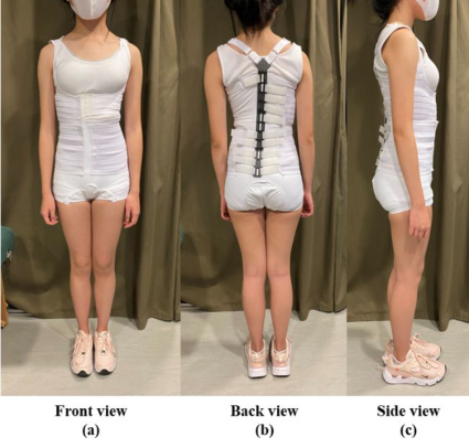
In this study, the anisotropic textile brace (ATB) in Wong (2021) is discus- sed as an alternative choice for AIS patients which would resolve the problem of low wear compliance as is the case with rigid braces (Figure 1). The mechanism for spinal correction is similar to that of the Boston brace - a conventional rigid brace, which utilizes the three-point pressure system by exerting lateral corrective forces onto the spine. The design concept is mainly based on the function intimate apparel designed by Fok (2020). To evaluate the effectiveness of the ATB, Coillard et al. (2003) defined successful bra- cing treatment with a reduction or stabilization of the spinal curvature of 5° or more. Treatment failure is an increase of more than 5° of the spinal curvature. Therefore, the initial rate of reduction with the ATB is 26.7%, versus the 26.1% with the functional intimate apparel and 21.3% with the
Preliminary Wear Trial of Anisotropic Textile Brace Designed 119
SpineCor (Wong et al., 2021). However, there are no supine radiographs con- ducted in studies of the ATB. Therefore, there is the lack of details regarding the inherent flexibility of the spine.
Figure 1: Design of the ATB in (a) Front view, (b) Back view and (c) side view.
According to Luk et al. (1998), spinal flexibility can be estimated by using an index that compares the change in Cobb angle based on standing and supine radiographs. The equation is: Spinal flexibility index = [standing Cobb angle – supine Cobb
angle]/standing Cobb angle x 100%)
Cheung et al. (2018) also suggested that supine radiographs can effectively determine the flexibility of the spine and are reliable enough to determine the ideal achievable amount of correction of the bracing treatment. The effective- ness of a brace is dependent on the inherent flexibility of the spine. A strong correlation can be found between supine and immediate in-brace Cobb angles (r = 0.740). The regression model is therefore: In-brace Cobb angle = 0.809 x supine Cobb angle
Validation of the above model has been performed in Cheung and Cheung (2020). The experimental data are in good agreement with the modelled results for the in-brace Cobb angle values. A flexibility of more than 28% is more likely that the curvature progresses to a lesser degree during bracing intervention. They also suggested that the flexibility of the spine can be used as a reference to determine the success of bracing treatment.
METHODOLOGY
Participant The subject recruited for the wear trial in this study is a 14-year-old female, with a Risser Grade 2. She has a C-curve, which is a single thoracic curve with a Cobb angle of 34.8 degrees. The apex of her spinal curvature is between T9-T10. She has never received any spine-related treatment before, including bracing intervention or physiotherapy. According to Table 1, the likelihood that her curvature will increase is 70%.
Procedures The wear trial mainly focused on evaluating the immediate effect of wearing the ATB after 2 hours of wear. Li et al. (2014) stated that the maximum effect of a spinal orthosis cannot be understood unless donned for at least 2 hours. In order to evaluate the immediate effect of the brace, it is important to measure the Cobb angle from a standing anterior radiograph to obtain the angle of the scoliotic spine according to Negrini et al. (2012). To compare the result between pre- and post-intervention, an in-brace standing radiograph was taken right after the subject had worn the ATB for 2 hours. In this study, a supine radiograph is also required in the interest of investigating the spinal flexibility of the subject so as to predict the in-brace Cobb angle.
To obtain the pre-intervention in-brace standing radiograph and a radi- ograph without donning the brace, the subject was referred to a medical imaging service centre called Hong Kong Advanced Imaging (HKAI). Low- dose x-rays were obtained by using the EOS™imaging system. To obtain the supine radiograph, the subject was referred to another professional centre cal- led ProCare Medical Imaging and Laboratory Centre. Apart from the Cobb angle, the coronal and sagittal balances were also compared and measured based on the radiograph images by using RadiAnt DICOM Viewer (64-bit) software.
RESULTS AND DISCUSSION
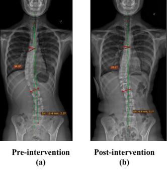
Immediate Effect on Cobb Angle and Coronal Balance: Pre-Intervention vs Post-Intervention Radiographs In view of the degree of correction of the spinal curvature, Figure 2 shows the results of the in-brace spinal curvature changes of the subject after she wore the ATB for at least 2 hours. According to the radiograph, her Cobb angle is reduced from 34.8° to 25.1°. A reduction of 9.7° was measured on the thoracic curve, which is a 27.9% reduction of the spinal curvature. This can be classified as a success in terms of bracing intervention since the spinal curvature is reduced by more than 5 degrees (Collard et al., 2003). Thus, a positive effect is realized with the intervention of the ATB since the reduction of the curvature exceeds the measurement error (Morrissy et al., 1990).
In view of the coronal balance, the value is closer to 0 which represents reduced coronal imbalance. If the value is more than 20 mm, the subject has coronal imbalance. In this case, although no coronal imbalance can be found before intervention with the ATB, the coronal imbalance is reduced from
Preliminary Wear Trial of Anisotropic Textile Brace Designed 121
Figure 2: Radiographic images of subject: (a) pre-intervention and (b) in-brace standing radiographs.
16.4 mm at pre-intervention to 4.9 mm post-intervention after 2-h of donning the ATB, which approximates coronal balance. The result demonstrates that the ATB effectively reduces the spinal curvature when comparing pre- and post-intervention radiographs.
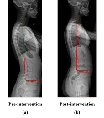
Immediate Effect on Sagittal Balance: Pre-Intervention vs. Post-Intervention Radiographs Apart from the reduction of the Cobb angle and increase in coronal bala- nce, the sagittal balance is also increased in this case study with the ATB donned for 2 hours. According to Yang et al. (2018), measuring the sagit- tal vertical axis (SVA) is one of the ways to determine the sagittal imbalance. They defined SVA as the horizontal distance between the center of C7 and the posterosuperior corner of the sacrum. A positive value indicates that the C7 plumb line is anterior to the posterior corner of the sacrum, and vice versa. An SVA value that is 40 mm or more is classified as sagittal imbalance. Alth- ough there is no sagittal imbalance found before treatment in this case, the SVA is reduced from −15.1 mm to −2.3 mm which demonstrates increased body alignment of this subject after 2-hours of intervention with the ATB (Figure 3).
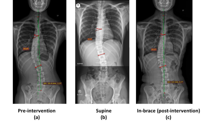
Correlations Among Pre-Intervention, Supine, and Post-Intervention Radiographs The supine Cobb angle has been used as an indicator to predict the degree of spinal curvature resultant of bracing intervention. The angle can also be used to estimate the spinal flexibility which can affect the result of the bracing treatment. Using the formula for calculating the spinal flexibility index in Luk et al. (1998), the spinal flexibility index for this case study is 36.8%. Therefore, the subject is less likely to experience progression of the curvature during bracing intervention with the ATB since the index is more than 28%. The index also shows that this subject has a very flexible
Figure 3: Sagittal balance of subject: (a) pre-intervention and (b) in-brace standing radiographs.
spine, so that the effectiveness of the bracing treatment might be conside- red more successful in terms of an immediate effect. The regression model in Cheung et al. (2018) predicts that the in-brace Cobb angle of this subject would be 17.8°. However, it is actually 25.1° according to Figure 4, which is a 7.3° difference from the predicted value. Therefore, the ATB might not provide the most ideal result in spinal correction according to the supine radiograph.
Figure 4: Comparison of Cobb angle: (a) pre-intervention, (b) supine and (c) in-brace radiographs.
Preliminary Wear Trial of Anisotropic Textile Brace Designed 123
CONCLUSION
In this case study, the immediate effect after a 2-hour intervention with don- ning of the ATB can be considered as positive based on a reduction of 9.7° in her Cobb angle. Besides, both coronal and sagittal imbalances are reduced with the use of the ATB. Both values are comparatively closer to 0 (thereby reaching coronal and sagittal balances) after the subject has donned the brace for 2 hours. However, since the wear trial is conducted in a rather short period of time i.e. 2 hours, the effectiveness of the brace treatment in the ATB can- not be considered as effective yet but further investigation can be conducted for a longer term of wear trial. With the used of the 2-hour preliminary wear trial, it might be useful to evaluate whether a subject is suitable in partici- pating long-term wear trial. By observing the change in the in-braced Cobb angle with the pre-intervention Cobb angle, it can be utilised as a standard to determine whether a subject should commence the long-term wear trial. Also with the use of supine radiograph, it can be used to predict the ideal achievable amount and observe the spinal flexibility of the patients.
Furthermore, by using the supine radiograph to predict the in-brace Cobb angle, the result has indicated that the ATB cannot offer an optimal redu- ction since there is a 7.3° difference. Since the ATB is still under investigation and being modified, its effectiveness might be less comparable to a traditio- nal hard brace. Further improvement on the corrective mechanism has to be made so as to improve its effectiveness of the ATB. The limitation in this study is the sample size since there is only one subject. Future studies can consider a larger sample size with long periods of wear trial to further investigate the in-brace spinal corrective effect of the ATB.
ACKNOWLEDGMENT
This project is supported by funding from the Lee Hysan Foundation entitled “Evaluation and enhancement on the performance of the Anisotropic Textile Brace for moderate Adolescent Idiopathic Scoliosis (AIS)”.
REFERENCES
Altaf, F., Gibson, A., Dannawi, Z. and Noordeen, H. (2013). “Adolescent idiopathic scoliosis.” BMJ : British Medical Journal, 346, f2508. Cheung, J. P. Y., Yiu, K. K. L., Vidyadhara, S., Chan, P. P. Y., Cheung, P. W. H., &
Mak, Cheung, J. P., and Cheung, P. W. (2020). “Supine flexibility predicts curve progression for patients with adolescent idiopathic scoliosis undergoing underarm bracing.” The bone & joint journal, 102(2), 254–260. Coillard, C., Leroux, M. A., Zabjek, K.F. and Rivard, C. (2003). “SpineCor a
non-rigid brace for the treatment of idiopathic scoliosis: post treatment results.” European Spine Journal, 12, 141–148. Fok, L. H. (2020). “Functional intimate apparel for adolescents with scoliosis.” Greiner, A. K. (2002). “Adolescent idiopathic scoliosis, radiographic decision making.” American family physician, 65, 1817. K. C. (2018). “Predictability of supine radiographs for determining in-brace corre-
ction for adolescent idiopathic scoliosis.” Spine, 43(14), 971–976.
Lenssinck, M. L. B., Frijlink, A. C., Berger, M. Y., Bierma-Zeinstra, S. M., Verkerk, K. and Verhagen, A. P. (2005). “Effect of bracing and other conservative interven- tions in the treatment of idiopathic scoliosis in adolescents: a systematic review of clinical trials.” Physical therapy, 85, 1329–1339. Lonstein, J. E. (2006). “Scoliosis: surgical versus nonsurgical treatment.” Clinical
Orthopaedics and Related Research, 443, 248-259. Luk, K. D., Cheung, K. M. Lu, D. and Leong, J. C. (1998). “Assessment of scoliosis correction in relation to flexibility using the fulcrum bending correction index.” Spine, 23, 2303–2307. Morrissy, R. T., Goldsmith, G. S., Hall, E. C., Kehl, D., and Cowie, G. H. (1990).
“Mesaurement of the Cobb angle on radiographs of patients who have.” J Bone Joint Surg Am. 72(3), 320–327. Nachemson, A. L., Peterson, L. E., Bradford, D. S., Burwell, R. G., Duhaime, M., Edgar, M. A., … and Willner, S. V. (1995). “Effectiveness of treatment with a brace in girls who have adolescent idiopathic scoliosis. A prospective, controlled study based on data from the Brace study of the Scoliosis Research Society.” Journal of Bone and Joint Sugery-Series A, 77(6), 815–822. Negrini, S., Aulisa, A. G., Aulisa, L., Circo, A. B., De Mauroy, J. C., Durmala, J.,
Grivas, T. B., Knott, P., Kotwicki, T. and Maruyama, T. (2012). “2011 SOSORT guidelines: orthopaedic and rehabilitation treatment of idiopathic scoliosis during growth.” Scoliosis, 7, 1-35. Prujis, J. E., Hageman M. A., Keessen, W., van der Meer R., van Wieringen J. C. (1994). “Variation in Cobb angle measurements in scoliosis.” Skeletal Radiol.;23:517 20. Weinstein, S. L., Dolan, L. A., Cheng, J. C., Danielsson, A. and Morcuende, J. A.
(2008). “Adolescent idiopathic scoliosisi.” The lancet, 371(9623), 1527–1537. Wong, C. S.-H., Yip, J., Yick, K.-L. and Ng, Z. S.-P. (2021). “A Case Study of Initial In-Brace Spinal Correction of Anisotropic Textile Brace and Boston Brace. International Conference on Applied Human Factors and Ergonomics.” Springer, 109–115. Wong, S. H. (2021). “Design and development of anisotropic textile brace for
adolescent idiopathic scoliosis (AIS).” Yang, X., Hu, B., Song, Y., Liu, L., Zhou, C., Zhou, Z., and Feng, G. (2018). “Coronal and sagittal balance in Lenke 5 AIS patients following posterior fusion: impor- tant role of the lowest instrument vertebrae selection.” BMC musculoskeletal disorders, 19(1), 1–10.
LISTED IN THE AHFE DIGITAL BOOKSTORE CATALOGUE
Explore the complete digital volume
Healthcare and Medical Devices
Read this individual research paper free through the original publisher, or explore the full digital proceedings book.
ISBN 978-1-958651-27-8
Explore this ISBN in the AHFE Bookstore ↗Listed in the July 2026 bookstore catalogue; search by ISBN to confirm current availability · Digital book purchase optional