David Edquilang, Jeff Feng (2022). A Novel Headset System Synchronizing Vision and EEG testing for a Rapid Assessment and Diagnosis of Concussions and Other Brain Injuries. In: Healthcare and Medical Devices. AHFE Open Access, vol. 51, pp. 256–260. https://doi.org/10.54941/ahfe1002125
Millions of concussions happen each year in the US alone. A proportionally large number of these concussions are due to high impact sports injury. Currently, there exists no solution to quickly monitor brain functions and test the oculomotor functions of individuals who have suffered a traumatic brain injury in order to diagnose them as having suffered a concussion. What is presently done to diagnose concussions is a CT scan or MRI, which are lengthy procedures to schedule, set up, and conduct; and furthermore, takes additional time to analyze the results in order to arrive at a diagnosis. This prolongation of the diagnosing process is inherently problematic since the longer time it takes between time of injury and time of diagnosis, there is greater risk of decisions and actions which can worsen damage to the brain. The sooner a concussion can be diagnosed, the sooner and better the treatment can be performed for recovery. In order to ameliorate this issue, we seek to develop a device to perform the function of diagnosis and monitoring of brain activity in a more rapid and timely manner. Literature review into the anatomy of vestibular and ocular brain functions was performed; as well as research into various testing and monitoring methodologies of these vestibular and ocular functions. One such method that has proven to be a reliable method for diagnosis is Vestibular Ocular Motor Screening (VOMS), which is a visual and balance test performed by a doctor with a patient. Further research was also done into existing technologies whose functionalities would allow the device in order to perform brain monitoring, visual testing, and ultimately diagnosis; namely EEG, VR, and infrared eye tracking. Currently, very few devices on the market take advantage of these technologies together for medical uses. A device incorporating these technologies together allows would allow for more consistent administering of visual tests and real-time monitoring of brain activity. With a functional prototype, user testing is to be performed in order to assess the function and viability of the device.
Concussions occur as a result of impacts or rapid acceleration or decelera- tion dealt to the head, with inertia causing the brain to experience physical
A Novel Headset System Synchronizing Vision and EEG Testing 257
trauma within the skull. In the US, millions of concussion cases happen each year; with many of them being caused by contact sports, such as football and soccer. Other common causes are by falls or by vehicle accidents. The physical trauma dealt to the brain by a concussion often causes patients to experience impairment in their vestibular and oculomotor functions (Mucha et al., 2014). It is imperative to be able to diagnose a concussion as soon as possible from the time of injury in order to prevent the risk of improper care or treatment of the patient (Kutcher et al., 2014). In the context of contact sports, one such risk is the decision to allow an athlete to return-to-play after incurring a possible concussion (Kutcher et al., 2014). The danger in that choice lies in the possibility of a subsequent concussion occurring, which leads to second impact syndrome (SIS), causing brain swelling and can often lead to death. Even if the victim of a concussion does not display any obvious symptoms, such as loss of consciousness at the time of injury, it does not rule out a concussion diagnosis. In fact, less than 10% of diagnosed concussion cases exhibited a loss of consciousness (Kutcher et al., 2014). Thus, a means to quickly verify and diagnose on the field that a concussion has occurred, when such uncertainties exist, is highly necessary in order to prevent further injury.
DIAGNOSIS Current neuroimaging methods of diagnosing concussions are performed most commonly via CT scan, or less commonly through MRI in order to detect brain bleeding. These methods are accurate at determining a diagno- sis, however they take a considerable amount of time to schedule, set up, and conduct. Furthermore, analysis of the imagery collected takes further time in order to arrive at a confident diagnosis. The time from injury to diagnosis through these methods can take hours to days.
An alternative method of diagnosing concussions is through EEG monito- ring of the patient’s brain wave frequency bands. If the patient’s brain waves deviate from a baseline reading, then the deviation can serve as an indica- tor that the patient has experienced a concussion or other traumatic brain injury. Irregularities in delta, alpha, beta, and gamma frequency bands are most indicative of a concussion (Munia et al., 2017).
Detecting signs of a concussion can also be performed through videony- stagmography (VNG). VNG directly monitors a patient’s eye movements via infrared eye tracking in order to identify abnormal eye motions known as nystagmus (Kelly et al., 2019). Since vestibular and oculomotor functions are closely linked together in the vestibulo-ocular reflex, assessing oculomotor acuity for irregularities can reveal impairment (Somisetty et al., 2020).
Another effective method for concussion diagnosis is through neurophy- siological screening tests conducted by a doctor with the patient. One such test is Vestibular Ocular Motor Screening (VOMS), which involves a series of visual and balance tests performed by the patient under observation by a doctor. The VOMS assessment involves five domains of visual testing, including smooth pursuit, horizontal and vertical saccades, convergence, horizontal vestibular ocular reflex, and visual motion sensitivity (Mucha
258 Edquilang and Feng
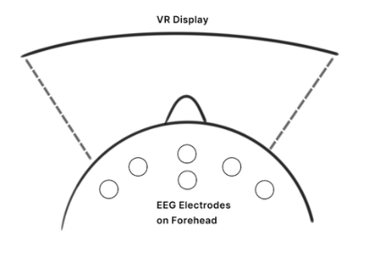
Figure 1: Depicts an orthographic top-down layout of the proposed system. A VR display situated in front of the user’s eyes fills their field of view. Dry EEG skin electrodes placed frontally collect EEG data.
et al., 2014). Since concussions often cause impairment to a patient’s vestibu- lar and oculomotor functions, VOMS testing can identify such impairments through close observation of the patient’s eye movements and determine a diagnosis.
METHOD
Hypothetically, integration of the aforementioned diagnosis methods and emerging technologies together would allow for rapid and accurate concus- sion diagnosis. A headset with a VR display serves as an optimal method of administering the visual tests in VOMS screening, as testing can be condu- cted consistently, devoid of any surrounding visual distractions which may interfere with results. Infrared eye tracking in tandem with VR visual testing would allow for observation and recording of the patient’s eye movements as they perform the tests. Concurrent with VOMS visual testing through VR, EEG monitoring of the patient’s brain waves can be performed in order to record real time brain activity and identify any deviations from the patient’s baseline.
To examine and identify the correlations between the EEG signal chan- ges and VOMS domain testing scores, a study has been conducted through a MUSE 2 device. The participants performed VOMS domain tests while wearing a MUSE 2 device to collect the raw EEG data. Based on a compa- rable analysis conducted between the participant’s EEG baseline recording and VOMS, the findings show relatively consistent characteristic brain acti- vity patterns dependent upon what VOMS domain is being tested (Tristan B., Jeff, F., 2021).
DESIGN DEVELOPMENT Initial concept and modeling of a headset device for rapid concussion diagno- sis is as outlined below. The backbone of the device is a VR headset, which serves to deliver the VOMS five domains of visual tests directly to the user’s field of vision. Dry EEG electrodes integrated into the padded face seal of the
A Novel Headset System Synchronizing Vision and EEG Testing 259
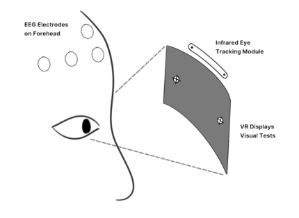
Figure 2: Depicts the functional theory behind the design of the proposed system. With the VR display filling the user’s field of view, it will display the VOMS five domains of visual tests (horizontal saccades depicted) in virtual space. Infrared eye tracking cameras situated above the VR display will monitor the user’s eye movements during the visual tests to identify nystagmus. The EEG electrodes on the forehead collect EEG data of the user’s brain wave frequencies as they perform the visual tests.
Figures 3 and 4: Depict a conceptual design of the proposed headset system. A head mounted VR display with integrated infrared eye tracking is combined with dry EEG skin sensors in the padded face seal to produce an all-in-one system capable of multimodal concussion diagnosis.
headset are situated to make contact with the forehead in order to scan and collect EEG data of the user’s brain wave frequencies. Infrared eye tracking within the headset allows for monitoring and analysis of the user’s eye move- ments as they proceed through the VOMS visual tests. The headset having an adjustable harness would ensure fitment on a wide variety of head sizes and shapes.
DISCUSSION AND FUTURE WORK Further design development of the headset is to be performed, culminating in the production of a functional prototype in order to evaluate the viability of the proposed headset device. An Oculus Quest 2 VR headset is to serve as the basis of the functional prototype, while a Muse EEG headband will be used in conjunction to collect EEG data. The timings of visual tests and collected
260 Edquilang and Feng
EEG data will be synchronized in post-processing for analysis. Data collected in this study will verify the correlation pattern between the EEG signals and VOMS doman testing procedures. The findings of the prototype testing will likely shed light on concussion diagnosis through such a system.
ACKNOWLEDGMENT
We are grateful for the funding provided by the REU Site at the University of Houston Cullen College of Engineering BRAIN Center (National Science Foundation (NSF) REU Site award #1650536 (REU site), NSF PFI award #1827769, and NSF IUCRC BRAIN Award # 1650536) which was contri- buted to pursue the research of this study. Involvement with the REU program granted us valuable experience and knowledge in research and design dealing with neurotechnologies and human factors.
REFERENCES
Kelly, K. M., Kiderman, A., Akhavan, S., Quigley, M. R., Snell, E. D., Happ, E., Synowiec, A. S., Miller, E. R., Bauer, M. A., Oakes, L. P., Eydelman, Y., Gallagher, C. W., Dinehart, T., Schroeder, J. H., & Ashmore, R. C. (2019). Oculomotor, Vestibular, and Reaction Time Effects of Sports-Related Concussion: Video-Oculography in Assessing Sports-Related Concussion. The Journal of head trauma rehabilitation, 34(3), 176–188. https://doi.org/10.1097/HTR.0000000000000437 Kutcher, J. S., & Giza, C. C. (2014). Sports concussion diagnosis and manage-
ment. Continuum (Minneapolis, Minn.), 20(6 Sports Neurology), 1552–1569. https://doi.org/10.1212/01.CON.0000458974.78766.58 Mucha A, Collins MW, Elbin RJ, et al. A Brief Vestibular/Ocular Motor Screening (VOMS) Assessment to Evaluate Concussions: Preliminary Fin- dings. The American Journal of Sports Medicine. 2014;42(10):2479-2486. doi:10.1177/0363546514543775 Munia, T.T.K., Haider, A., Schneider, C. et al. A Novel EEG Based Spectral Analysis of Persistent Brain Function Alteration in Athletes with Concussion History. Sci Rep 7, 17221 (2017). https://doi.org/10.1038/s41598-017-17414-x Somisetty S, M Das J. Neuroanatomy, Vestibulo-ocular Reflex. [Updated 2020 Aug
10]. In: StatPearls [Internet]. Treasure Island (FL): StatPearls Publishing; 2021 Jan-. Available from: https://www.ncbi.nlm.nih.gov/books/NBK545297/ Tristan B., Jeff, F. Establishing a Relationship Between VOMS Performance and Changes in EEG Signatures for a Rapid Assessment and Diagnosis of Concussions. AHFE 2021, LNNS 263, pp. 101–108, 2021. https://doi.org/10.1007/978-3-030- 80744-3_13
LISTED IN THE AHFE DIGITAL BOOKSTORE CATALOGUE
Explore the complete digital volume
Healthcare and Medical Devices
Read this individual research paper free through the original publisher, or explore the full digital proceedings book.
ISBN 978-1-958651-27-8
Explore this ISBN in the AHFE Bookstore ↗Listed in the July 2026 bookstore catalogue; search by ISBN to confirm current availability · Digital book purchase optional