Masami Matsushima, Shunpei Kiuchi, Keiichi Watanuki (2026). Feasibility study of estimating visuospatial cognition and mental states using eye movement and brain activity during domain-specific tasks. In: Affective and Pleasurable Design. AHFE Open Access, vol. 204, pp. 50–61. https://doi.org/10.54941/ahfe1007331
1 Je respire Co., Ltd. 8-8-10-101 Akasaka, Minato-ku, Tokyo 107-0052 Japan; Advanced Institute of Innovative Technology, Saitama University 255 Shimo-okubo Sakura-ku, Saitama-shi, Saitama 338-8570 Japan ↗
2 Graduate School of Science and Engineering, Saitama University 255 Shimo-okubo Sakura-ku, Saitama-shi, Saitama 338-8570 Japan ↗
3 Advanced Institute of Innovative Technology, Saitama University 255 Shimo-okubo Sakura-ku, Saitama-shi, Saitama 338-8570 Japan; Graduate School of Science and Engineering, Saitama University 255 Shimo-okubo Sakura-ku, Saitama-shi, Saitama 338-8570 Japan ↗
Visual cognitive abilities contribute to learning and occupational performance. However, individuals with high cognitive abilities do not necessarily exhibit adaptive psychological functioning. Building on our previous findings (Matsushima et al., 2025), this study examined the differences in arousal–performance relationships and psychosocial adaptation across four visual cognitive domains: visual spatial cognition, visual linguistic processing, visual working memory, and visual attentional control. Physiological indices (pupil diameter, near infrared spectroscopy activity, and autonomic measures) and psychological indicators (stress responses, emotion regulation, and resilience) were assessed. Participants in the high visual spatial cognition group exhibited heightened physiological arousal, including pupil dilation and increased right dorsolateral prefrontal cortex activation, accompanied by elevated stress responses and reduced cognitive reappraisal, indicating a profile characterized by high performance and a substantial psychological load. In contrast, the high visual attentional control group showed an adaptive profile characterized by parasympathetic dominance, effective emotion regulation, and greater resilience, demonstrating that cognitive strength does not uniformly predict psychological adaptation and that arousal patterns differ qualitatively across cognitive domains. Domain-specific assessments integrating physiological arousal and psychosocial adaptation may facilitate the early identification of individuals with high cognitive ability who are susceptible to psychological difficulties and provide a foundation for domain-specific screening and early preventive intervention.
Visual cognitive abilities play critical roles in learning and occupational performance. However, individuals with high cognitive abilities do not necessarily exhibit adaptive psychological functioning; some demonstrate the coexistence of high performance and substantial psychological burdens. Such Received April 14, 2026; Revised May 29, 2026; Accepted June 13, 2026; Available online July 20, 2026
Feasibility Study of Estimating Visuospatial Cognition 51 dissociations are difficult to detect using performance indices alone and may increase the risk of overlooking latent psychological vulnerabilities. Visual cognitive function is not a unitary construct but consists of several subdomains. Moreover, an inverted U-shaped relationship between the arousal level and performance, in which optimal performance is achieved at moderate arousal, has been consistently reported in the literature. In our previous study, we integrated multiple visual cognitive tasks and demonstrated the associations between arousal-related indices and task performance. However, the domain- specific differences in arousal responses and psychosocial characteristics remain poorly understood. Therefore, the purpose of the present study was to compare high- and low-performance groups across four visual cognitive domains: visual spatial cognition, visual linguistic processing, visual working memory, and visual attentional control. A key objective was to elucidate domain-specific differences in arousal-related physiological indices and psychosocial measures. Through this approach, we aimed to establish a multidimensional framework for characterizing domain-specific arousal performance relationships and patterns of psychological adaptation.
METHOD
Participants Ten healthy males in their twenties took part in the study.
Experimental Tasks To examine the construct validity, established comparison tasks were selected for each of the four cognitive domains assessed using DiabiEye (Table 1).
Psychological Scales The psychological scales used in this study were as follows:
The experimental procedure consisted of the following steps:
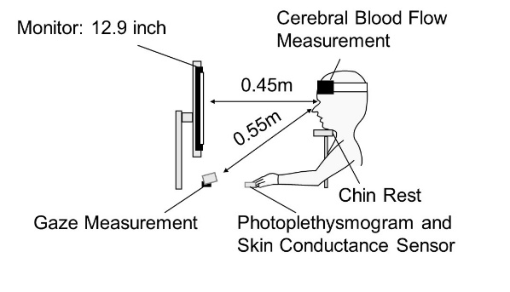
1. Attachment of physiological measurement devices (eye-tracking, cerebral blood flow, finger photoplethysmography, and skin conductance) 2. Completion of the DiabiEye test 3. Completion of the comparison tasks for validating the DiabiEye 4. Completion of the questionnaires for assessing psychological characteristics The duration of the experiment was approximately 50 min. The order of presentation and the environment (lighting, noise, and desk height) were standardized (Fig. 1).
Table 1: Comparison tasks used for validating the DiabiEye.
Table available in the original PDF paper.
Cognitive Domains Comparison Tasks Visual linguistic processing (V-LP) Words Per Minute (WPM) Test Cloze Test
Visual attentional control (V-AC) Visual Search Task Useful Field of View (UFOV) Test
Figure 1: Experimental setup.
Analysis Methods The analysis methods are as follows.
1. For the analysis, the following behavioral indices (performance on comparison and DiabiEye tasks), psychological scale scores, and physiological indices were used: eye tracking (Tobii Pro Fusion), near infrared spectroscopy (NIRS; WOT-220), and pulse wave/ skin conductance (NeXus10 Mark II). Validation was performed by determining correlations (Spearman’s ρ) between DiabiEye scores and the corresponding task performance and psychological scales. 2. Participants were divided into high- and low-performance groups based on their scores in four domains: visual spatial cognition, visual linguistic processing, visual working memory, and visual attentional control, using the mean score as the threshold. 3. Physiological associations were detected in terms of point-biserial correlations between binary accuracy (1 = correct, 0 = incorrect) and physiological indices. 4. A machine learning paradigm was adopted where a binary classification model was built to predict the DiabiEye accuracy from representative physiological indices, interpreted using the SHapley Additive exPlanations (SHAP).
5. Eye movements were detected using Brunner–Munzel tests comparing the total eye movement distances between full and lower score responses. 6. Pupil diameter is affected by multiple factors, including cognitive load, illumination, individual differences, and task order. To isolate the effect of cognitive load, we used a hierarchical Bayesian model in which the mean pupil diameter was predicted by cognitive load and illumination, with participant- and item-level random effects. This model allowed us to estimate cognitive load–related pupillary responses while controlling for luminance, baseline differences, and task order. 7. To evaluate training-related physiological changes, cerebral blood flow indices (NIRS) and finger photoplethysmography–derived indices (pulse wave measures) were compared between pre- and post-training sessions using paired statistical tests.
RESULTS
Pupil Diameter
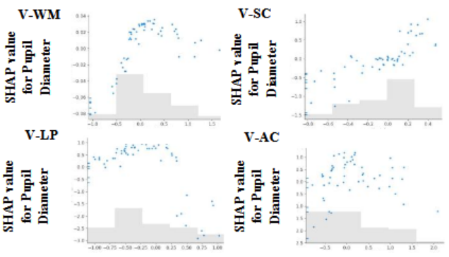
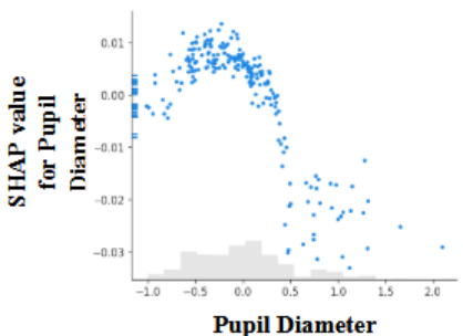
Figure 2 shows the SHAP values for pupil diameter obtained from the machine learning model predicting DiabiEye accuracy. Group comparison revealed that, in cognitive tasks, the high-performance group exhibited significantly larger pupil diameters than the low-performance group. SHAP analysis of the relationship between pupil diameter and performance revealed a nonlinear association between the two, such that the influence on incorrect responses was high when pupil diameters were either extremely small or extremely large (Fig. 3). This suggests that accuracy is highest at moderate pupil diameters.
Figure 2: Standardized pupil diameter and SHAP values score.
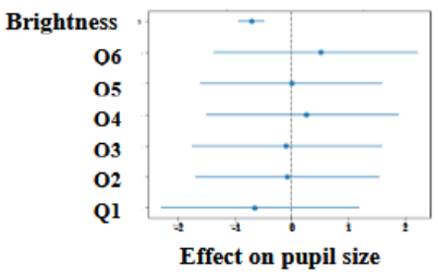
Hierarchical Bayesian Modeling of Pupil Diameter A hierarchical Bayesian regression model was constructed with the cognitive load of the task, presentation order, individual differences, and illumination
brightness as explanatory variables and pupil diameter as the dependent variable. Fixed and random effect regression coefficients were estimated. As shown in Fig. 4, the pupil diameter decreased with increasing brightness, which is consistent with the known physiological responses to luminance. This result validates the proposed model. Furthermore, pupil diameter varied not only with lighting conditions but also across problems, reflecting differences in cognitive load between the tasks.
Figure 3: Pupil diameter and SHAP values score (Matsushima et al., 2025).Figure 4: Hierarchical Bayesian modeling of pupil diameter.
NIRS a. Prefrontal Activation Patterns Related to Visual Spatial Cognition Performance
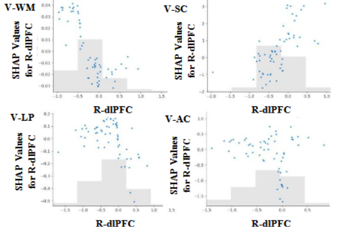
Figure 5 shows the results of a binary classification model predicting the DiabiEye accuracy based on physiological indices and illustrates the relationship between response accuracy and oxygenated hemoglobin (HbO2) activation in the right dorsolateral prefrontal cortex (R-dlPFC) using SHAP. In cognitive tasks, a linear trend was observed where higher HbO2 activation in the R-dlPFC was associated with a higher probability of correct responses. In the visual spatial cognition domain, the high-performance group showed
Feasibility Study of Estimating Visuospatial Cognition 55 a tendency toward higher HbO2 activation in the R-dlPFC (CH3) compared with the low-performance group (p = 0.0509). Conversely, in the left dorsolateral prefrontal cortex (L-dlPFC, CH18), deoxygenated hemoglobin (deoxyHb) activation was significantly higher in the low-performance group (p = 0.0123). No significant intergroup differences were observed in these channels in the other domains.
Figure 5: R-dlPFC and SHAP values score.
b. Right Dorsolateral Prefrontal Cortex Activation and Pupil Diameter Only in the visual spatial cognition domain was a tendency toward a positive association observed between HbO2 activation in the R-dlPFC (CH3) and pupil diameter (p = 0.0509).
Autonomic Nervous System Indices
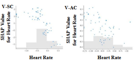
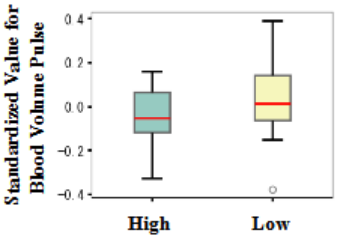
Figure 6 shows the results of a binary classification model predicting the DiabiEye accuracy from physiological indices and illustrates the relationship between response accuracy and heart rate using SHAP. Figure 6 shows that a higher heart rate tended to be associated with incorrect responses. In addition, Fig. 7 shows the differences in the fingertip blood volume pulse (BVP) amplitude between groups with high and low visual spatial cognition task scores.
a. Autonomic Indices Across Domains In the visual spatial cognition domain, the high-performance group showed significantly lower BVP amplitudes than the low-performance group (p = 0.0272). In the visual attentional control domain, the high-performance group exhibited a significantly lower heart rate (p = 0.0210).
Figure 6: Heart rate and SHAP values score.Figure 7: Comparison of blood volume pulse between high and low.
b. Integrated Physiological Profile By integrating and comparing the pupil diameter, R-dlPFC activation (CH3), and BVP, the high visual spatial cognition group exhibited a simultaneous pattern of increased pupil diameter, increased R-dlPFC activation, and decreased BVP amplitude. Simultaneous changes across these three indices were not observed in the other domains.
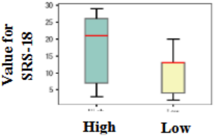
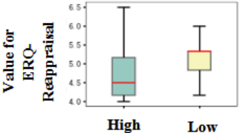
Psychosocial Indices The high visual spatial cognition group showed significantly higher SRS-18 scores and significantly lower ERQ reappraisal scores than the low visual spatial cognition group (p = 0.00362 and p = 0.0393, respectively; Figs. 8 and 9). Conversely, the high-VA control group showed significantly lower SRS-18 scores and significantly higher ERQ reappraisal scores (p = 0.0278 and p = 0.0101, respectively).
Comparison of Physiological Indices Before and After Training After two weeks of training, the participants’ DiabiEye cognitive scores increased overall (a mean increase of nine points across the four domains; for example, the total score increased from 112 to 148). Among the two participants who completed both pre- and post-training measurements
Feasibility Study of Estimating Visuospatial Cognition 57 of cerebral blood flow and psychological indices, the heart rate showed a significant increase, along with a decrease in the standard deviation of prefrontal blood flow. Simultaneously, SRS-18 scores increased, CD-RISC scores decreased, ERQ reappraisal scores decreased, and ERQ suppression scores increased. Changes in the physiological indices before and after training were examined using the Wilcoxon signed rank test for paired data (significance level = 0.05). The physiological indices showing statistically significant differences are listed in Table 2.
Figure 8: Comparison of SRS-18 between high and low.Figure 9: Comparison of ERQ-Reappraisal between high and low.
DISCUSSION
Pupil Diameter: Domain-Specific Differences in the Arousal– Performance Relationship in Visual Spatial Cognition The high-performance group exhibited pupil dilation only in the visuospatial cognition task, whereas an inverted U-shaped relationship was observed in the other three domains. This difference is likely attributable to the nature of these tasks. In reading and memory tasks, information is efficiently compressed and processed through linguistic encoding.
Attentional control tasks primarily involve passive monitoring of external stimuli. In contrast, visual spatial cognition requires continuous active manipulation of visual images. During mental rotation, objects must be continuously rotated, and during perspective taking, one’s viewpoint must be dynamically shifted. As these processes are difficult to verbalize and automatize, they require sustained attentional resources and high levels of arousal. Furthermore, the application of a hierarchical Bayesian model
confirmed that pupil diameter varied not only as a function of illumination- related physiological changes but also reflected differences in cognitive load across tasks. This finding suggests that the cognitive load may be estimated from the pupil diameter, even under varying environmental conditions.
NIRS: A Domain-Specific Neural Network in Visual Spatial Cognition In the visual spatial cognition task, the high-performance group showed significantly greater HbO2 activation in the R-dlPFC (CH3) than the low- performance group, indicating right hemispheric dominance in visuospatial information processing (Cona & Scarpazza, 2019; Reuter—Lorenz et al., 2000). In contrast, the low performance group exhibited a significant increase in deoxyHb levels in the L-dlPFC (CH18). Previous studies have reported that during visuospatial cognition tasks, HbO2 increases, whereas deoxyHb decreases (Causse et al., 2017; Liu et al., 2024). The opposite response pattern observed in the low-performance group suggests that oxygen supply may be insufficient relative to the amount required for task execution. This finding may reflect the reduced neural processing efficiency in the low-performance group. In addition, a significant positive correlation was observed only in the high- performance group between HbO2 activation in the R-dlPFC (CH3) and pupil diameter during the visual spatial cognition task. This association indicates an appropriate arousal level and concentrated allocation of attentional resources (Wainstein et al., 2017). Although widespread prefrontal activation was observed during visual linguistic processing, visual working memory, and visual attentional control tasks, the co-occurrence of selective R-dlPFC activation and pupil dilation was specific to the visual spatial cognition domain.
As visual spatial cognition relies less on linguistic or semantic processing than on other domains, this pattern may involve a combination of neural networks specialized for visual-imagery manipulation and heightened arousal.
Autonomic Nervous System: A Visual Spatial Cognition-Specific Physiological Profile Only the high visual spatial cognition group exhibited a simultaneous pattern of decreased BVP amplitude, increased pupil diameter, and increased activation in
Feasibility Study of Estimating Visuospatial Cognition 59 the R-dlPFC, indicating a maintained high-arousal state. Because the R-dlPFC is involved in spatial working memory and active manipulation of visuospatial information, the selective activation of this region observed in the present study is considered to reflect these processing operations (Nagel et al., 2013). In contrast, the high visual attention control group showed a decreased heart rate, suggesting a physiological preparatory state suitable for the sustained monitoring of external stimuli (sensory intake state). This finding indicates that physiological response patterns differ qualitatively between visuospatial cognition, which involves the active manipulation of internal information, and attentional control, which primarily involves the observation of external stimuli.
Psychosocial Characteristics: Domain-Specific Profiles Across Cognitive Domains The high visual spatial cognition group exhibited elevated stress responses and reduced cognitive reappraisal scores. No between-group differences were observed in the other cognitive domain scores between the high and low visual spatial cognition groups, suggesting that visual spatial ability may be partially independent of cognitive function. Furthermore, higher visual spatial cognition was not associated with higher cognitive reappraisal scores. The high visual linguistic processing group showed higher stress responses and lower resilience, whereas the high visual working memory group exhibited higher emotional suppression and lower resilience. In contrast, the high visual attention control group demonstrated an adaptive profile characterized by higher cognitive reappraisal, lower stress responses, and greater resilience. This pattern supports a functional association between attentional control and emotion regulation. In the high visual spatial cognition group, the coexistence of high arousal indicators with elevated stress responses and reduced cognitive reappraisal was consistent with the neural and physiological characteristics observed in this domain. While previous studies have largely focused on cognitive performance or its association with physiological indices, the present study adopted an integrative approach that combined physiological measures, cognitive assessments, and psychosocial adaptation. This approach reveals a dissociation between high cognitive ability and psychological adaptation. Notably, in the visuospatial cognition domain, neural processing styles associated with sustained high arousal appear to be linked to higher levels of everyday stress. These findings suggest that individuals who demonstrate high cognitive capability but are vulnerable to psychological burdens can be identified at an early stage in educational and occupational settings, enabling preventive interventions. However, because this analysis employed a cross- sectional design, longitudinal research is required to establish the causal relationships between cognitive abilities and psychological adaptation.
Temporal Dissociation Between Neural Efficiency and Psychological Adaptation Four participants completed approximately two weeks of intensive training with the goal of reaching DiabiEye difficulty level A, resulting in an average increase of 9 points (out of 48) in cognitive scores (n = 4). This finding
suggests that goal-oriented training contributes to short-term improvements in cognitive function. In the two participants who completed both pre- and post-training measurements of cerebral blood flow and psychological indices, a significant decrease in the standard deviation of prefrontal blood flow was observed, suggesting improved task-processing efficiency (neural efficiency; Liu et al., 2024). However, increased stress responses (SRS-18), decreased resilience (CD-RISC), decreased ERQ reappraisal, and increased ERQ suppression were also observed. These results suggest temporal dissociation in which neural efficiency emerges earlier than psychological adaptation. Specifically, although the resolution of visual cognition (i.e., the ability to detect fine-grained information) improved, participants remained in a transitional state in which automatic processing and reappraisal of this information had not yet been established. At this stage, because reappraisal skills are not fully developed, individuals may rely more on suppressive coping strategies, and this psychological cost may manifest physiologically as an increased heart rate. According to skill acquisition theory (Fitts & Posner, 1967), this pattern is characteristic of the cognitive stage of learning and may diminish as training continues, and individuals progress to the associative and autonomous stages. A major limitation of this study is that only two participants provided post-intervention physiological and psychological data and no control group was included. Future studies with larger sample sizes, control groups, and longitudinal designs are required to examine whether temporal dissociation is resolved with continued training.
LIMITATIONS AND FUTURE DIRECTIONS This study was limited by its small sample size (n = 2 with complete intervention data), uncontrolled pre-post design, and short training period. Future research should replicate these findings with larger samples, employ longitudinal controlled designs to examine causal relationships and the time course of cognitive–psychological adaptation, and develop domain-specific intervention programs.
CONCLUSION
The present study demonstrated that the relationships between performance on visual cognitive tasks and physiological and psychological responses differ qualitatively across cognitive domains. Specifically, high performance in visual spatial cognition tasks was accompanied by a high-arousal state (indexed by pupil dilation and R-dlPFC activation) and an elevated psychological load, whereas visual attentional control tasks were characterized by an adaptive state with parasympathetic dominance. Furthermore, analyses of short-term training effects suggest the presence of temporal dissociation, in which improvements in neural processing efficiency precede subsequent psychological adaptations. These results indicate that even when cognitive abilities are enhanced, the underlying physiological costs and psychological characteristics are domain-specific. The domain-specific response patterns identified in this study provide important foundational evidence for the
Feasibility Study of Estimating Visuospatial Cognition 61 early identification of individuals who exhibit high ability yet are prone to psychological burdens as well as for the development of appropriate learning support and mental health care tailored to individual characteristics.
REFERENCES
Causse, M., Chua, Z., Peysakhovich, V., Del Campo, N., and Matton, N. (2017), Mental workload and neural efficiency quantified in the prefrontal cortex using fNIRS, Scientific Reports, Volume 7, No. 1, p. 5222. Cona, G. and Scarpazza, C. (2019), Where is the “where” in the brain? A meta—
analysis of neuroimaging studies on spatial cognition, Human Brain Mapping, Volume 40, No. 6, pp. 1867—1886. Connor, K. M. and Davidson, J. R. T. (2003), Development of a new resilience scale: The Connor—Davidson Resilience Scale (CD—RISC), Depression and Anxiety, Volume 18, No. 2 pp. 76–82. Fitts, P. M. and Posner, M. I. (1967), Human performance, Brooks/Cole. Gross, J. J. and John, O. P. (2003), Individual differences in two emotion regulation
processes: Implications for affect, relationships, and well—being, Journal of Personality and Social Psychology, Volume 85, No. 2, pp. 348–362. Liu, Q., Zhu, H., Liu, C., Ge, Y., Sun, H., Chen, X., and Wang, K. (2024), Exploring the dynamics of prefrontal cortex in the interaction between orienteering experience and cognitive performance by fNIRS, Scientific Reports, Volume 14, pp. 15234. Matsushima, M., Sakata, T., Kiuchi, S., Watanuki, K. (2025), Visual-Cognitive
Profiling Using Eye Movements and Brain Activity: Validation of a Novel Assessment Tool, AHFE Open Access, Vol. 199, pp. 1393–1403, http://doi. org/10.54941/ahfe1006951 Nagel, B. J., Herting, M. M., Maxwell, E. C., Bruno, R., and Fair, D. (2013), Hemispheric lateralization of verbal and spatial working memory during adolescence, Brain and Cognition, Volume 83, No. 2, pp. 167—174. Powers, W. T. (1973), Behavior: The control of perception, Aldine. Reuter—Lorenz, P. A., Jonides, J., Smith, E. E., Hartley, A., Miller, A., Marshuetz, C.,
and Koeppe, R. A. (2000), Age differences in the frontal lateralization of verbal and spatial working memory revealed by PET, Journal of Cognitive Neuroscience, Volume 12, No. 1, pp. 174—187. Suzuki, S., Shimada, H., Miura, M., Katayanagi, K., Umano, R., and Sakano, Y.
(1997), Development and validation of a new self—referenced psychological stress response scale (SRS—18), Japanese Journal of Behavioral Medicine, Volume 4, No. 1, pp. 22–29. Wainstein, G., Rojas—Líbano, D., Crossley, N. A., Carrasco, X., Aboitiz, F., and Ossandón, T. (2017), Pupil size tracks attentional performance in attention— deficit/hyperactivity disorder, Scientific Reports, Volume 7, No. 1, p. 8228.
LISTED IN THE AHFE DIGITAL BOOKSTORE CATALOGUE
Explore the complete digital volume
Affective and Pleasurable Design
Read this individual research paper free through the original publisher, or explore the full digital proceedings book.
ISBN 978-1-964867-80-9
Explore this ISBN in the AHFE Bookstore ↗Listed in the July 2026 bookstore catalogue; search by ISBN to confirm current availability · Digital book purchase optional