Kosuke Okano, Hideyuki Okano, Hiromi Ishiwatari, Keiichi Watanuki (2022). Evaluation of the Effects of AC Magnetic Field Exposure on Muscle Fatigue Using Surface Electromyography. In: Affective and Pleasurable Design. AHFE Open Access, vol. 41, pp. 138–146. https://doi.org/10.54941/ahfe1001790
4 Graduate School of Science and Engineering, Saitama University, Saitama 338-8570 Japan; Advanced Institute of Innovative Technology, Saitama University, Saitama 338-8570 Japan ↗
Although magnetic field therapy has been applied for various physical conditions, clinical evidence and physiological mechanisms are not sufficiently clear to support the effectiveness of the therapeutic approach of alternating current magnetic fields (AC MFs) for muscle fatigue recovery. This study investigated the acute effects of AC MF exposure (50 Hz, Bmax = 180 mT for 15 min) on muscle fatigue in healthy humans. Healthy right-handed subjects (12 males, 20–27 years, mean = 22.83 years, SD = 1.57 years) participated after signing an informed consent form approved by the university’s institutional review board. Sham control (sham) and MF exposure were conducted using a randomized, double-blind, crossover study design. Handgrip training was performed by repeated 30 kg loading with a handgrip device up to the limit for each trial, and handgrip strength was measured before and after handgrip training. MF was applied to the left forearm for 15 min before handgrip training in a seated position. The conditions of localized muscle fatigue in the left forearm were analyzed using surface electromyography (EMG). Four electrodes were attached to the distal part of the flexor carpi ulnaris muscle (intensity 1), flexor carpi radialis muscle (intensity 2), proximal part of the flexor carpi ulnaris muscle (intensity 3), and brachioradialis muscle (intensity 4). The EMG sampling rate was 1,000 Hz. The raw data were bandpass filtered between 15 and 500 Hz. The energy inclusion line of the EMG waveform during repeated handgrip training was obtained, and the EMG signal during handgrip training was extracted for approximately 2 s. The Welch spectrum was obtained from the EMG signals measured during the handgrip exercise, and the intermediate frequencies for estimating muscle fatigue were calculated using frequency analysis. The results showed that the intermediate frequencies in the EMG power spectrum, which indicate muscle fatigue, were significantly reduced after handgrip training from the baseline values for intensities 1–4. MF exposure significantly suppressed the reduction of these frequencies in intensity 1, and there were significant differences between MF and sham exposure were observed intermittently. However, there was no significant difference between the MF and sham exposure for intensities 2-4. Thus, the effect of MF on intermediate frequencies varied depending on the muscle region. We speculate that the MF-induced electric fields caused eddy currents and thereby induced changes in EMG, suppressing the reduction of intermediate frequencies. Moreover, the maximum number of handgrip repetitions with MF exposure was significantly larger than that with sham exposure. Furthermore, the mean values of handgrip strength immediately after handgrip training were significantly reduced from the baseline values after both MF and sham exposure. Although the mean values of handgrip strength in the MF exposure group were higher than those in the sham exposure group, there was no significant difference between them. For both exposures, these values did not return to the baseline 5 min post-training. Overall, these results indicate that MF may prevent muscle fatigue.
Evaluation of the Effects of AC Magnetic Field Exposure on Muscle Fatigue 139
INTRODUCTION
Many types of studies have explored the biological and health-hazardous effects of extremely low-frequency magnetic fields (ELF-MFs) in the range of 1–300 Hz. Notably, several studies have evaluated exposure systems for medical therapeutic applications (Pilla, 2007; Ueno and Okano, 2011; Ohkubo and Okano, 2015).
ELF-MF therapy has been applied by practitioners of alternative medi- cine for various purposes, including cell-growth promotion, pain reduction, improved blood circulation, bone repair, increased wound healing, sedative effects, enhanced sleep, and arthritic relief (Begue-Simon and Drolet, 1993). However, clinical evidence and physiological mechanisms are not sufficiently clear to support the effectiveness of the therapeutic approach of alternating current magnetic fields (AC MFs) for blood circulation and the recovery of muscle fatigue and pain. Therefore, we investigated the hemodynamic effe- cts of nonthermal AC MF exposure. It was recently reported that forearm blood flow velocity in the ulnar artery is significantly increased by forearm exposure to an AC MF (50 Hz, Bmax = 180 mT for 15 min) (Okano et al., 2017, 2021; Kondo et al., 2019). However, the effects of AC MF exposure on muscle fatigue in the forearm flexor muscles have not been investigated. In this study, we comprehensively investigated the acute effects of AC MF exposure on muscle fatigue in healthy human subjects.
METHODS
Subjects Healthy volunteer subjects (12 males, 20–27 years, mean = 22.83 years, SD = 1.57 years) participated after signing an informed consent form appro- ved by the university’s institutional review board. All participants were right-handed. During the study period, the participants did not use any form of physical therapy and were not taking any medication. The subjects’ hei- ght (163–189 cm, mean = 171.50 cm, SD = 7.41 cm), weight (48–85 kg, mean = 64.39 kg, SD = 9.88 kg), body-mass index (BMI; 17.7–26.0 kg/m2, mean = 21.79 kg/m2, SD = 2.15 kg/m2), body temperature, and systolic and diastolic blood pressure were all within normal ranges.
Study Protocol In a randomized, double-blind, crossover study design, sham control (sham) and 50 Hz MF exposure were performed. A crossover trial was performed on different days for each participant. All trials were performed during daytime (11:00–17:00). The operation switch was turned on and off remotely by the investigator using an extension cord. Technicians were not allowed to check the switch in the MF exposure device (Soken MS; Soken Co., Ltd., Toride, Japan). The allocation of MF or sham exposure was blinded to both the participants and technicians, except for one investigator who was an admi- nistrative controller/investigator of the MF exposure device and did not have any contact with the participants or technicians. None of the participants
Figure 1: Experimental protocol.
were informed of when the MF exposure device was switched on or off. The measurements were compared between the two types of exposure (Figure 1).
In this study, we used electromyography (EMG), which is a common method for assessing localized muscle fatigue that can evaluate a wide range of muscle conditions (De Luca, 1984; Bonato et al., 1996, 2001). Frequency analysis was used to analyze muscle fatigue from EMG data based on the report that muscle fatigue causes a decrease in intermediate frequencies (IFs) (De Luca, 1984; Bonato et al., 1996, 2001). However, EMG measurement is difficult because of the electromagnetic noise caused by AC MFs, which is why the effects of MF on muscle fatigue had not been evaluated using EMG so far.
Handgrip training of the left hand (the non-dominant hand) was perfor- med by repeated 22 kg loading with a handgrip device (NST-90-25, Marushin Sangyo, Sapporo, Japan). The real loading of the handgrip device, 22 kg was checked using a digital force gauge meter (model: YST-500 N, CNYST, Anhui East Electronic Technology Co., Ltd., Chizhou, China). The left hand was chosen under the assumption that muscle loading on the non-dominant hand is easier than on the dominant hand. Handgrip training was repeated up to the limit for each trial. That is, the 22 kg handgrip loading was performed every 3 s until exhaustion was reached. The maximum number of handgrip repetitions varied depending on the individual; therefore, the training dura- tion for the individuals was ad libitum. The skeletal muscle mass in the left forearm ranged from 2.27 to 3.44 kg (mean = 2.64 kg, SD = 0.39 kg), which was measured using a bioelectrical impedance analyzer (InBody770; InBody Japan Inc., Tokyo, Japan).
Handgrip strength was measured at the start of the EMG measurement before exposure (baseline, time point I), after exposure, before handgrip trai- ning (post-exposure, time point II), after handgrip training (post-training, time point III), and 5 min after handgrip training at the end of the EMG measurement (5 min post-training, time point IV). Handgrip strength was measured using a digital handgrip strength meter (Jammer Type, MG-4800; AS ONE Corporation, Osaka, Japan). The measurement was performed three times at each time point, and the average value was used as the handgrip strength at each time point.
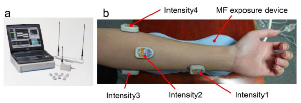
EMG Measurement Surface EMG measurements were performed noninvasively in the left fore- arm continuously for at least 20 min using a multichannel telemetry system (WEB-1000; Nihon Kohden, Tokyo, Japan) (Figure 2a).
Evaluation of the Effects of AC Magnetic Field Exposure on Muscle Fatigue 141
Figure 2: (a) An EMG monitoring device and (b) four electrodes and an MF exposure device. attached to a forearm.
As shown in the photograph of EMG measurement (Figure 2b), the left forearm was placed on the AC MF exposure device in a seated position, and four electrodes were attached to the distal part of the flexor carpi ulna- ris muscle (intensity 1), flexor carpi radialis muscle (intensity 2), proximal part of the flexor carpi ulnaris muscle (intensity 3), and brachioradialis muscle (intensity 4). During the EMG measurement, the dorsal side of the left forearm was exposed to an MF for 15 min (Figure 2b).
The EMG sampling rate was 1,000 Hz. The raw data were bandpass filte- red between 15 and 500 Hz. The energy inclusion line of the EMG waveform during repeated handgrip training was obtained, and the EMG signal during handgrip training was extracted for approximately 2 s. The Welch spectrum was obtained from the EMG signals measured during the handgrip exercise, and the IFs for estimating muscle fatigue were calculated using frequency analysis.
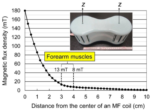
AC MF Exposure Device The AC MF exposure device was covered with a specially designed thin cover cloth (P27Mar15; Soken Co., Ltd., Toride, Japan) made of cotton to absorb perspiration (Figure 2b). Two separate electromagnetic coils were set hori- zontally inside the MF exposure device, and Bmax was measured as 180 mT at 50 Hz using a single-axis teslameter with a suitable transverse probe (F41 teslameter, FP-2X-250-TS15 probe, Lake Shore Cryotronics Co., Westerville, OH, USA) on the surface of the MF exposure device above the centers of the coils. Figure 3 shows the spatial distribution of Bmax on the surface of the MF exposure device.
The magnetic flux density of the MF exposure device decreased expo- nentially with distance. The estimated Bmax values in forearm muscles were approximately 8–13 mT, considering a distance of approximately 3–4 cm from the surface (Bmax = 180 mT) of the MF exposure device (Figure 3).
The room temperature and the temperature at the surface of the MF expo- sure device during the MF exposure period were maintained at 25±0.5°C. The relative humidity was maintained at 50±10%.
Statistical Analyses The differences in mean values for the sham control and MF exposure groups were statistically analyzed using the Wilcoxon rank-sum test (between
Figure 3: Spatial distribution of the peak magnetic flux density Bmax values along z-direction.
groups) and Wilcoxon paired signed-rank test (within a group) for non- parametric data. For parametric data, statistical analyses were performed using the Student’s t-test (between groups) and paired t-test (within a group). In addition, post-hoc differences were analyzed using two-way ANOVA for repeated measures (experimental groups and time points). For all compari- sons, a P value less than 0.05 was considered significant.
RESULTS AND DISCUSSION
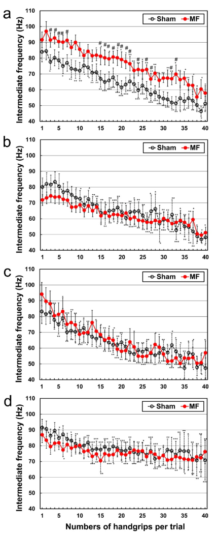
EMG Twelve individuals were assessed on different days after the sham or MF exposure. The effects of MF on the IFs in the EMG power spectrum are shown in Figure 4. The mean IF values in the EMG power spectrum, indica- ting muscle fatigue, were significantly reduced from the baseline values after handgrip training for intensities 1–4 (Figure 4a–d). When muscles become fatigued, the power spectrum shifts to a lower frequency band (De Luca, 1984). This is thought to be due to a decrease in the propagation velo- city of the action potential along the muscle fibers (De Luca, 1984). The MF exposure significantly suppressed the reduction of IFs for intensity 1, and significant differences between MF and sham exposures were observed intermittently (P < 0.05, Figure 4a). However, no significant difference was observed between MF and sham exposures for intensities 2–4 (Figure 4b–d). Thus, the effect of MF on IFs varied depending on the muscle region.
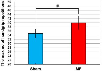
The effect of MF on the maximum number of handgrip repetitions per trial is shown in Figure 5. The maximum number of handgrip repetitions in the MF exposure group was significantly higher than that in the sham exposure group (P < 0.05, Figure 5).
Evaluation of the Effects of AC Magnetic Field Exposure on Muscle Fatigue 143
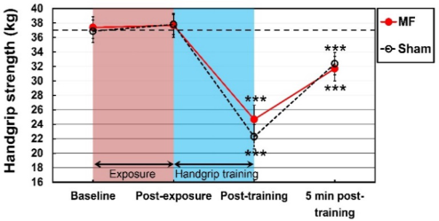
Figure 4: Effect of MF on the IFs in the EMG power spectrum. These figures show the changes in IFs with respect to the maximum number of handgrip repetitions per trial (up to 40) during handgrip training (repeated 22 kg loading) in the MF and sham exposure groups from the following four electrodes: (a) intensity 1, attached to the distal part of the flexor carpi ulnaris muscle; (b) intensity 2, attached to the flexor carpi radialis muscle; (c) intensity 3, attached to the proximal part of the flexor carpi ulnaris muscle; and (d) intensity 4, attached to the brachioradialis muscle. Values are expres- sed as mean ± SEM (n = 12 in each group). #P < 0.05 was considered statistically significant between groups. *P < 0.05, **P < 0.01, and ***P < 0.001 were considered statistically significant within a group.Figure 5: Effect of MF on the maximum number of handgrip repetitions (repeated 22 kg loading) per trial in the MF and sham exposure groups. Values are expressed as mean ± SEM (n = 12 for each group). #P < 0.05 was considered statistically significant between groups.Figure 6: Effect of MF on handgrip strength. The graph shows the handgrip streng- ths before and after handgrip training (repeated 22 kg loading) in the MF and sham exposure groups. ***P < 0.001 was considered statistically significant within a group.
Handgrip Strength The effects of MF on the handgrip strength are shown in Figure 6.
The mean values of handgrip strength immediately after handgrip training (post-training) were significantly reduced from the baseline values in both the MF and sham exposure groups (P < 0.001, Figure 6). The mean values of handgrip strength in the MF exposure group seem to be higher than those in sham exposure group, but no significant difference was observed between the two groups (0.05 < P < 0.10, Figure 6). The handgrip strength in neither exposure group returned to the baseline values 5 min post-training (P < 0.001, Figure 6).
Plausible Mechanisms of the Effects of MF on Muscle Fatigue The estimated Bmax values in the forearm flexor muscles are approximately 8–13 mT. In a previous study, computer simulations were performed to obtain order-of-magnitude estimates (Okano et al., 2021). In the simulations, the order-of-magnitude estimates of the peak induced electric fields (EFs) in the forearm were up to approximately 320 mV/m depending on the exposure
Evaluation of the Effects of AC Magnetic Field Exposure on Muscle Fatigue 145
conditions (Okano et al., 2021). The induced EFs were the highest under- neath the coils, with minor diffusion to other regions (Okano et al., 2021). The peak values of induced EFs were 319.3 mV/m in the skin, 282.3 mV/m in the fat, 141.9 mV/m in the muscles, and 142.8 mV/m in the blood vessels (Okano et al., 2021). We speculate that these induced EFs caused eddy cur- rents and thereby induced changes in EMG, suppressing the reduction of IFs in the present experiment.
In addition, the hemodynamic mechanisms of 50 Hz MFs have been reviewed (McNamee et al., 2009; Ueno and Okano, 2011; Ohkubo and Okano, 2015), and we have proposed the mechanisms by which 50 Hz MFs could alter acetylcholine (ACh) and nitric oxide (NO) signaling pathways (Okano et al., 2017, 2021; Kondo et al., 2019). Concerning the physiologi- cal mechanisms of NO, it has been reported that NO formed via eNOS and neuronal NOS (nNOS) causes vasodilatation, hypotension, and increased blood flow (Toda et al., 2009). Therefore, in this context, 50 Hz MF-induced NO activation/production could help promote blood circulation. When considering the physiological significance of the non-thermal MF effects, the present results imply that MF-enhanced vasodilation and microcircula- tion might help eliminate metabolic waste products and endogenous pain- producing substances that induce muscle stiffness and pain as well as muscle fatigue.
CONCLUSION
MF exposure significantly suppressed the reduction in IFs, indicating decrea- sed muscle fatigue. Moreover, the maximum number of handgrip repetitions with MF exposure was significantly larger than those with sham exposure. The mean values of handgrip strength in the MF exposure group seemed to be higher than those in the sham exposure group, but no significant diffe- rence was observed between the two groups. Overall, these results suggest that MF decreases muscle fatigue. One of the limitations of this study is that the type of muscle loading was only one type with a small number of parti- cipants. Multiple muscle loading trials with a larger number of participants are needed in order to draw firm conclusions.
REFERENCES
Begue-Simon, A.M., Drolet, R.A. (1993). Clinical assessment of the RHUMART system based on the use of pulsed electromagnetic fields with low frequency, Int J Rehabil Res. Vol. 16, No. 4. pp. 323–337. Bonato, P., Gagliati, G., Knaflitz, M. (1996). Analysis of surface myoelectric signals
recorded during dynamic contractions, IEEE Eng Med Biol. Vol. 15, No. 6. pp. 102–111. Bonato, P., Roy, S.H., Knaflitz, M., De Luca, C.J. (2001). Time-frequency parameters of the surface myoelectric signal for assessing muscle fatigue during cyclic dynamic contractions, IEEE Trans Biomed Eng. Vol. 48, No. 7. pp. 745–753. De Luca, C.J. (1984). Myoelectric manifestations of localized muscular fatigue in
Kondo, T., Okano, H., lshiwatari, H., Watanuki, K. (2019). “Evaluation of the hemodynamic effects of AC magnetic field exposure by measurement of an FMD and a microscope,” in: Advances in Human Factors and Ergonomics in Healthcare and Medical Devices, Lightner, N.J. (Ed.). pp. 68–79. Basel, Switzerland: Springer International Publishing. McNamee, D.A., Legros, A.G., Krewski, D.R., Wisenberg, G., Prato, F.S., Tho-
mas, A.W. (2009). A literature review: the cardiovascular effects of exposure to extremely low frequency electromagnetic fields, Int Arch Occup Environ Health. Vol. 82, No. 8. pp. 919–933. Ohkubo, C., Okano, H. (2015). “Magnetic field influences on the microcirculation,” in: Electromagnetic Fields in Biology and Medicine, Markov, M.S. (Ed.). pp. 103–128. Boca Raton, FL: CRC Press. Okano, H., Fujimura, A., Ishiwatari, H., Watanuki, K. (2017). “The physiological
influence of alternating current electromagnetic field exposure on human subje- cts,” IEEE International Conference on Systems, Man, and Cybernetics (SMC). pp. 2442–2447. Okano, H., Fujimura, A., Kondo, T., Laakso, I., Ishiwatari, H., Watanuki, K.
(2021). A 50 Hz magnetic field affects hemodynamics, ECG and vascular endoth- elial function in healthy adults: A pilot randomized controlled trial, PLOS ONE. Vol. 16, No. 8. e0255242. Pilla, A.A. (2007). “Mechanisms and therapeutic applications of time-varying and static magnetic fields,” in: Handbook of Biological Effects of Electromagnetic Fields, 3rd Edition, Barnes, F., Greenebaum, B. (Eds.). pp. 351–411. Boca Raton, FL: CRC Press. Toda, N., Ayajiki, K., Okamura, T. (2009). Cerebral blood flow regulation by nitric
oxide: recent advances, Pharmacol Rev. Vol. 61, No. 1. pp. 62–97. Ueno, S., Okano, H. (2011). “Static, low frequency and pulsed magnetic fields in biological systems,” in: Electromagnetic Fields in Biological Systems, Lin, J.C. (Ed.). pp. 115–196. Boca Raton, FL: CRC Press.
LISTED IN THE AHFE DIGITAL BOOKSTORE CATALOGUE
Explore the complete digital volume
Affective and Pleasurable Design
Read this individual research paper free through the original publisher, or explore the full digital proceedings book.
ISBN 978-1-958651-17-9
Explore this ISBN in the AHFE Bookstore ↗Listed in the July 2026 bookstore catalogue; search by ISBN to confirm current availability · Digital book purchase optional