Kelly Fernandes, Cláudia Sá Dos Reis, Florentino Serranheira (2022). Activity simulation in Radiology: Chest X-Ray in bed-ridden patients. In: Safety Management and Human Factors. AHFE Open Access, vol. 64, pp. 16–24. https://doi.org/10.54941/ahfe1002623
Work-related musculoskeletal disorders (WRMSDs) symptoms are frequent in healthcare workers. In Radiology Departments, demanding postures assumed by radiographers frequently occurred. Chest plain radiography to bed-ridden patients is one of the most demanding ones. To evaluate postural strain in radiographers’, professional activity was simulated during bedside chest plain radiography performance.Bed-ridden chest X-ray examinations are generally performed by two radiographers. The simulations scenarios were recorded using photogrammetry methods, assessing postural variations of the main body segments (head/neck, upper arm, and trunk) (Kapitaniak et al. method, 2001). The main body angles of observed body segments were measured with dedicated software (Kinovea, version 0.8.15). The postures were classified into three categories according to European Standards (EN 1005-4:2005 + A1:2008): “acceptable”, “conditionally acceptable”, and “not acceptable” (British Standard, 2018).Results show demanding postures assumed by radiographers occurred during the X-ray tube manipulation requiring arm flexion, being more evident for radiographers that were shorter. Regarding patient handling, upper arm flexion was “not acceptable” when holding and pushing the patient by the shoulders. This arm posture may increase the risk of injuries especially since it is associated with high exertion of force and is repeated during the examination. In contrast, by supporting patients by scapula passing under the axilla, the constraint in the upper arm was reduced and the trunk flexion, even a bit more important, remain “acceptable”. This observation indicates that the need to train radiographers in manual patient handling. Unacceptable neck extensions can also be observed in the medium and smallest radiographers while handling the patient to probably have an overview (Cernean et al., 2017; Giger et al., 2008) and/or maintain a certain physical distance from the patient.Since bedside chest radiography is one of the most performed examinations requiring repeated movements and awkward posture, particular attention should be paid to biomechanical load. One strategy to reduce this physical constraint is the collaboration of both radiographers to lift the patient. Another one is changing the bedside radiography procedure, for instance raising the upper side of the bed to 45º, which will reduce the main risk factors doing the X-ray.Most of the radiographers self-associated conventional radiography practice with low back complaints, which is in line with previous studies in X-ray radiographers (Lorusso et al., 2007). This result was not surprising, even though the trunk posture was never classified as “not acceptable”, because the repetitive truck flexion with exertion of force to lift the patient may increase the risk of WRMSDs symptoms. Further research is needed to identify the demanding tasks responsible for symptoms.There is a lack of literature on the impact of anthropometric characteristics of radiographers performing X-rays, but mammography related studies showed that not adjustable radiological equipment to anthropometrics characteristics, is a physical risk factor, requiring radiographers to assume awkward postures possibly leading to WRMSDs symptoms (Cernean et al., 2017; Costa et al., 2014a). We dare to say there is a need to improve communication between users, equipment manufacturers, and designers to fit the needs of a wider range of radiographers' anthropometrics characteristics.
Work-related musculoskeletal disorders (WRMSDs) are frequently observed in radiographers (Daniel et al., 2018), however, prevalence, and symptoms are slightly dependent on studies and imaging modalities included (Griffin, 2018; Lorusso et al., 2007; Pompeii et al., 2008; Siewert et al., 2013; Tinetti & Thoirs, 2019). Previous studies dedicated to diagnostic radiographers sho- wed a prevalence of WRMSDs symptoms ranging from 67% (Lorusso et al., 2007) to 93% (Daniel et al., 2018) with back, neck and upper limbs as the most affected anatomical regions (Bright Ofori-Manteaw et al., 2015; Kumar et al., 2004b; Lorusso et al., 2007).
WRMSDs prevalence and symptoms are different in each radiographic modality and there is a need to understand the real work demands that con- tributes for WRMSDs symptoms. In a Lamar study (2004), low-back pain (LBP) is prevalent in magnetic resonance imaging (MRI) and interventional radiographers, while neck symptoms are more observed in mammography and computed tomography (CT) (Lamar, 2004). Nowadays, radiographers are confronted with high pressure from the organizations to face the patients
Activity Simulation in Radiology: Chest X-Ray in Bed-ridden Patients 17
diagnostic needs, impacting the WRMSDs symptoms, which can vary, accor- ding to the physical and psychosocial work demands (Bright Ofori-Manteaw et al., 2015; Kumar et al., 2004b).
Main ergonomic risk factors threaten radiographers health in Conventio- nal Radiology are the adoption of awkward joints angles while positioning the detector under the patient, contributing to upper extremity and low-back pain (Kumar et al., 2004b). This issue was also identified in mammography, where awkward postures are adopted such as twisting the body and using unacceptable joints angles due to technical requirements for breast positio- ning and equipment handling (Costa, Oliveira, Reis, Viegas, & Serranheira, 2014). Radiographers frequently manipulate obese and elderly patients, that can promote or aggravated muscular disorders due to the increase low-back load and lack of patient participation (Augner & Kaiser, 2019; European Occupational Safety and Health Administration, 2020; Griffin, 2018). High exposure to physical load was also identified in emergency rooms, having plain and mobile radiography as main responsible for back and upper limbs discomfort (Kumar et al., 2004a). Chest plain radiography performed to the patients in bed, is one of the most demanding X-ray exams.
The study aimed evaluate postural strain in radiographers doing chest plain radiography to bed-ridden in the Radiology department at Centre Hospitalier Universitaire Vaudois (CHUV). The professional activity performance was simulated to identify postural risk factors related to WRMSDs symptoms to take in consideration by stakeholders to promote an improvement in working conditions, and design occupational health and musculoskeletal prevention strategies.
MATERIAL AND METHODS The targeted population was the diagnostic radiographers currently working in conventional radiology at CHUV and practicing bedside chest examina- tions for, at least, one year. Radiographers suffering from chronic or acute disease or being pregnant at the time of the study were excluded. For the simulations, two radiographers with extreme heights (shorter and taller) were identified. Another radiographer (weight ≥80 kg) was invited to simulate a passive patient. All participants gave their agreement to participate in this study.
Radiographers’ prescribed work regarding equipment manipulation and patient handling for bedside chest X-ray examinations were examined through an internal document of CHUV (DIAG, 2020). The real work was also analyzed through a sequential observation of the radiographers in clini- cal context. Due to the presence of patients in real practice, simulations were required to record the performance of radiographers during bedside chest X-rays acquisition (Costa et al., 2014). The data collected during observati- ons were used to ensure the similarity between the simulations and clinical activity. The simulations were performed with a radiography device from Philips (Philips Bucky Diagnost TH X-ray, Phillips Healthcare, Guildford, United-Kingdom). Chest X-ray examinations are generally performed by two
Table 1. References for postural assessment (European Standard BS EN 1005- 4:2005 + A1:2008).
Table available in the original PDF paper.
Body segments References values Acceptable Conditionally
Not acceptable acceptable
0° to 40° < 0° or > 40°
Head/neck upward/downward bending Trunk forward/ backward bending 0° to 20° < 0° or 20° to 60° > 60° Upper arm flexion/extension 0° to 20° 20° to 60° < 0° or > 60°
radiographers, one to position the detector and manipulate the X-ray tube (designated as “performing radiographer-P”), and the other to handle the patient (called “helping radiographer-H”). Permutations of heights and radi- ographers’ roles were simulated, resulting in 2 scenarios. The simulations were recorded using photogrammetry methods to assess postural variations of the main body segments (head/neck, upper arm, and trunk) according to Kapitaniak et al. method (2001). Three cameras (one camera Canon EOS 90D and two cameras Canon EOS 1300D, Tokyo, Japan) were placed in order to record simultaneously posterior and lateral views of radiographers. The videos were visualized by the observer and the most demanding and/or persistent postures were selected by two raters. The main body angles of observed body segments were measured with a dedicated software (Kinovea, version 0.8.15).
The joint angles were measured in simulated situations and classified into three categories according to European Standards (EN 1005-4:2005 + A1:2008): “acceptable”, “conditionally acceptable”, and “not accepta- ble” (Table 1) (British Standard, 2018).
The research project was submitted and accepted by two Ethics Commissi- ons (EC): SwissEthics of Canton of Vaud (Reference: 2020-011774) and the EC of Centre Hospitalier Universitaire Vaudois (CHUV).
RESULTS
Two radiographers were asked to simulate a bedside chest X-ray perfor- med in a bed in a conventional radiography room. Two scenarios were played according to the radiographers’ anthropomorphic characteristics and radiographers’ roles (performer-P/helper-H).
Main tasks for chest X-ray in bed-ridden patients were: i) Preparation to position the detector under the patient’s back; ii) Patient handling to position the detector under the patient’s back; iii) Control of detector’s position; iv) X-ray tube positioning; v) Preparation for removing the detector from under the patient’s back; vi) Patient handling to remove the detector from under the patient’s back.
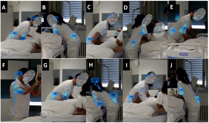
Scenario 1(Fig. 1) - Taller radiographer (performer-P) & shorter radiogra- pher (helper-H) i) Preparation to position the detector under the patient’s back (A&B). The P and H radiographers prepared to lift the patient by performing a slight
Activity Simulation in Radiology: Chest X-Ray in Bed-ridden Patients 19
Figure 1: Postures assumed by the taller (performer) and shorter (helper) radiograph- ers during bedside chest X-ray examination in scenario 1 for main tasks (A-J).
trunk flexion. The performer slightly bent downward the head/neck, while the helper tended to extend it. Both radiographers’ visible arms assumed a sli- ght flexion by placing the forearm under patient’s back. The trunks (P = 48°; H = 42°) and arms angles (P = 20°; H = 38°) were classified “conditionally acceptable”. The flexion of the head/neck of the performer was classified as “acceptable” (11°), and the neck extension of the helping radiographer was “not acceptable” (−7
).
ii) Patient handling to position the detector under the patient’s back (C&D). Trunk flexion of the performer was more critical than those of the helper during patient’s lift. The arms supporting the patient’s back remained in a neutral position for the performer and flexed for the helper. Perfor- mer slid the detector under the patient keeping the same posture as before. The trunk angle (37°) position of the performer was classified as “conditi- onally acceptable”, while the upper arm (0°) and head/neck position (10°) were rated as “acceptable”. The helper trunk angle (20°) was considered as “acceptable”, but the arm’s flexion was classified as “conditionally accepta- ble” (31°). The head/neck for the helper was not measurable due to a slight rotation.
iii) Control of detector position (E). During this task the performer bent the trunk to overview the detector position. Both arms were flexed to verify and reposition when necessary. The trunk (44°) and arm flexion (40°) were rated as “conditionally acceptable” while the head/neck was classified as “acceptable” (0°).
iv) X-ray tube positioning (F) required the adopting an orthostatic posture from the performer with the trunk aligned with the mid-sagittal plane of the body. The posture of the trunk was “acceptable”, the head/neck (22°) and arms angles (52°) were classified as “conditionally acceptable”.
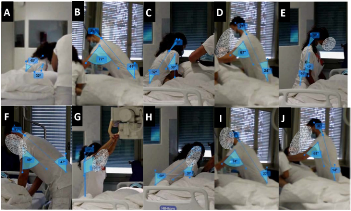
Figure 2: Postures assumed by the shorter (performer) and taller (helper) radiograph- ers during bedside chest X-ray examination in scenario 2 for main tasks (A-J).
v) Preparation to remove the detector from under the patient’s back (G&H) obliged both radiographers to bend the trunk and flex the arms to place their hands under the patient’s back and prepare to exert force to lift the patient. The head/neck of performer stayed aligned with the rest of the vertebral spine in contrast to helper, who extended her head/neck during this procedure. The angles of the trunk (P = 48°; H= 50°) and arms (P = 28°; H= 36°) were considered as “conditionally acceptable” for both radiograph- ers. The segment of head/neck for performer was “acceptable” (0°), while the helper adopted a head/neck extension (−24°) considered as “not acceptable”.
vi) Patient handling to remove the detector from under the patient’s back (I&J) required a slightly flexion of the trunk from both radiographers. The arm pulling the patient was in a neutral position (0°) for the performer and in flexion for the helper arm. The performer slightly bent downward the hea- d/neck segment, while the helper has extended. The angles formed by trunk (30°), arms (0°) and the head/neck (16°) segments for the performer were considered “acceptable”. The helper adopted postures classified as “conditi- onally acceptable” for trunk (31°) and arms (28°), while head/neck was not measurable.
Scenario 2 (Fig. 2) - Shorter radiographer (performer-P) & taller radiogra- pher (helper-H) i) During the preparation to position the detector under the patient’s back (A&B), the radiographers bended over to place their hands to lift the pati- ent. The performer put the right arm under the scapula passing by under the axilla, which required the upper arm’s flexion. The helper also flexed the upper arms to put the hands on the patient’s shoulders. Besides, the head/neck of the taller radiographer was bending downward to observe the patient. The
Activity Simulation in Radiology: Chest X-Ray in Bed-ridden Patients 21
trunk angles (P = 48°; H = 41°) were determined as “conditionally acce- ptable” in both radiographers. The upper arm flexion for performer (32°) was considered “conditionally acceptable”, while the arm flexion in helper (77°) was considered as “not acceptable”. The head/neck angle of the helping radiographer (21°) was classified as “acceptable”.
ii) Patient handling and detector position under the patient’s back (C&D) required from the helper to apply force to pull the patient back to allow the performer to slide the detector under the patient. Their trunks were less flexed when compared to the previous situation. The visible arms were flexed to support the pulled patient. Both radiographers flexed the head/neck to observe the position of the detector. The performer slid the detector under the patient. This action did not induce a change in the posture. Radiographers’ trunks (P = 32°; H = 24°) were in a flexion considered as “acceptable”. The performer arm’s angle was considered as “conditionally acceptable” (30°) while the helper (67°) was considered as “not acceptable”. The angles of the head/neck were both classified as “acceptable” (P = 9°; H = 21°).
iii) Control of detector position (E&F). In this scenario, both radiograph- ers controlled and adapted the position of the detector under the patient. The head/neck, trunk and upper arm segments were in flexion. These segments’ angles were more critical in the taller radiographer due to the difference between his height and the patient height. The body segments measured in performer radiographer were all rated as “acceptable” (trunk = 14°; arm = 13°; head/neck = 30°). The posture adopted by the helper required to assume a “conditionally acceptable” angle in the trunk (47°) and arms (49°), only the flexion of the neck (13°) was “acceptable”.
iv) X-ray tube positioning (G) required from the performing radiographer to raise the arms above the head, in hyperflexion, due to a need for sufficient distance between the source (tube) and the detector. The head/neck was fle- xed, allowing observation of patient position, the centering and diaphragms verification. The trunk was in an orthostatic posture aligned with the body’s mid-sagittal plane. The radiographer’s posture was considered as “accepta- ble”regarding measured angles of trunk (0°) and head/neck (31°), while arms flexion (119°) was classified as “not acceptable”.
v) During the preparation to remove the detector from under the patient’s back (H&I), the performer flexed the trunk to reach the patient’s back pas- sing under the axilla and extended the head/neck. By taking the patient by the shoulders, the helper was less bent downward than the performer. The visi- ble arms of both radiographers were flexed to support the patient’s back. The postural assessment of the performer revealed that angles of the trunk (50°) and right arm (38°) were “conditionally acceptable”, but “not acceptable” for head/neck (-27°). The helper’s head/neck (17°) and trunk (33°) positions were “acceptable” but the arm (74°) was in a “not acceptable” position.
vi) Patient handling to remove the detector from under the patient’s back (J) required force from both radiographers while maintaining a flexion of the trunk. The arms and head/neck were also in flexion to support the patient’s back and keep the patient lifted to remove the detector, observing the patient at the same time. Helper’s trunk (24°) was considered as “conditionally acce- ptable”. The patient handling required to the helper adopting an arm flexion
(61°) considered as “not acceptable”. The flexion of the head/neck (P = 20°; H = 20°) was “acceptable” for both radiographers.
DISCUSSION
Postural strain in radiographers’ occupational activity, such as reported during patient and equipment handling (Kumar et al., 2003; Pompeii et al., 2009), were also observed in this study during bedside chest plain X-ray simulations. The most demanding postures assumed by radiographers as “performers-P” mainly occurred during the X-ray tube manipulation requi- ring arm flexion, being more evident for shorter radiographers. Smaller is the radiographer, more flexion of the arm is required due to the need to respect a certain distance between the source (tube) and the detector. There is a lack of literature on the impact of anthropometric characteristics of radiographers performing X-rays, but mammography related studies showed that when the medical imaging equipment is not adaptable to the anthropometrics characte- ristics, there exists a physical risk factor, requiring radiographers to assume awkward postures leading possibly to WRMSDs symptoms (Cernean et al., 2017; Costa et al., 2014a). It seems critical to improve communication betw- een users, equipment manufacturers and designers to fit the needs of a wider range of anthropometrics characteristics.
From “helping radiographer” viewpoint, patient handling required upper arm flexion that was “not acceptable” when holding and pushing the patient by the shoulders. This arm posture may increase the risk of injuries especially since it is associated with high exertion of force and it is repeated during the examination. In contrast, by supporting patients by scapula passing under the axilla, the constraint in the upper arm was reduced and the trunk flexion, even being slightly more important, remained “acceptable”. Unacceptable neck extensions could be also observed for the medium and shortest radiogra- phers while handling the patient to have probably a full overview (Cernean et al., 2017; Giger et al., 2008) and/or maintain a certain physical distance from the patient.
Most of the radiographers self-associated conventional radiography pra- ctice with low back complaints, which is in line with previous studies considering X-ray radiographers (Lorusso et al., 2007). This result was not surprising, even though the trunk posture was just once classified as “not acceptable”, because the repetitive truck flexion with exertion of force to lift the patient may increase the risk of WRMSDs symptoms. Preventive action for this specific imaging modality needs to be considered for low back complaints as a priority. This action could improve physical well-being, but further research is needed to identify the tasks/causes responsible for the symptoms, even it can be hypothesized that they are related to patient handling.
CONCLUSION
Observation of clinical activity permitted to characterize the real work per- formed during bedside chest plain radiography, and simulations allowed the
Activity Simulation in Radiology: Chest X-Ray in Bed-ridden Patients 23
identification of “not acceptable” postures for upper arms and/or necks, especially during patient handling and the X-ray tube manipulation. During patient handling, the collaboration of radiographers with anthropometric dif- ferences did not significantly affect the postures classification, but it affected manual handling techniques and radiographers’ practice. However, anthro- pometric characteristics directly impacted the constrained postures of the arm during X-ray tube positioning.
ACKNOWLEDGMENT
The authors would like to acknowledge to CHUV and all the Radiographers for the support.
REFERENCES
Augner, C., & Kaiser, G. (2019). Predictors of musculoskeletal symptoms in radiology technologists in Austria, Europe. Work, 64(4), 853–858. https://doi.org/10.3233/WOR-193047 Bright Ofori-Manteaw, B., Kwadwo Antwi, W., & Arthur, L. (2015). Ergonomics and occupational health issues in diagnostic imaging: a survey of the situation at the Korle-Bu teaching hospital. Journal of Health, Medecine and Nursing, 19, 93–101. British Standard (2018). Safety of machinery: Human physical performance. BS
EN 1005–4:2005 + A1:2008. Swiss National Report on Quality and Safety in Healthcare. Cernean, N., Serranheira, F., Gonçalves, P., & Sá dos Reis, C. (2017). Ergonomic strategies to improve radiographers’ posture during mammography activities. Insights into Imaging, 8(4), 429–438. https://doi.org/10.1007/s13244-017-0560-7 CHUV - service radiodiagnostic et radiologie interventionnelle (2020). Conceptuali-
sation prise en charge d’un patient au DIAG. Costa, S., Oliveira, E., Reis, C., Viegas, S., & Serranheira, F. (2014). Mammography equipment design: impact on radiographers’ practice. Insights into Imaging, 5(6), 723–730. https://doi.org/10.1007/s13244-014-0360-2 Daniel, S. V., Umar, M. S., Ahmad, N. M., & Joseph, Z. D. (2018). Work-related
musculoskeletal disorders : rrevalence among clinical radiographers in teach- ing hospitals in North-Western Nigeria. Journal of Radiography & Radiation Sciences, 32(1), 57–63. European Occupational Safety and Health Administration (2020). Discussion paper musculoskeletal disorders in the healthcare. European Agency for Safety and Health at Work, 1–23. https://osha.europa.eu/en/publications/musculoskeletal- disorders-healthcare-sector/view Giger, M. L., Chan, H. P., & Boone, J. (2008). Anniversary paper: History and status
of CAD and quantitative image analysis: The role of Medical Physics and AAPM. Medical Physics, 35(12), 5799–5820. https://doi.org/10.1118/1.3013555 Kapitaniak, B., Péninou, G., & Samuelson, B. (2001). Assistance ergo- nomique pour la conception de mammographe - GE Medical Systems. https://streaming-canal-u.fmsh.fr/vod/media/canalu/documents/universite_pa ul_verlaine_metz_sam/histoire.s.de.l.ergonomie.3.7.la.pratique.le.d.veloppement .d.un.m.tier_11649/33.kapitaniak.mammographe.pdf
Kumar, S., Moro, L., & Narayan, Y. (2003). A biomechanical analysis of loads on x-ray technologists: A field study. Ergonomics, 46(5), 502–517. https://doi.org/10.1080/0014013031000061659 Kumar, S., Moro, L., & Narayan, Y. (2004a). Morbidity among X-ray technologists. International Journal of Industrial Ergonomics, 33(1), 29–40. https://doi.org/10.1016/j.ergon.2003.06.002 Kumar, S., Moro, L., & Narayan, Y. (2004b). Perceived physical stress at work and
musculoskeletal discomfort in X-ray technologists. Ergonomics, 47(2), 189–201. https://doi.org/10.1080/00140130310001617958 Lamar, S. L. (2004). Investigation of factors associated with prevalence and severity of musculoskeletal symptoms among the workers in clinical specialties of radio- logic technology: an ergonomic and epidemiological approach [North Carolina State University]. https://repository.lib.ncsu.edu/handle/1840.16/143 Lorusso, A., Bruno, S., & L’abbate, N. (2007). Musculoskeletal com- plaints among Italian X-ray technologists. Industrial Health, 45(5), 705–708. https://doi.org/10.2486/indhealth.45.705 Pompeii, L. A., Lipscomb, H. J., & Dement, J. M. (2008). Surveillance of muscu-
loskeletal injuries and disorders in a diverse cohort of workers at a tertiary care medical center. American Journal of Industrial Medicine, 51(5), 344–356. https://doi.org/10.1002/ajim.20572 Pompeii, L. A., Lipscomb, H. J., Schoenfisch, A. L., & Dement, J. M. (2009). Musculoskeletal injuries resulting from patient handling tasks among hospital workers. American Journal of Industrial Medicine, 52(7), 571–578. https://doi.org/10.1002/ajim.20704 Siewert, B., Brook, O. R., Mullins, M. M., Eisenberg, R. L., & Kruskal, J. B. (2013). Practice Policy and Quality Initiatives: Strategies for Optimi- zing Staff Safety in a Radiology Department. RadioGraphics, 33(1), 245–261. https://doi.org/10.1148/rg.331125174 Tinetti, C. J., & Thoirs, K. (2019). Prevalence, risks, underlying mechanisms, preven-
tative guidelines, and interventions of sonographer work-related injuries: A lite- rature review. Sonography, February, 1–14. https://doi.org/10.1002/sono.12187
LISTED IN THE AHFE DIGITAL BOOKSTORE CATALOGUE
Explore the complete digital volume
Safety Management and Human Factors
Read this individual research paper free through the original publisher, or explore the full digital proceedings book.
ISBN 978-1-958651-40-7
Explore this ISBN in the AHFE Bookstore ↗Listed in the July 2026 bookstore catalogue; search by ISBN to confirm current availability · Digital book purchase optional