Makiko Aoki, Satoshi Suzuki (2023). Characteristics of Cerebral Blood Flow During Working Memory Tasks: Comparison of the Follicular and Luteal Phases Among Sexually Mature Females and Males. In: Emerging Technologies in Healthcare and Medicine. AHFE Open Access, vol. 116, pp. 372–378. https://doi.org/10.54941/ahfe1004391
Publisher statistics will load when you open Metrics.
AHFE OPEN ACCESS OPEN ACCESS CONFERENCE PROCEEDINGS PAPER
Characteristics of Cerebral Blood Flow During Working Memory Tasks: Comparison of the Follicular and Luteal Phases Among Sexually Mature Females and Males
In this study, we aimed to clarify the characteristics of cerebral blood flow during the N-back task for males and for females in the follicular and luteal phases. Near infrared spectroscopy (NIRS) was used to measure Oxyhemoglobin (Oxy-Hb) in the prefrontal cortex during the N-back task. In the analysis, the prefrontal cortex was divided into right and left regions, and the integrated Oxy-Hb value, center of gravity value, and activation rate (initial activation) in the first 5 seconds of the task were calculated for each region. The percentage of correct responses to the N-back task was also calculated. Differences in each representative value among the three groups (follicular phase, luteal phase, and male) were examined. The task correct response rate was lowest in the luteal phase group for males and the luteal phase group (p<.05) and in the follicular phase group and the luteal phase group (p<.05). There were no significant differences between groups in integral and center-of-gravity values, and there were significant differences between groups in the initial activation of CH10-13 (left area) during the 2-back task (p<.05), with the lowest in the luteal phase group among males (p<.05), follicular phase group (p<.05) and luteal phase group (p<.05). A decrease in working memory is suggested in luteal phase women. This may be due to the presence of women with premenstrual syndrome symptoms or to sex hormone effects.
The ability to prioritize multiple tasks and work efficiently is necessary to perform one’s duties in diverse societies. One such ability is working mem- ory. Working memory is future-oriented and often refers to memory involved in the planning and execution of actions and cognition. Working memory is active memory for preparing and executing actions and plans (Osaka et al., 2007), as well as a foundation for higher-order cognition, such as thinking
Characteristics of Cerebral Blood Flow During Working Memory Tasks 373
and comprehension; it is a function that simultaneously retains and processes information (Osaka, 2012). Therefore, working memory is an important function for people in a diversified society.
Previous studies on working memory show that cerebral blood flow in the prefrontal cortex region increases during the N-back task, which is a working memory task, using functional magnetic resonance imaging (fMRI) (Mar- quand et al., 2008; Owen et al., 2005). Another report correlates N-back task performance with testosterone and shows that N-back task performance decreases during the luteal phase among females with premenstrual syndrome (Akishita, 2014). There have also been reports of decreased performance in N-back tasks during the luteal phase in women with premenstrual syndrome (Aoki et al., 2020). However, there are no studies on the differences in work- ing memory between men or females during their menstrual cycles. However, to coexist in modern society, it is necessary for males and females to under- stand each other’s characteristics and cooperate. Therefore, we examined the differences in working memory according to sex and female menstrual cycles.
Recently, functional brain imaging has attracted attention; near-infrared spectroscopy (NIRS) is one such technique. Although NIRS can measure only a shallow brain region (20 mm from the skin surface), the prefrontal region, which is one of the regions controlling working memory, is located behind the forehead. Therefore, brain activity during working memory tasks using NIRS has been studied (Aoki et al., 2022). Consequently, we hypothesize that it would be possible to understand the characteristics of working mem- ory by evaluating the cerebral blood flow in the prefrontal cortex during a task involving working memory. Therefore, this study aims to clarify the characteristics of cerebral blood flow during the N-back task among males and females in the follicular and luteal phases.
MATERIAL AND METHODS Participant Selections
The participants included 11 males and 13 females between 20 and 25 years.
All participants were healthy and without systemic diseases. The inclusion criteria included:
1) Not currently pregnant 2) Body mass index of 18–25 3) Non-smoker 4) No psychiatric history 5) Self-Rating Depression Scale (SDS) scores <50 Procedure
The females were asked to measure their basal body temperature for two cycles. We confirmed that the basal body temperature was biphasic. Mea- surements of cerebral blood flow during the performance of working memory tasks were conducted during the luteal phase (the period between ovulation and menstruation) and the follicular phase (the period between the start of menstruation and ovulation). The follicular phase is within 5–12 days of
374 Aoki and Suzuki
menstruation onset, and the luteal phase is within 21–30 days of menstrua- tion onset. Males had their cerebral blood flow evaluated on one convenient day. Before the cerebral blood flow measurement, a questionnaire (self-rating depression scale [SDS]) was completed. The SDS is a depression severity scale, developed by Zung (1965), which is used for screening depressed patients. Cerebral blood flow was measured during the performance of the working memory task.
Cerebral Blood Flow Measurement Cerebral blood flow measurements were performed using NIRS (OEG16- SpO2, Spectratech Inc.) at a room temperature of 24
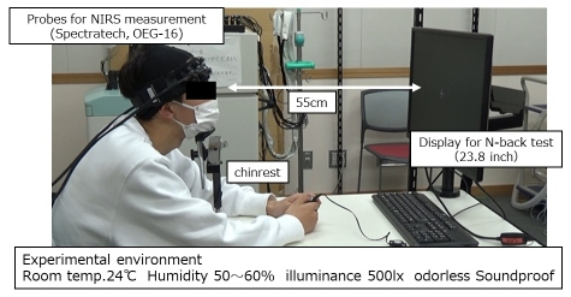
C, humidity of 50–60%, illumination of 500 lx, and in an odor-free environment. The probe for eval- uating changes in cerebral blood flow consisted of a light source unit and a light-receiving sensor unit; each of the 12 units was arranged in a grid with 3 cm spacing and a measurement area of 150 mm × 60 mm. Figure 1 shows a photograph of the cerebral blood flow measurements. The subject was seated in a chair with a measurement probe attached to their forehead and the sole was grounded 55 cm away from the LCD monitor (23.8 inches). A chinrest was used to prevent head vibrations. To allow the subjects to perform the cog- nitive function task in a relaxed state, they were asked to place their upper limbs on the desk and use a cushion. The subjects were asked to perform the cognitive task displayed on the LCD monitor while wearing the mea- surement probe. The cognitive task used was the N-back task; NIRS signals were sampled at 0.65 second intervals. Sample human systems integration test parameters (Folds et al., 2008).
Figure 1: Experimental set up for.
Task and Design In the N-back task, letters were displayed randomly, and the user was asked to answer the letter displayed before N. Letters were displayed every 1.5 seconds for 0.5 seconds. Twelve letters including b, d, g, p, q, t, B, D, G, P, Q, and T, were displayed. Upper- and lower-case letters were separated from each other
Characteristics of Cerebral Blood Flow During Working Memory Tasks 375
(Harvey et al., 2005). The subjects were instructed to click the ring mouse when the correct letter was display.
Analysis of NIRS Data The subjects in this study were mainly females in their early 20s with small faces. Because some of the NIRS measurement probes (ch1-3, ch14-16) were placed on the temporal side, there was poor adherence between the light- receiving units and the forehead in these areas. Therefore, the NIRS data acquired from ch4 to ch13 of the measurement probes were used for the analysis. NIRS data acquired from each channel from 5 seconds before the start of the N-back task to the end of the task presentation (5 seconds before the task presentation and 60 seconds after the task presentation) were ana- lyzed. The average value of oxy-Hb during the 5-second period before task presentation was calculated and used as the cerebral blood flow in the base state. The difference between the baseline and oxy-Hb values during the task was used to calculate the amount of change. Using this amount of change, the integral and center of gravity values, which were representative of the NIRS data for each subject, were calculated (Suto et al., 2004). The integral value indicates the degree of brain activation and the center-of-mass value indicates the speed of brain activation. For each channel, the oxy-Hb values were linearly connected from the beginning of the task until 5 s, when the slope was calculated (initial activation).
Data Analysis The Kruskal-Wallis test, Mann-Whitney U test, and Bonferroni’s adjustment were performed in the male, follicular, and luteal phase groups for the correct response rate to the N-back task, integrated oxy-Hb change, center of gravity value, and initial activation value.
Ethical Considerations Participants were provided a written and verbal explanation of the study and asked to sign a written informed consent accordingly. This study was conducted in accordance with the principles of the Declaration of Helsinki. This study was approved by the Human Ethics Committee of the Kanagawa Institute of Technology (approval no. 20191011-01).
RESULTS
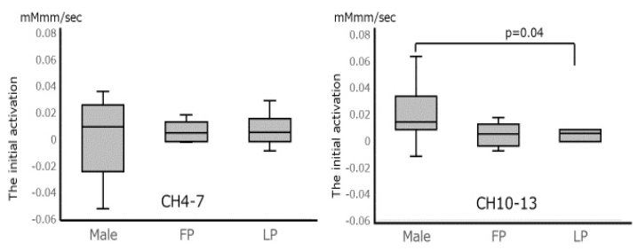
Table 1 shows the basic information for males and females. The three groups differed significantly in terms of the percentage of correct responses on the 2-back task (p=.04). Additionally, multiple comparisons between the male and luteal phase groups (p=.04) and the follicular and luteal phase groups (p=.04), females in the luteal phase had the lowest correct response rate. No significant differences were found between the groups in terms of inte- gral and central gravity values, and the initial activation of CH10-13 (left region) during the 2-back task (p=.04) was significantly lower in the luteal phase group than in the male group (p=.04). Figure 2 shows a comparison of the initial activation of the male group, the follicular phase group, and the
376 Aoki and Suzuki
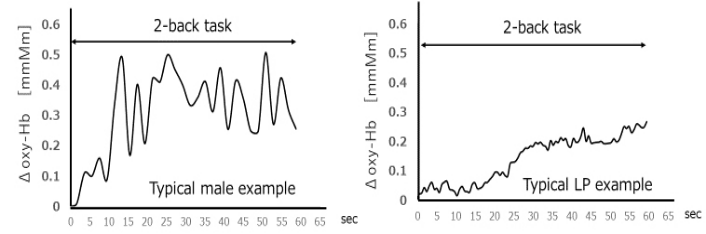
luteal phase group. Figure 3 shows the change in oxy-Hb at CH10-13 in one male and one luteal phase group over time. In the male group, Oxy-Hb was activated at the onset of the task; however, in the luteal phase, Oxy-Hb did not increase until approximately 25 seconds later, after which it continued to increase slowly.
Table 1. Participant characteristics.
Table available in the original PDF paper.
All participants Malea) Femaleb) Mean SD Mean SD Mean SD
Age, Year 21.5 0.8 29.2 6.5 21.6 1.1 BMI, kg m−2 20.3 2.8 19.7 2.1 20.6 3.1 SDS 36.4 4.8 37.6 7.7 37.6 5.0 a) n = 13. b) n = 11; body mass index. SDS; Self-Rating Depression Scale score
Figure 2: Comparison of the initial activation during 2-back in male, follicular phase (FP), and luteal phase (LP).Figure 3: Charge in 1Oxy-Hb in CH10-13 over time.
DISCUSSION
Estrogen levels have been reported to correlate with cognitive task perfor- mance (Baller et al., 2013; Jacobs & D’Esposito, 2011). Therefore, the correct response rate to the task was expected to increase during the luteal phase
Characteristics of Cerebral Blood Flow During Working Memory Tasks 377
when estrogen concentrations are elevated. However, in this study, the cor- rect response rate was lower than in the male and follicular phase groups. One reason may be the presence of premenstrual syndrome (PMS) or premen- strual dysphoric disorder (PMDD), wherein mental and physical discomfort occurs before menstruation. Aoki et al. reported that the N-back response rate among women with PMS was lower in the luteal phase than in the fol- licular phase (Aoki et al., 2020). They further stated that the oxy-Hb changes decreased in the 2-back task. Although PMS and PMDD were not examined among the female subjects in this study, future studies should include sex hormones, PMS, and PMDD.
The results of this study suggest that working memory may be different between males and females in the luteal phase.
CONCLUSION
In the male, follicular, and luteal phase groups, the correct response rate to the 2-back task was lowest in the luteal phase group. The initial activation of Oxy-Hb during the 2-back task was greater in the male group and lower in the luteal phase.The study did not take into account the physical and mental modulation during the female menstrual cycle, and thus could not consider the characteristics of PMS and PMDD. In the future, we would like to further increase the number of subjects and examine the relationship with the female menstrual cycle and sex hormones.
ACKNOWLEDGMENT
We thank all participants involved in this study. This research was partially supported by JSPS KAKENHI Grant Number JP 22K12950.
REFERENCES
Adrian M. Owen Kathryn M. McMillan Angela R. Laird Ed Bullmore. (2005).
N-back working memory paradigm: A meta-analysis of normative functional neuroimaging studies. Human Brain Mapping, 25(1), 46–59. Retrieved from https://search.proquest.com/docview/67789675 Baller, E. B., Wei, S., Kohn, P. D., Rubinow, D. R., Alarcón, G., Schmidt, P. J., & Berman, K. F. (2013). Abnormalities of dorsolateral pre- frontal function in women with premenstrual dysphoric disorder: A multi- modal neuroimaging study. American Journal of Psychiatry, 170(3), 305-314. Doi:10.1176/appi.ajp.2012.12030385. Harvey, P., Fossati, P., Pochon, J., Levy, R., LeBastard, G., Lehéricy, S.,... Dubois, B. (2005). Cognitive control and brain resources in major depression: An fMRI study using the n-back task. NeuroImage (Orlando, Fla.), 26(3), 860–869. Doi:10.1016/j.neuroimage.2005.02.048. Jacobs, E., & D’Esposito, M. (2011). Estrogen shapes dopamine-dependent cognitive
processes: Implications for women’s health. The Journal of Neuroscience, 31(14), 5286–5293. Doi:10.1523/JNEUROSCI.6394–10.2011. Makiko M, Masato S, Hisayo O. (2020). Assessing n-back task performance of menstrual adult women: Comparison with and without premenstrual syndrome. Journal of Nursing Science and Engineering, 8, 47–57.
378 Aoki and Suzuki
Makiko, A, Masato S, Satoshi S, Hidenobu T, Hisayo O (2022), “Cognitive function evaluation in premenstrual syndrome during the follicular and luteal phases using near-infrared spectroscopy”, Comprehensive Psychoneuroendocrinology, 10. Marquand, A., Mourão-Miranda, J., Brammer, M., Cleare, A., & Fu, C. (2008). Neu-
roanatomy of veral working memory as a diagnostic biomarker for depression. Neuroreport, 19(15), 1507–1511. Doi:10.1097/WNR.0b013e328310425e. Osaka, M., Komori, M., Morishita, M., et al. (2007). Neural bases of focusing attention in working memory : An fMRI study based on group differences. Cog. Affect Behav. Neurosci., 7 : 130 – 139. Osaka, N (2012). Prefrontal cortex and Working memory: Higher brain function
research, 32(1), 7–14. Suto, T., Fukuda, M., Ito, M., Uehara, T. & Mikuni, M (2004), “Multichannel nearinfrared spectroscopy in depression and schizophrenia: cognitive brain activation study”, Biological psychiatry, 55(5), 501–511.
LISTED IN THE AHFE DIGITAL BOOKSTORE CATALOGUE
Explore the complete digital volume
Emerging Technologies in Healthcare and Medicine
Read this individual research paper free through the original publisher, or explore the full digital proceedings book.
ISBN 978-1-958651-92-6
Explore this ISBN in the AHFE Bookstore ↗Listed in the July 2026 bookstore catalogue; search by ISBN to confirm current availability · Digital book purchase optional