Rosangela Santos, Ademir Marques Jr, João Silva, Flávio Cardoso Ventura, Célio Favoni, Isabel Cristina Buttignon, Rosemeire Dos Santos Almeida, Marcelo Oliveira (2024). Analysis of Procedures and Instruments for the Digitization of the Foot and Ankle With the Aim of Orthosis Development. In: Healthcare and Medical Devices. AHFE Open Access, vol. 130, pp. 180–187. https://doi.org/10.54941/ahfe1004851
Orthoses are products considered assistive technology for maintaining or expanding the skills of people with physical disabilities (Pwd), promoting greater social integration. All Pwd's who have sequelae of hemiplegia/hemiparesis need to use an orthosis. Generally, such an artifact has characteristics that interfere negatively during its use. The design of products (orthesis and footwear) separately often meets the functional requirements but has limitations regarding the conditions to facilitate usability and offer attractive aesthetic attributes that can promote a pleasant experience for people with disabilities. Such aspects influence the way people move around and interfere negatively in the physical rehabilitation process. Rapid prototyping can contribute to the development of these products in a personalized way and provide better patient satisfaction. For this, it is necessary to develop a digital model with reliable dimensions and formats based on three-dimensional scanning. The objective of this study was to demonstrate the quality of the digital model through image capture by different instruments and image processing procedures to obtain the three-dimensional model. Three-dimensional scanning was performed in a hemiparetic woman, patient at APAE-Jaú. The procedures performed were Kinect device and Skanect software; tomography scanning, and digital photogrammetry technique using a professional camera and the software Agisoft Metashape. All scans were aimed at the development and production of orthoses. It was noted that the most efficient digital model for the development of a customized orthosis was the tomography procedure. The software for developing the three-dimensional model is free, however, data collection to obtain the image (CT scanner) is expensive.
Analysis of Procedures and Instruments for the Digitization of the Foot and Ankle 181
INTRODUCTION
Assistive Technology (AT) is the term used to define the group of resources and services developed to enhance the functional abilities of people with disabilities, promoting independence and inclusion (Hensen, 2019). The pri- mary goal of AT products is to improve the user’s quality of life, either by increasing motor skills, maintaining, or fully restoring movement. These products can be mass-produced or customized, offering numerous possibili- ties for adaptations to provide user comfort (Fernandes, 2015).
Orthoses are an example of assistive products designed to enhance the structural and functional characteristics of the neuromuscular and skeletal systems. They are mechanical devices or orthopedic appliances that apply forces to a specific region to provide support, correction, alignment, or prevent deformities (Arce and Foggiatto, 2017). Despite common prescrip- tions, orthoses are often abandoned for various reasons, primarily due to no user involvement during fabrication, lack of comfort, and no anatomical fit (Rosenmann, 2017).
Conventional products available in the market are standardized and come with disadvantages, particularly, regarding anatomical aspects that impact usability. Paterson (2013) suggests utilizing Additive Manufacturing (AM) and three-dimensional (3D) scanning in orthoses development and produc- tion. AM enables the creation of personalized products for each user, and the 3D scanning step is crucial as the scanned region serves as a refer- ence for building the three-dimensional model for subsequent manufacturing (Rosenmann, 2017).
Mikolajewska et al. (2014) mention that the combination of 3D printing and scanning can contribute to the future of rehabilitation since devices based on the patient’s anatomy tend to be more comfortable and effective during treatment (Hensen, 2019). Hence, it is crucial to identify 3D scanning equip- ment and software that can capture the human body’s geometry, ensuring the 3D model’s quality in the process. Moreover, considering that most available equipment comes with a high cost, this could hinder the dissemination of orthoses manufacturing methods in a developing country like Brazil (Rosenmann, 2017). Three-dimensional scanning allows capturing various models, obtaining curves, textures, and surface details. However, it has drawbacks such as the time demand and pre- cision required for scanning parts of the human body, especially due to patient movements during the process.
Dessery and Pallari (2018) suggest that low-cost scanners may have clin- ical applications due to the simplicity of the process. Still, they may offer lower precision compared to high-cost equipment for extensive scanning areas. The application of these methods should be analyzed according to the body segment’s size (Hensen, 2019).
Considering the above, this study aimed to scan and demonstrate the quality of digital models using different instruments and image processing procedures for obtaining a three-dimensional model.
Theoretical Framework Three-Dimensional Scanning of the Human Body In the 3D printing stage, a three-dimensional model of the part is required. The modeling can be entirely developed in Computer-Aided Design (CAD) software, or from a model obtained by three-dimensional scanning. In the case of assistive products, the physical model to be scanned is the human body itself, or parts of it (Hensen, 2019).
3D scanning obtains data from physical objects, subsequently generating three-dimensional models with software assistance, capturing surface details with precision. Information obtained from scanning parts of the human body can be used in projects requiring accurate and personalized data (Brendler et al., 2016).
For such applications, advanced technology systems are generally used. Despite their growing popularity, these systems demand a significant invest- ment when considering the Brazilian market, making them impractical for low-cost projects (Silva, 2011). Data acquisition through human body scan- ning poses challenges, including comfort issues, as humans do not feel comfortable standing in a specific position for an extended period. Contin- uous movement and vulnerability to shape variations due to external and internal factors make human body scanning a challenge (Jones and Riouxb, 1997). Silva (2011) notes that while small errors individually may fall within the necessary tolerance for many applications, they can lead to significant losses in model assembly and greater loss of surface precision.
In the context of scanning human body parts, Ciobanu et al. (2013) rec- ommend the use of equipment based on laser triangulation, structured light, and digital photogrammetry (Rosenmann, 2017).
Laser Triangulation-Based Scanning According to Gazziro (2011), the triangulation-based scanning process involves projecting a vertical line beam produced by a laser emitter onto the object’s surface to be captured. This projection is then captured by a camera, and the distance to the object is calculated through geometric calculations (Moreira, 2016).
Triangulation-based laser scanning is well-established, with most systems focusing on high precision, resulting in higher costs. This system offers the possibility of capturing color during scanning using an RGB color filter posi- tioned in front of the sensor. After scanning, the surface point cloud of the object is obtained, which, after triangulation, generates three-dimensional surfaces or meshes (Silva, 2011).
White Light Scanning (Structured Light) According to Silva (2011), this system works by projecting a known pattern of light (parallel lines or grids) onto an object. The light lines deform when they hit the object’s surface and are captured by a calibrated camera. Based on the pattern distortion, the system calculates the object’s three-dimensional coordinates.
Analysis of Procedures and Instruments for the Digitization of the Foot and Ankle 183
Unlike laser systems that project only a thin line, multiple lines are pro- jected simultaneously, enabling an increase in scanning speed. The use of multiple cameras can enhance scanning accuracy, and higher the equipment resolution, more points can be obtained per image (Silva, 2011).
Photography-Based Scanning (Digital Photogrammetry) Photography-based scanning is founded on photogrammetry principles. The algorithm identifies the cameras’ positions during capture, and the scanning software compares the photos, seeking the best overlap. If the capture is suc- cessful, the orientations of the photos are used to calculate key point locations in a three-dimensional space, resulting in a point cloud representing the object (Silva, 2011).
Data accuracy is influenced by the camera’s resolution and lens, the dis- tance between the model and the equipment, and the object surface texture pattern. This is a passive scanning method, as no radiation other than natu- ral light external to the camera is projected onto the model. However, it has limitations concerning areas with low light, reflexive surfaces, point density, and processing time (Silva, 2011).
Methodology
A female patient with hemiparesis from APAE (Association of Parents and Friends of Exceptional Children) in Brazil, participated in the research, having signed the Informed Consent Form as required by the Brazilian Ethics Committee. The following procedures were performed to obtain three-dimensional scans: Digital scanning using tomography: Image capture by computed tomogra- phy, using continuous slices or overlaps of a helical scanner, with a thickness of 1 mm and a spacing of 1 mm between slices, generating images of 512x512 pixels. Image processing was performed using a Python script and open3D and OpenCV libraries, with visualization done in the Cloud Compare software.
Digital scanning using motion sensor - image capture using the Kinect (motion sensor) that wasfirst developed for the Xbox 360 video game, which has RGB, infrared, and depth sensors. The 3D processing and final modeling was carried out with the software Skanect. For the foot scanning, the patient positioned herself seated, supporting the foot on a glass table to achieve an angle between the foot and leg close to 90 degrees. The orthosis design requires adaptation to the foot’s shape and anatomical positioning to balance the anterior and posterior regions of the right foot. The glass table where the patient put her foot was necessary to facilitate the scanning of the plantar region.
Digital scanning using digital photogrammetry: Image capture using CANON EOS 600D RGB photographic camera, with 36 MPixel resolution, and image processing of 100 images using the Agisoft Metashape software.
RESULTS AND DISCUSSION
The following are the resulting digitized models:
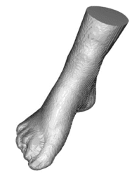
Figure 1 illustrates the three-dimensional digital model developed by the first procedure. In this scanning method, the patient needed to lie down with the foot supported on the plantar region and the posterior region of the ankle, favoring the positioning of this segment, to simulate the standing position.
Figure 1: Tomography-based digital scanning.
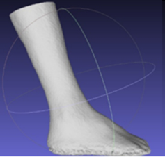
Digital scanning using motion sensor (Kinect): The digital model devel- oped with the Kinect method (Figure 2) was efficient, producing good quality, albeit with a smooth surface. However, the participant’s positioning was not entirely comfortable during scanning, and due to the pedal deformity of hemi- paresis and paraplegia in the entire left lower limb, scanning in the ankle joint region was impaired, as the angle was much greater than 90 degrees between the foot and leg. It is also noteworthy that the placement of markers in the scanned region contributed to obtaining more accurate measurements.
Figure 2: Kinect motion sensor digital scanning.
Microsoft’s Kinect is a low-cost 3D scanner compared to conventional three-dimensional scanning scanners on the market. According to some authors, Kinect has been used in virtual reality applications for physical spaces and in computer graphics, requiring realism in 3D models of human bodies. The Kinect can be used as a low-cost 3D scanner for generating 3D models and aiding in the development of product projects, especially for
Analysis of Procedures and Instruments for the Digitization of the Foot and Ankle 185
personalized products. As the Kinect for Xbox and Windows have been dis- continued, the newer Azure Kinect needs to be considered for future works. Regardless, the Kinect method provides instantaneous visualization of the modelling outcome during scanning, which contributes to making fewer scanning errors.
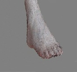
Digital scanning using digital photogrammetry: Figure 3 illustrates the dig- ital model (point cloud) of the participant’s right foot (hemiparetic foot). One hundred images were collected from the plantar, dorsal, ankle, hindfoot, midfoot, and forefoot regions.
Figure 3: Photographic camera scanning and agisoft metashape software.
The digital photogrammetry method experienced interference and pre- sented some flaws in the digital model generated, likely due to differences in brightness, especially between the plantar and upper foot regions, hinder- ing perfect image acquisition in these parts. Also, the patient had to hold her foot still during image capture, as images with different positions of the foot were impossible to align with other images during the photogrammetry pro- cessing. However, this method is easy to replicate and have low-cost as the image acquisition can be made with portable camera or smartphones easily accessible, For consideration, all three methods presented good results when consid- ering cost limitations and are applicable to the limb reconstruction to later model the orthosis, while the tomography method was the best regarding geometry accuracy.
CONCLUSION
After analyzing the three scans, the best scanning procedure was selected based on two criteria: 1 - better image quality and 2 - better foot positioning angle during scanning. The selected procedure was tomography scanning. In addition to offering the best image quality, it also provides the best ankle positioning. During the examination, the patient supported the plantar and posterior regions of the leg, allowing the most suitable ankle joint angle, simulating the standing position.
After analyzing the three scans, the tomography scanning procedure was selected as the best method for offering the best model quality and more foot positioning comfort during scanning.
It is emphasized that obtaining the anatomical model from tomography images was performed free of charge, and the model processing did not require paid software. However, the most viable way to perform tomography is in specialized laboratories, but the cost is high.
REFERENCES
Booher, Harold, ed. (2003). Handbook of human systems integration. New Jersey: Wiley. Honour, Eric C. (2006) “A Practical Program of Research to Measure Systems Engi-
neering Return on Investment (SE-ROI)”, proceedings of the Sixteenth Annual Symposium of the International Council on Systems Engineering, Orlando, FL. Castilho, K.; Martins, M. (2005). Fashion Discourses: Semiotics, design, and body.
Chap. 1. Fashion communication through design. Ed. Anhembi Morumbi, São Paulo. Fernandes, B. O.; Fogiatto, J. A.; Poier, P. H. (2015). Use of 3D printing in orthosis manufacturing - A case study. IN: Fourth International Conference on Integration of design, engineering, and management for innovation. Florianópolis. Hensen; J. C. D. (2019). Development of a procedure for the manufacturing of non-
articulated ankle and foot orthoses through Additive Manufacturing. 2019. 172f. Dissertation – Postgraduate Program in Mechanical and Materials Engineering, Federal Technological University of Paraná. Curitiba. Jenett, B.; Cameron, C.; Tourlomousis F.; Rubio, A. P.; Ochalek, M.; Gershenfeld, N.
(2020). Discretely assembled mechanical metamaterials. Science Advances, v. 6, n. 47. Mavroids, et al. (2011). Specific patient ankle-foot orthosis using Rapid prototyping.
Journal of neuroengineering and Rehabilitation. V. 8, n. 1. Mikolajewska, E.; Macko, M.; Ziarnecki, L.; Stanczak, S.; Kawalec, P.; Mikolajewski, D. (2014). 3D printing Technologies in Rehabilitation Engineering. Journal of Health Sciences, v. 4, n. 12, p. 78–83. Paterson, A. M. J. (2020). Digitization of the Splinting Process: Exploration and Eval-
uation of a Computer-Aided Design Approach to Support Additive Manufacture. 2013. 368 f. Doctoral Thesis – Loughborough University, Loughborough, 2013. Rodgers, H. Risk factors for the first-ever stroke in older people in the North East of England: a population-based study. Stroke, 35: 7–11. Roncoletta, M. R. (2011). The desire of women with physical disabilities in footwear
design. IN: I National Meeting of Fashion Research. Goiânia, Federal University of Goiás. Rosenmann, G. C. (2017). Evaluation of low-cost 3D scanning systems applied to the development of orthoses by additive manufacturing. 2017. 113f. Disserta- tion - Postgraduate Program in Mechanical and Materials Engineering, Federal Technological University of Paraná. Curitiba. Santos, R. M, Ventura, F. C.; Hernandes, F.; junior-marques, A; Guarnetti, J. E.;
Paschoarelli, L. C. (2018). Use Perception Analysis in Custom Made Footwear for People with Physical Disability In: Advances in Intelligent Systems and Computing.588 ed.: Springer International Publishing, pp. 581–590. Silva, A. D. M. M; Alves, F. J. L. (2015). Study of the potentialities of 3D printing in supporting the manufacture of shoes for people with cerebral palsy. Dissertation [master’s], Postgraduate Program in Mechanical Engineering, University of Porto. Porto, Portugal.
Analysis of Procedures and Instruments for the Digitization of the Foot and Ankle 187
Trombly, C. A. (2005). Occupational therapy for physical dysfunctions. 5th ed, São Paulo, Santos Livraria Publisher. White, H.; Jenkins, J.; Neace, W. P.; Tylkowski, C.; Walker, J. (2000). Clinically
prescribed orthoses demonstrate an increase in the velocity of gait in children with cerebral palsy: A retrospective study. Developmental Medicine & Child Neurology. v. 44.
LISTED IN THE AHFE DIGITAL BOOKSTORE CATALOGUE
Explore the complete digital volume
Healthcare and Medical Devices
Read this individual research paper free through the original publisher, or explore the full digital proceedings book.
ISBN 978-1-964867-06-9
Explore this ISBN in the AHFE Bookstore ↗Listed in the July 2026 bookstore catalogue; search by ISBN to confirm current availability · Digital book purchase optional