Yongkyun Lee, Hyesu Park, Seunghoon Yoo, Sungho Kim, Joonghoon Ko, Jongwoo Ha, Dongsoo Kim (2024). Detecting High-Risk Fatigue: Tracking with Alertness and Physiological Metric Pattern. In: Physical Ergonomics and Human Factors. AHFE Open Access, vol. 147, pp. 62–68. https://doi.org/10.54941/ahfe1005178
Fatigue is a recognized factor contributing to safety incidents and reduced task performance. However, classifying fatigue levels among workers in the workplace is a challenging task due to its subjective nature. This study aimed to establish universal criteria for identifying high-risk levels of fatigue. Industrial settings commonly use risk management checklists for this purpose. This study analyzed the correlation between fatigue levels and physiological indicators associated with fatigue, including alertness levels. The alertness levels were measured by the psychomotor cognition test (PCT), and the physiological indicators included salivary C-reactive protein related to cumulative fatigue, blood lactate concentration related to physical fatigue, and salivary cortisol associated with mental fatigue. Subjective fatigue levels were categorized into five levels, with Level 1 representing optimal mental and physical conditions and Level 5 indicating severe fatigue. Results showed that the mean reaction time in PCT at Level 5 significantly increased, and the success rate sharply decreased, demonstrating a significant difference compared to Levels 1 and 2 (p<0.01). Blood lactate levels were exhibited a positive correlation with fatigue levels, sharply increasing from Level 4. Blood lactate levels in Levels 4 and 5 were significantly higher than those of Levels 1 and 2 (p<0.01). Salivary C-reactive protein (CRP) levels were also showed a rapid increase from Level 4, significantly higher than those of Levels 1 and 2 (p<0.001). Salivary cortisol concentration at Level 5, however, was relatively lower compared to those of Levels 1-4. In conclusion, cumulative fatigue and high-risk levels of fatigue due to excessive workload are reflected in vigilance tests and physiological indicators. These indicators can be valuable in reducing subjective biases when assessing fatigue levels. The study suggests that profiling vigilance tests and physiological indicators associated with fatigue can be employed to screen for high-risk fatigue levels.
Detecting High-Risk Fatigue: Tracking With Alertness and Physiological Metric Pattern 63
INTRODUCTION
Fatigue is widely acknowledged as a significant factor contributing to safety incidents and diminished task performance (Williamson et al., 2011). Yet, classifying fatigue levels among workers poses a considerable challenge due to its subjective nature. Therefore, the evaluation of fatigue levels pre- dominantly depends on individual judgment, and systematic intervention is typically uncommon. Nevertheless, monitoring the fatigue levels of task performers, particularly in high-risk occupations such as pilots and long- haul drivers, proves to be a valuable risk management tool, not only for enhancing the qualitative aspects of task execution but also for preventing safety incidents (Aghdam et al., 2019; Kim & Lee, 2015). A more objective classification of fatigue levels is highly valuable for task-rest management, leading to accident prevention and cost savings. Accuracy in classifying fatigue levels should reduce dependence on individual subjective judgments. This can be achieved by comparing and analysing subjective fatigue levels with physiological indicators associated with fatigue (Koo et al., 2018). The physiological indicators emphasized in this study include blood lactate con- centration, which rises cumulatively with physical fatigue (Nozaki et al., 2009), salivary CRP associated with cumulative fatigue such as sleep depriva- tion (Strawbridge et al., 2019), and salivary cortisol associated with mental fatigue. Additionally, an acute fatigue level was assessed using a vigilance test. The Psychomotor Vigilance Test (PCT) was employed as the vigilance test, and PCT results were represented by mean reaction time and success rate, indicating attention maintenance (Lee et al., 2023). The goal of this study is to investigate patterns indicating severe fatigue and exhaustion based on discontinuous points represented by these indicators according to fatigue levels. The refined fatigue level data obtained from this study will be uti- lized as training data for a deep learning fatigue level classifier along with concurrently collected biological information.
Participant and Methods Subjects: The study enlisted a total of 170 participants, comprising indi- viduals from high-risk fatigue categories such as firefighters and nurses, alongside a control group composed of office workers. Within this partic- ipant pool, seventy-two were male, and ninety-eight were female, with an average age of 40.9 years. The Institutional Review Board at the Repub- lic of Korea Air Force Aerospace Medical Center granted approval for all experimental procedures involving human subjects (ASMC-21-IRB-005).
Table 1. The criteria for classifying subjective fatigue levels.
Table available in the original PDF paper.
Fatigue levels The state of awakening Level 1 The optimal state where work can be sustained for over 2 hours without a break. Level 2 A state where focus on work is possible, though not optimal. Level 3 A usual state, capable of handling work without difficulty, but easily influenced by environmental factors. Level 4. A tired state, able to work but feeling the need for rest. Level 5 Exhausted state, very tired but working out of necessity, both physically and mentally drained
Fatigue levels classification: The participants reported their fatigue lev- els according to the criteria presented in Table 1 and completed the Daily Multidimensional Fatigue Inventory (DMFI). The daily fatigue levels of the participants were determined based on the fatigue levels indicated in the DMFI. Fatigue level data where the participant’s self-reported fatigue lev- els differed by two or more levels from the fatigue levels in the DMFI were excluded from the fatigue analysis.
Alertness Test: The alertness test was performed through the PCT embed- ded in the Bio-Signals Collecting System for fatigue level classification (Lee et al., 2023). The PCT involves random presentation and disappearance of visual stimuli on a touch screen. Participants are required to touch the left screen as quickly as possible when two stimuli of the same colour appear, and the right screen when they are of different colours. After 20 repetitions of stimuli and responses, the mean reaction time and success rate are measured.
Physiological Indicator Analysis: The blood lactate concentration was measured on-site using Lactate Pro-2 (Arkray Inc, Japan). Salivary samples for cortisol and CRP level measurement were collected on-site using salivettes and stored at −20
C. Salivary CRP and cortisol concentrations were mea- sured using the Enzyme-Linked Immunosorbent Assay (ELISA) after thawing the frozen saliva samples. The quantification of CRP and cortisol levels through ELISA followed the procedures provided by the kit manufacturer, Salimetrics (USA).
RESULTS
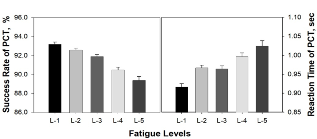
The mean reaction time and success rate in PCT: As represented in Figure 1, the success rate decreased as the fatigue level increased. Conversely, the mean reaction time increased with the rise in fatigue level. However, classifying the fatigue level based solely on the success rate and mean reaction time in the PCT proved insufficient. The most significant difference in success rate between neighbouring fatigue levels was observed between levels 3 and 4, while reaction time differences were notable between levels 1 and 2. Notably, the mean reaction time and success rate in PCT at Level 5 were found to be significantly different from those at levels 1 (p<0.01).
Figure 1: Success rate and mean reaction time in the PCT according to fatigue levels.
Detecting High-Risk Fatigue: Tracking With Alertness and Physiological Metric Pattern 65
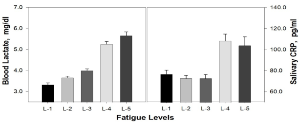
Blood lactate and salivary CRP concentrations: Figure 2 represents the changes in blood lactate and salivary CRP concentrations according to fatigue levels. Blood lactate concentration increased concomitantly with the elevation of fatigue levels, notably showing a significant rise between fatigue levels 3 and 4 (p<0.01). Salivary CRP concentration did not exhibit signif- icant differences based on fatigue levels. However, as observed in the right panel of Figure 2, it demonstrated a discontinuous upward trend between fatigue levels 3 and 4 (p<0.001). Ultimately, both blood lactate and salivary CRP, while not individually sufficient as physiological indicators for classify- ing fatigue levels, were confirmed as potential tools for binary distinguishing between absolute fatigue and non-fatigue states.
Figure 2: Blood lactate and salivary CRP concentrations according to fatigue levels.
Salivary cortisol concentrations: Salivary cortisol is a circadian rhythm- dependent arousal hormone that also functions as a stress hormone (Elverson & Wilson, 2005). Additionally, it can serve as an indicator of mental fatigue in situations where the distinction between mental stress and fatigue is ambiguous.
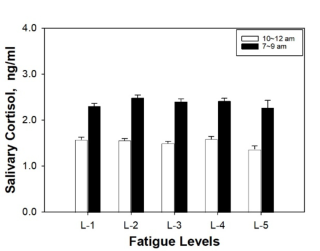
The concentration of salivary cortisol was analysed by separating the cor- tisol level changes between 7 and 9 in the morning, considering the rise immediately after waking from early morning, and the cortisol level changes during the rest of the day (see Figure 3).
Figure 3: Differential salivary cortisol concentrations according to fatigue levels.
The salivary cortisol level did not serve as a physiological indicator classi- fied by the level of fatigue. However, it relatively decreased at level 5, where fatigue levels were worst, both in the morning and throughout the day. This may be ultimately considered a reflection of the impairment in arousal due to extreme fatigue-induced exhaustion (Rothe et al., 2020).
CONCLUSION
Fatigue is a psychophysiological phenomenon that individuals frequently experience in their daily lives. People engage in various experiences and learn- ing during their daily routines, and the differences in experiences and learning contribute to create individual differences. These differences among indi- viduals also affect the perception of fatigue and the awareness of its levels. Dependence on subjective evaluation for fatigue levels may be insufficient for managers who must ensure fair and accurate mission-rest management and risk management. This study investigated whether fatigue levels that could affect task performance could be distinguished through physiological indica- tors. Alertness tests indicating acute fatigue measure the ability to maintain attention on tasks. The attention indicators obtained from alertness tests were not able to classify fatigue levels into 5 stages but were useful tools for classifying them into 2 or 3 levels. Blood lactate indicating physical fatigue and salivary CRP indicating cumulative fatigue were found to be useful tools for determining the presence of severe fatigue. Lastly, salivary cortisol, investigated as an indicator of mental fatigue, was insufficient as a fatigue classification indicator. Ultimately, it was concluded that fatigue classifica- tion based on a single physiological indicator could lead to another error. Therefore, this study suggests that the combination of these indicators can be used to classify fatigue levels accompanied by physiological phenomena to a limited extent.
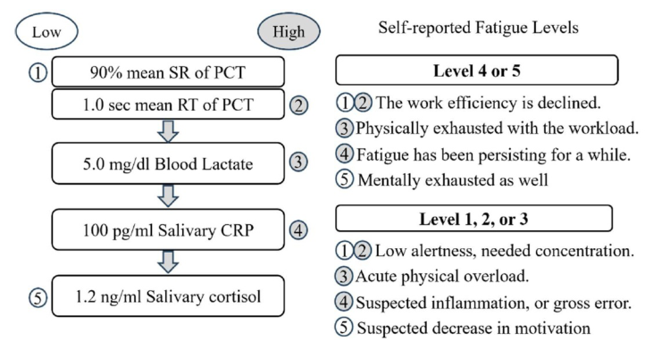
Figure 4: The pattern and meaning of physiological indicators depending on fatigue levels.
Detecting High-Risk Fatigue: Tracking With Alertness and Physiological Metric Pattern 67
In cases where the success rate in PCT is below 90% and the mean reac- tion time exceeds 1 second, it indicates a situation of acute fatigue where task efficiency decreases. Additionally, if the blood lactate concentration exceeds 5.0 mg/dl, it signifies a state of physical overload requiring rest. Moreover, if salivary CRP exceeds the level of 100 pg/ml, it indicates that the current fatigue is not acute but has been persistent for a long time, while salivary cortisol below 1.2 ng/ml indicates mental exhaustion. In cases where rest is necessary, the pattern of physiological indicators is shown as PCT’s SR down, longer RT, increased blood lactate, increased salivary CRP, and decreased salivary cortisol. The patterns and measurement values of alertness tests and physiological indicators can complement and aid in interpreting self-reported fatigue levels, as illustrated in Figure 4. In conclusion, cumulative fatigue and high-risk levels of fatigue due to excessive workload are reflected in vigilance tests and physiological indicators. These indicators can be valuable in reduc- ing subjective biases when assessing fatigue levels. The study suggests that profiling vigilance tests and physiological indicators associated with fatigue can be employed to screen for high-risk fatigue levels.
ACKNOWLEDGMENT
This research was supported by Civil Military Technology Cooperation Center, Republic of Korea (project code: 20-CM-BD-18).
REFERENCES
Aghdam, S. R. Alizadeh, S. S. Rasoulzadeh, Y. and Safaiyan, A. (2019). Fatigue Assessment Scales: A Comprehensive Literature Review. Archives of Hygiene Sciences, Volume 8, No. 3. Elverson, C. A., & Wilson, M. E. (2005). Cortisol: Circadian rhythm and response to
a stressor. Newborn and Infant Nursing Reviews, Volume 5, No. 4, pp. 159–169. Kim, D., & Lee, W. (2015). Development of Evaluation System for Aviation Mission Suitability Depending on Pilot’s Alertness and Physiological Stability Level. Jour- nal of the Korea Institute of Military Science and Technology, Volume 18, No. 6, pp. 789–796. Koo, H. Kim, C. Y. Kim, Y. Sin, S. J. Cheon, K. and Kim, D. (2018). Biomark-
ers for Combat-Related Stress and Fatigue-Mitigating Drugs Discovery. Journal of The Korea Institute of Military Science and Technology, Volume 21, No. 2, pp. 246–254. Lee, Y. Lee, Y. Yoo, S. Shin, S. Park, H. and Kim, D. (2023). The Psychomotor Cognition Test for Measurement of Sleepiness/Fatigue on a Touch Screen. In 2023 45th Annual International Conference of the IEEE Engineering in Medicine & Biol- ogy Society (EMBC). IEEE Open Access, pp. 1–4. IEEE Conference Publication, Australia. https://ieeexplore.ieee.org/document/10340988 Lee, Y. Lee, Y. and Kim, D. (2023). Bio-signals Collecting System for Fatigue
Level Classification. In 2023 45th Annual International Conference of the IEEE Engineering in Medicine & Biology Society (EMBC). IEEE Open Access, pp. 1–5. IEEE Conference Publication, Australia. https://ieeexplore.ieee.org/docu ment/10340350
Nozaki, S. Tanaka, M. Mizuno, K. Ataka, S. Mizuma, H. Tahara, T. Sugino, T. Shirai, T. Eguchi, A. Okuyama, K. Yoshida, K. Kajimoto, Y. Kuratsune, H. Kajimoto, O. and Watanabe, Y. (2009). Mental and physical fatigue-related biochemical alterations. Nutrition, Volume 25, No. 1, pp. 51–57. Rothe, N., Steffen, J., Penz, M., Kirschbaum, C., & Walther, A. (2020). Examination
of peripheral basal and reactive cortisol levels in major depressive disorder and the burnout syndrome: A systematic review. Neuroscience & Biobehavioral Reviews, Volume 114, pp. 232–270. Strawbridge, R. Sartor, M. L. Scott, F. and Cleare, A. J. (2019). Inflammatory proteins are altered in chronic fatigue syndrome—A systematic review and meta-analysis. Neuroscience & Biobehavioral Reviews, Volume 107, pp. 69–83. Williamson, A. Lombardi, D. A. Folkard, S. Stutts, J. Courtney, T. K. and Connor,
J. L. (2011). The link between fatigue and safety. Accident Analysis & Preven- tion, Volume 43, No. 2, pp. 498–515.
LISTED IN THE AHFE DIGITAL BOOKSTORE CATALOGUE
Explore the complete digital volume
Physical Ergonomics and Human Factors
Read this individual research paper free through the original publisher, or explore the full digital proceedings book.
ISBN 978-1-964867-23-6
Explore this ISBN in the AHFE Bookstore ↗Listed in the July 2026 bookstore catalogue; search by ISBN to confirm current availability · Digital book purchase optional