Yuki Mizuno, Shino Izutsu, Mayumi Nishikubo, Yoshihiro Hoshikawa (2025). Frontal Cortex Hemodynamics during measured by NIRS in a Rehabilitation Task of Cerebrovascular Disorders Using VR Technology. In: Accessibility, Assistive Technology and Digital Environments. AHFE Open Access, vol. 162, pp. 127–136. https://doi.org/10.54941/ahfe1006020
In recent years, digitization has spread in various medical fields, and there is growing interest in the introduction of VR technology, which can be enjoyed like a game, in the rehabilitation field. VR can train spatial cognition and attention to an object, and can also simultaneously train higher brain functions in people with cerebrovascular disorders. In this study, we investigated the brain activation state of people with cerebrovascular disorders when using a rehabilitation app. Based on the results, the characteristics of brain activity dynamics during VR use were examined. The subjects of the study were two people with cerebrovascular disorders (women in their 60s, returning to work, symptoms: right brain injury, left hemiparesis). Brain activity status was measured using NIRS (Near Infrared Spectroscopy) for Oxy-Hb, Deoxy-Hb, Total-Hb and StO2. Rehabilitation was performed using an app to train visual and cognitive functions, and before and after rehabilitation, a task to test attention function (TMT: Trail Making Test) was performed. Two devices were used: a tablet and VR. Oxy-Hb increased in most trials with the tablet device compared to the resting device, but decreased in half of the trials with the VR device compared to the resting device. Comparing tablet and VR, Oxy-Hb increased significantly for tablet (p>0.001), while Deoxy-Hb decreased significantly for VR (p>0.001) . In terms of StO2 results, both Tablet and VR conditions showed an increase in values compared to resting condition. During the TMT test, OxyHb and rSO2 after VR were significantly higher than those after Tablet use (p>0.05), suggesting that rehabilitation using VR may help maintain brain activation afterward.
In recent years, digitization using information and communication technologies has spread in various medical fields, and VR (Virtual Reality) has been actively utilized in telemedicine, education, and training (Hsieh and Lee, 2018; Sekiguchi and Makino, 2021). In the rehabilitation field, there is growing interest in the introduction of VR technology that can be enjoyed like a game, with the aim of improving the functional role of rehabilitation and patient motivation (Yoshioka, 2018; Kumita et al., 2020). In particular, although early start of rehabilitation is the shortest way
to recovery of ADL ability in cerebrovascular disorders, chronic pain and mental stress during rehabilitation and decreased motivation of patients for training are issues. Since VR is expected to alleviate pain and stress (Wong et al., 2021, Matsumoto et al., 2019), the introduction of VR in rehabilitation is considered more effective.
VR is able to present information to multiple senses, including visual, auditory, cognitive, and tactile, and is able to train spatial cognition (directional attention) and attention to necessary objects. It has also been reported that VR helps to recognize space better than PC screens, stimulating spatial awareness and having a positive effect on memory maintenance (Eric et al., 2018). Training with VR can be expected to improve higher brain function in people with cerebrovascular disorders.
In this study, we examined the state of frontal cortex hemodynamics during and after rehabilitation by using a VR and a tablet to manipulate a rehabilitation application that trains visual and cognitive functions in people with cerebrovascular disorders. Based on the results, the characteristics of hemodynamics responses in VR rehabilitation were discussed.
METHOD
Experiment Overview In this experiment, the participants were two women in their 60s with cerebrovascular disorders (Sub1 and Sub2). Both had damage to the right side of their brains and paralysis on the left side of their bodies. They were able to walk with a cane and had returned to work. After obtaining informed consent, the experiment began, and an occupational therapist accompanied the participants during the experiment.
A rehabilitation app was covering five areas of visual cognitive function (attention/memory/shape recognition/spatial recognition/motor integration) was used for approximately 10 minutes, and the Trail Making Test (TMT) was administered before and after the app use to assess attention function. During the experiment, NIRS (near-infrared spectroscopy) was used to measure the left and right frontal regions of the prefrontal cortex, and hemodynamics state during app use and task performance afterward was investigated.
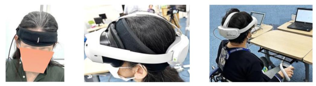
Used Devices Two types of devices were used in the experiment: the tablet (iPad) and the VR (PICO 4). The rehabilitation app used was “Visual Cognitive Balancer” (for iPad, PC) manufactured by Redix. To ensure that the same apps could be used on the tablet and in the VR, we installed the apps on a laptop computer and projected the PC screen onto the VR. VR screen simulated a theatre room-like setting in a movie theatre, with the rehabilitation app projected on the screen portion. The app was operated with the right hand on the non-paralyzed side, the tablet touch panel was operated with fingertips, and the VR was operated with the attached mouse (Figure 1).
Frontal Cortex Hemodynamics During Measured by NIRS in a Rehabilitation 129
Frontal Cortex Hemodynamics Data NIRS measurements were performed using Astem’s Hb134 (Figure 1), and the measurement sites were the prefrontal cortex corresponding to Fp 1 and Fp 2 in the international Electroencephalography 10–20 System.
The measurement indices were oxygenated hemoglobin (OxyHb), deoxygenated hemoglobin (DeoxyHb), total hemoglobin (TotalHb), and regional oxygen saturation (rSO2). TotalHb is the sum of OxyHb and DeoxyHb (TotalHb = OxyHb+ DeoxyHb), but since OxyHb and TotalHb sometimes have relatively similar waveforms, the results for TotalHb are omitted. rSO2 represents the percentage of oxygenated hemoglobin in total hemoglobin and is expressed as a percentage (rSO2 = OxyHb/ TotalHb).
Frontal cortex hemodynamics data were calculated as relative changes from the initial values at the start of measurement. In this experiment, the average values of the measurement indices measured at rest were used as reference values, and the increase or decrease trends from the reference values (1OxyHb, 1DeoxyHb) were used for evaluation. In VR conditions, the VR face cover may overlap with the NIRS, and securing the VR to the head may cause mild but temporary pressure on the head. In experiments where a head cuff was wrapped around the head to compress the superficial temporal artery, a decrease in skin blood flow in the prefrontal cortex and a decrease in 1OxyHb were reported (Hirasawa, 2016). Therefore, the evaluation of 1OxyHb and 1DeoxyHb during VR wear was based on hemodynamics data measured under Resting conditions immediately after VR wear.
Experimental Protocol The experimental protocol is shown in Figure 2. To eliminate the effect of order, the experiment was conducted twice on different days.
In the first experiment, after operating the tablet, the TMT was conducted, followed by a 15-minute repose, after which the VR was attached and the VR operations were performed. After completing the VR operations, the VR was disconnected and the TMT was conducted. In the second experiment, the order of the devices used was reversed, with the VR operation followed by the tablet operation. The interval between experiments was approximately one month.
Figure 2: The experimental protocol.
Result and Consideration Since NIRS data varies greatly among individuals, analysis was performed using differences from the reference values. However, in order to confirm the effects of frontal cortex hemodynamics due to order effects, data from each condition was analyzed individually rather than averaging data from two trials. In the second experiment of Sub2, data from one channel (left side) was missing, so the acquired data consisted of 8 data for Sub1 and 7 data for Sub2.
Regarding the significance test of hemodynamic data, since measurements were taken at 10 Hz per second, the amount of data became enormous, making it easy to obtain statistically significant differences even for minute differences. In this paper, we will not show the results of the significance test, but will describe what can be inferred from the trends in the magnitude of the hemodynamic data.
Frontal Cortex Hemodynamics During use of Rehabilitation App The app has 16 types of tasks, and four relatively simple tasks were performed in the same order under all conditions. From the start to the end of the tasks, Tablet condition took an average of 10.2 minutes (7.7-13.8 minutes) and VR condition took an average of 11.8 minutes (9.4-13.6 minutes). Since participants were observed to move their heads frequently before and after a rehabilitation task completion, we considered the possibility of effects different from activation (Yoshii and Takahashi, 2013) and analyzed hemodynamics responses during a rehabilitation task using data from the first 7 minutes after starting the app.
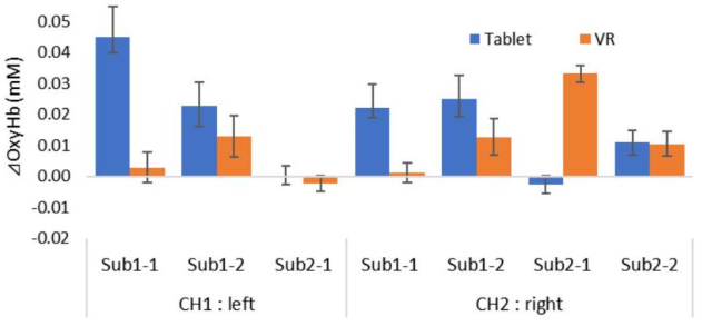
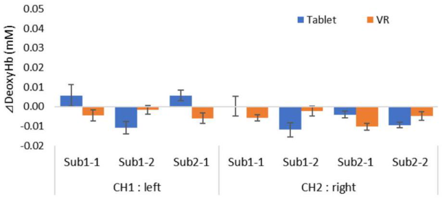
The results of hemodynamics response are shown in Figures 3-5. Compared to a resting state, the difference in hemodynamic response increased 1OxyHb in both Tablet and VR conditions. Except for the two Tablet conditions, 1 DeoxyHb showed decreasing trend.
Regarding frontal cortex hemodynamics, it is generally defined as an increase in OxyHb accompanied by a decrease in DeoxyHb, indicating activation in that region and suggesting neural activity within the brain. This is based on the phenomenon observed in fMRI, widely used in brain function research, where blood flow increases in regions where neurons are activated. When neurons are activated, blood flow increases in that area, and arterial blood containing a large amount of OxyHb flows in, diluting the amount of DeoxyHb from the capillary bed to the veins (Ogawa et al., 1990). The results of this experiment showed that the trials that corresponded to the activated state of frontal cortex hemodynamics were three trials in Tablet condition
Frontal Cortex Hemodynamics During Measured by NIRS in a Rehabilitation 131
(CH1: Sub1-2, CH2: Sub1-2, Sub2-2) and five trials in VR condition (CH1: Sub1-1, Sub1-2, CH2: Sub1-2, Sub2-1, Sub2-2).
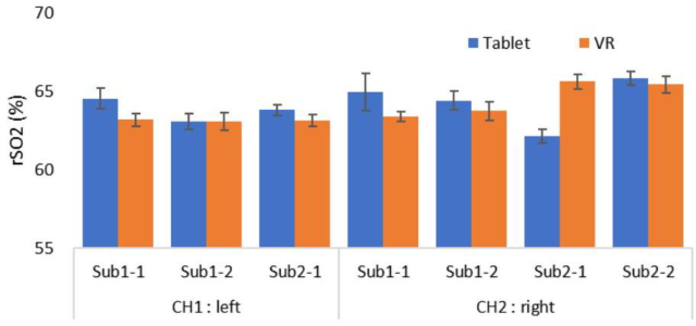
Comparing local oxygen saturation (Figure 5), rSO2 of Tablet and VR conditions showed higher values than that of Resting condition. In addition, comparing Tablet and VR conditions, rSO2 of Tablet condition showed higher values than VR condition in all conditions except CH 2 : Sub2 −1. It has been reported that during exercise, oxygen supply to the brain is necessary to meet the increased metabolic levels of neurons (Ide and Secher, 2000; Secher et al., 2008), and in experiments investigating exercise and cognitive function, brain tissue oxygen saturation increased during task performance compared to rest (Ando et al., 2015). Therefore, in this experiment, rSO2 values were higher in both Tablet and VR conditions than at Resting condition, indicating that frontal cortex hemodynamics were activated during use of the rehabilitation app.
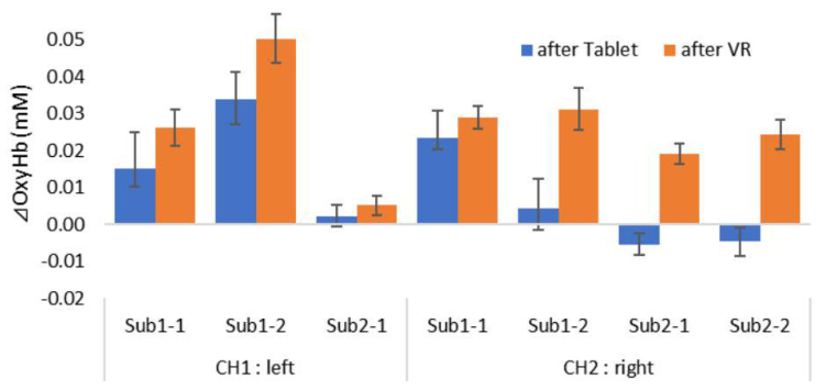
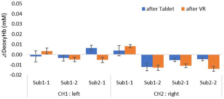
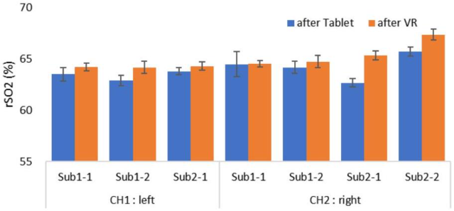
Figure 3: Changes in OxyHb during use of rehabilitation app.Figure 4: Changes in DeoxyHb during use of rehabilitation app.Figure 5: Changes in rSO2 during use of rehabilitation app.
Frontal Cortex Hemodynamics During TMT Performance There are two types of TMT tests: the TMT-A test (a test in which numbers are arranged in order) and the TMT-B test (a test in which numbers and letters are connected alternately). These two tests are administered as a set. This test provides a simple way to assess attention and executive function. In experiments where was administered using NIRS, it has been shown that activity in the prefrontal cortex is enhanced (Murata et al., 2008). From the start of the TMT-A task to the completion of the TMT-B task, the average time required was 3.47 minutes (3.433.58 minutes) after Tablet use and 3.35 minutes (3.02–3.90 minutes) after VR use. Brain activity during TMT performance after rehabilitation was analyzed using data from the first 3 minutes after the task began.
The results of hemodynamics response are shown in Figures 6-8. Comparing hemodynamics response differences relative to resting levels, 1OxyHb decreased in two trials under Tablet condition, but increased in all other trials, while it increased in all trials under VR condition. Comparing 1OxyHb between Tablet and VR conditions during TMT task performance, 1OxyHb in VR condition increased in all trials. Additionally, compared to 1OxyHb in VR condition when using the rehabilitation app (Figure 3), the values were clearly higher, and the absence of VR goggles is also thought to be a factor contributing to the higher values. 1 DeoxyHb showed a decreasing trend in many trials. It has been reported that cerebral blood flow increases during the performance of three-dimensional spatial cognition tasks (Murai et al., 2013), and it is possible that the VR screen has a wider field of view than the tablet and provides stronger stimulation for visual cognition.
The state where 1OxyHb increases simultaneously with a decrease in 1DeoxyHb (entering the negative range), which defines frontal cortex hemodynamics, was more commonly observed on the right brain side under VR conditions. In cases of cerebrovascular disease patients with right brain damage, there is a symptom called “left hemispatial neglect,” where the left side of the visual field is missing. The VR screen simulates a 100 -inch display, and participants must consciously expand their visual space while performing
Frontal Cortex Hemodynamics During Measured by NIRS in a Rehabilitation 133
the task. Therefore, during VR operation, the right brain side associated with spatial recognition ability may have responded more strongly, and activation of the right prefrontal cortex may have continued during subsequent tasks.
The results of local oxygen saturation (Figure 8), the rSO2 in VR condition was higher in all trials. In addition, it was demonstrated that the oxygen saturation ratio in the VR condition was the highest compared to the rSO2 in the pre-rehabilitation task, Tablet condition, and VR condition. Therefore, it is suggested that VR rehabilitation has the effect of maintaining the activation state of frontal cortex hemodynamics in the prefrontal cortex even after rehabilitation app use.
Figure 6: Changes in OxyHb during TMT after rehabilitation.Figure 7: Changes in DeoxyHb during TMT after rehabilitation.Figure 8: Changes in rSO2 during TMT after rehabilitation.
Limitations of this Study The NIRS device used in this experiment was a lightweight, bandana-type wearable device to minimize the burden on the participants. When wearing VR goggles, it is necessary to press the goggles against the face to adjust the interpupillary distance (focus). After focusing, the goggles are fixed to the head, causing moderate pressure on the head. For this reason, the values of OxyHb and DeoxyHb decreased when VR goggles were worn. Alternatively, the use of MR glasses that do not compress the head should be considered.
In addition, it is necessary to consider frontal cortex hemodynamics caused by factors other than neural activity related to task performance. For example, when operating equipment during task performance, the movement of the operating hand may stimulate the opposite side of the brain (hand moved: right hand, brain responding: left side). Furthermore, in cases of cerebrovascular disorders, it has been pointed out that even in a seated posture, left-right differences are likely to occur and posture is likely to be unstable (Kawate, 1997). It is possible that VRinduced head load and controller operation may cause fluctuations in the body’s center of gravity (Tsutsumi and Ishihara, 2021). When tasks to maintain balance in a seated position are added to VR operation tasks, multitasking may affect frontal cortex hemodynamics. In the future, we would like to examine the characteristics of frontal cortex hemodynamics in cerebrovascular disorder patients based on the location of brain damage, the degree of paralysis, and characteristics such as shifts in the center of gravity in a seated or standing position.
CONCLUSION
In this study, we investigated blood flow dynamics in frontal cortex hemodynamics during and after rehabilitation using a visual and cognitive function training app in cerebrovascular disorders. Based on the results, we examined the characteristics of hemodynamics response in VR rehabilitation.
During rehabilitation, compared to Resting state, oxygenated hemoglobin and oxygen saturation levels of Tablet and VR condition, indicating that
Frontal Cortex Hemodynamics During Measured by NIRS in a Rehabilitation 135
frontal cortex hemodynamics were activated during use of the rehabilitation app. During the TMT test, the levels of oxygenated hemoglobin and oxygen saturation after VR condition were higher than after Tablet condition, suggesting that VR rehabilitation maintains frontal cortex hemodynamics even after the rehabilitation task.
ACKNOWLEDGMENT
This work was supported by JSPS KAKENHI Grant Number 23K02017.
REFERENCES
Ando, S., Komiyama, T., Sudo, M. and Higaki, Y. (2015) Acute Exercise and Cognition: Effects of Cerebral Oxygenation. Fukuoka University review of sports and health science, 45.2: 25–34. Ide, K. and Secher, N. H. (2000) Cerebral blood flow and metabolism during exercise.
Progress in Neurobiology, 61(4), 397–414. Eric, K., Catherine, P. and Amitabh, V. (2019) Virtual memory palaces: immersion aids recall. Virtual Reality, Vol.23, 1–15. Kawate, N. (1997) The Sway Paths of the Centre of Gravity in the Static-standing
Posture, Static-sitting Posture and the Voluntary Shifting-sitting Posture of Hemiparetic Stroke Patients. The Japanese Journal of Rehabilitation Medicine, 997; 34: 121–128. Kumita, H., Simaka, N. and Imaizumi, K. (2020) Challenges of Virtual Reality Technology in Health Care. Journal of the Society of Instrument and Control Engineers, 59 (4), 269273. Hirasawa, A., Kaneko, T., Tanaka, N., Funane, T., Kiguchi, M., Sørensen, H.,
Secher, N. H. and Ogoh, S. (2016) Near-infrared spectroscopy determined cerebral oxygenation with eliminated skin blood flow in young males. Original Research. Journal of Clinical Monitoring and Computing, Vol.30, 243–250. Hsieh, M. C. and Lee, J. J. (2018). Preliminary study of VR and AR applications in medical and healthcare education. J Nurs Health Stud, 3(1), 1–5. Ogawa, S., Lee T. M., Kay A. R. and Tank, D. W. (1990) Brain magnetic resonance
imaging with contrast dependent on blood oxygenation. Proceedings of the National Academy of Sciences of the United States of America, 87: 9868–9872. Matsumoto, K., Kawachi, M., Morishige, Y., Shinagawa, A., Numata, M., Sugihara, T. and Yoshimura, K. (2019) Effects of virtual reality videos on relieving mental stress. Studies in Science and Technology, Vol.8, No.2, 137–143. Murai, Y., Takahashi, S., Kodama, N., Takeuchi, H., Kosugi, N. and Shimizu, S.
(2013) Measurement of the prefrontal cortex amount of brain blood task. Journal of the Japanese Society for Dementia Prevention, Vol.2, No.1, 14–17. Murata, S., Murata, J., Kodama, T. and Tanaka, S. (2008) Changes of Cerebral Oxygenation by Near Infrared Spectroscopy in Community-Dwelling Elderly Performing the Trail Making Test. Rigakuryoho Kagaku, 23(5): 561–565. Secher, N. H., Seifert, T. and Van Lieshout, J. J. (2008) Cerebral blood flow and
metabolism during exercise: implications for fatigue. J Appl Physiol (1985), 104(1), 306–314. Sekiguchi, T and Makino, M. (2021) A virtual reality system for dissecting vertebrates with an observation function. 2021 International Conference on Electronics, Information, and Communication (ICEIC). IEEE, 1–4.
Tsutsumi, K. and Ishihara, M. (2021) A Study on Impact of Binocular Parallax and Head Position/Posture Tracking on Vection in Immersive VR. The Virtual Reality Society of Japan, Proceedings of the 26th Annual Conference, 2D3-8. Wong, M. S., Brennan, MR S. and Kimberly D. G. (2021) Virtual reality reduces
pain in laboring women: a randomized controlled trial. American journal of perinatology, 38.S 01: e167–e172. Yoshii, K. and Takahashi, M. (2013) Experimental study on the influence of head movement during ultra-small NIRS device measurement. The Society of Instrument and Control Engineers (SICE), Tohoku Chapter, Proceedings of the 285th Research Meeting, Document Number 285-4. Yoshioka, K. (2018) Development and Psychological Effects of a VR Device
Rehabilitation Program: Art Program with Feed Back System Reflecting Achievement Levels in Rehabilitation Exercises. Proceedings of the 7th International Conference on Kansei Engineering and Emotion Research 2018, 358–364.
LISTED IN THE AHFE DIGITAL BOOKSTORE CATALOGUE
Explore the complete digital volume
Accessibility, Assistive Technology and Digital Environments
Read this individual research paper free through the original publisher, or explore the full digital proceedings book.
ISBN 978-1-964867-38-0
Explore this ISBN in the AHFE Bookstore ↗Listed in the July 2026 bookstore catalogue; search by ISBN to confirm current availability · Digital book purchase optional