Wiktoria Spikowska-Pawelec, Michal Rychlik, Agata Rzepnicka (2018). Biomechanical Evaluation of Professional Violinists Musculoskeletal System Overloading – Motion Capture, Electromyography and Thermovision Study. In: Advances in Human Factors and Sustainable Infrastructure. AHFE Open Access, vol. 3. https://doi.org/10.54941/ahfe100102
The work of professional instrumentalists requires a great effort and many hours of daily practice. The musculoskeletal system is under a considerable strain, which is often excessive and taking the form of overload. In the specialist literature conditions of this type are referred to as "playing-related musculoskeletal disorders (PRMDs)" of instrumental musicians. In the violinists these dysfunctions or diseases affect mostly the areas of the right shoulder and elbow, left hand and fingers, and cervical spine. This article presents some biomechanical methods of measuring the movement of violinists during the play. The study was performed using exoskeleton Motion Capture system. The authors present also the electromyography as a method for evaluating muscle tension. It enables the reference to ergonomic standards, applied to repetitive or monotype physical activity, and makes it possible to compare the involvement of individual muscles during play. Additionally, interesting observations concerning an instrumentalist’s body at work can be made using thermal imaging equipment. This method of measurement reflects physiological processes that accompany the effort. Research methods used in the article may help to determine the etiology of the most common ailments in string instrumentalists, provide accurate and objective biomechanical evaluation of the motion loads of musicians and may also pave the way for an effective prevention of diseases in this professional group.
The work of professional instrumentalists is a kind of activity that largely involves the load on human motoric system. Daily practice of musicians and frequent performances are a significant strain for their muscles and joints, which may contribute to ailments and diseases. In the specialist literature they are referred to as “playing-related musculoskeletal disorders (PRMDs)” (Zaza Ch., 1998, Bodnar A., 2006). It is a term that corresponds to term used in the ergonomics, in much broader sense: “work-related musculoskeletal disorders (WRMDs)”. Medicine defines overstrain as the consequences of excessive or repeated, or unusually long-lasting strain on bones, joints and muscles (Strzyzewski W., 2006). Such overstrain is observed primarily in three situations: – among intensively training athletes, – among individuals who include in their schedule unusual or seasonal types of activities, – among workers with a significant physical load and having primary deformities in their locomotor system. A typical example of such disorders of the upper limb is painful shoulder syndrome, tennis elbow (inflammation of muscle attachments of the lateral epicondyle of the humerus), and golfer’s elbow – inflammation of the muscle attachment of the medial epicondyle (Strzyzewski W., 2006). From the ergonomic point of view it is important to analyze the overstrain in terms of its etiology. Some authors (Roman-Liu D., Konarska M., 1997) list the following factors as the causes of overloads of the locomotor system: – incorrect position during work, – excessive forces to which the human body is subjected, such as heavy lifting, – high frequency of repetition, – long isometric muscle tension, not necessarily of high value. According to the authors, the physical condition of workers may deteriorate as a result of monotony of activities, automatic movements performed at a significant speed and insufficient intellectual commitment. Wagrowska-Koski (Wagrowska-Koski E., 2008) describes in her article a relation between the method of work and diseases of the musculoskeletal system. The author indicates the significance of normative breaks between repetitive work activities.
During the last 30 years an interest in health issues of artists-performers increased. The branch of medicine dealing with them is called “performing arts medicine”. An important step in its development was the publication of “Music and the Brain: Studies in the Neurology of Music” by Henson R.A. & Critchley M.D. in 1977 and “Tension in the Performance of Music” by Grinde C. one year later (Harman S.E., 2010, Bodnar A.., 2006, Bejjani F.J., et al., 1996).
In many countries epidemiological studies of diseases among orchestral musicians are carried out. With the use of questionnaires very large populations are studied, from hundreds to thousands of instrumentalists (Lederman R.J., 2003, Bodnar A., 2006). The Lederman (Lederman R.J., 2003) reports that of 1353 examined musicians 64% reported musculoskeletal disorders, the majority of whom were string musicians (52,3%). The author confirms the possibility of returning to the professional activities of these musicians, if their diagnosis was correct and accurate treatment was applied. According to Bejjani (Bejjani F.J., et al., 1996) – recalling their own research from 1984, and works of Dawson from 1988, Fry'a from 1986 – violinists are a group of instrumentalists experiencing most of all overstrains, ailments, disorders and dysfunctions. Bodnar (Bodnar A., 2006) citing Heming’s work entitled “Occupational injures suffered by classical musicians through overuse” confirms a particularly high risk of overstrain in string instrumentalists. Brandfonbrener (Brandfonbrener A.G., 2010) reports that 65-70% of violinists, have musculoskeletal disorders, and that is the highest scale of the problem among all orchestral musicians.
Modern epidemiological research in such countries as Germany (Gembris H., Heye A., 2012), Spain (Lopez T.M., Martinez J.F. 2013), Greece (Fotiadis D.G., at all., 2013), or Republic of South Africa (Ajidahun A.T., Phillips J., 2013) proves that the problem is common and on the increase, despite the growing scientific knowledge and regular conferences in the area of “performing arts medicine”.
In 2010-2011 a study was carried out among string musicians (violinists, violists, cellists and double bass players) in major orchestras of the Wielkopolska region in Poland. The results of this work were presented at the international symposium “Gesund Musizieren 2013” in Vienna (Pawelec W., Sierszenska-Leraczyk M., 2013). Ailments in the locomotor system related to work were reported by 73.6% of several dozen examined violinists. Polish musicians who experienced overstrain rarely referred for treatment to a doctor (22.7%) or physiotherapist (24%); usually (53.3% of the respondents) they used painkillers as the sole form of treatment. Nearly half of the participants (43%), from the persons which despite experienced pain and continuing job, is willing to change the technique of playing to incorrect (use motion compensation) if only to keep the correct sound track execution. Such a result of the study persuaded the authors of this paper to carry out detailed measurements of kinematic parameters of the movement of the right upper limb violinist while playing the instrument. The results of the study and analyses are presented and discussed in sections below.
MOVEMENT MEASUREMENTS OF VIOLINISTS' Motion Capture System
One of the relatively new techniques which has been used in biomechanics, medical diagnostic and rehabilitation is the technique connected with motion capture systems (Rychlik M., 2010, Rychlik M., Stanowski A., 2012). The Motion Capture systems (also known as Motion Tracking or MOCAP) provide digital recording of the motion parameters of a real person. These systems altogether with the applications of computer analysis allow for a full interaction between the user and the computer model in a virtual space (Rychlik M., 2009, Branowski B., et al., 2011). Four basic equipment groups are distinguished in the MOCAP technique: mechanical, optical, magnetic and inertial. The main difference between them is the data acquisition method, complexity level of the equipment, accuracy and data acquisition speed.
In the presented biomechanical study a mechanical motion capture system (Gypsy 5) was used for the measurement of human body movements. The measuring system of Gypsy 5 consists of 37 potentiometers and two electronic gyroscopes placed on the so-called exo-skeleton. The position of a person is analyzed and calculated on the basis of data from the sensors (rotation) in each module of the skeleton. Gyroscopes are responsible for the orientation of the body in the global space. Moreover, they are responsible for defining the torso rotation relative to the base point (the center of the pelvis - the so-called "root"). The structure of the device is made of rigid elements connected together by joints, respective to the number degrees of freedom of the joint (Figure 1.a). Potentiometers measuring the angle of rotation of the joints are installed in each joint.
Figure 1. Mechanical motion capture system Gypsy 5: a) musician with the exoskeleton on, b) hierarchical structure used to describe the human model (Meredith M. & S. Maddock, 2009)
Figure available in the original PDF paper.
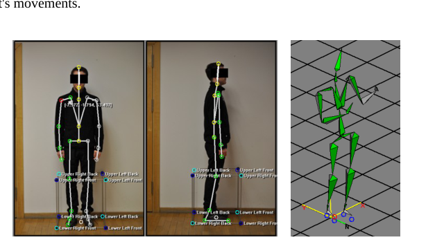
The data obtained from the measurements have a hierarchical structure (Figure 1.b) and consist of a 23-24 segments defining individual parts of the human body (Meredith M. & S. Maddock, 2009). Data files (saved as BVH file format) contain information on the structure and movement of individual segments relative to the segment located higher in the hierarchy. The main component is called node "root", which has full 6 degrees of freedom. Towards him are wired further segments, wherein one end is located on the beginning of the next, situated below in the hierarchy. For a full definition of the system it is necessary to determine the length of each segments, according to the anthropometric dimensions of a person equipped with a skeleton. The first phase of research procedure is to calibrate the system for adapting the device to the dimensions of the examined person. For calibration it is necessary to take two photographs (from profile and an en face) Sustainable Infrastructure (2018) with a special element calibration (Jig) as a cube composed of rods of known dimensions. The next step is to put the images to the special calibration program (Figure 2). This program defines the reference points on a cube (the corners) and then specific nodes exo-skeleton corresponding to real human skeleton joints. This way prepared calibration data give possibility to determine the necessary coefficients required to properly interpretation the results of measuring the subject's movements.
Figure 2. Calibration procedure (from left): calibration images with marked reference points of Jig and skeleton nodes, visualization of the skeleton in the recording program
Motion measurements of the Violinists In order to enable the analysis and the comparison of results obtained from measurements taken for various violists (different anatomical structure, individualized apparatus of play), measurements of all participants during performed of the same musical composition Rodolphe Kreutzer's Etude no. 2 with the forced upon them rate of 120 beats per minute. The use of a unified rate of the game, allows you to compare the obtained data: values of angles, accelerations, and other kinematic and dynamic parameters. Each person had to perform a given composition in five basic techniques of bowing: Lower detache, Upper detache, Spiccato, Sautille, Slurring four notes (Legato in 4).
Detache - this is the easiest form of the string play, includes all smooth changes of direction of the bow, across the entire range of bow. Bow carried out without stopping the hand, when changing direction and without accents. This technique can be played in different tempos and different parts of the bow, are distinguished: lower detache, upper detache, middle detache. In this technique, the movement of the bow is responsible arm and forearm, and the hand is a passive relay movement. Legato - is the combination of sounds (tunes) into a curve, its mean one dragging the bow. Most important is to bow smoothly and fluently touched the strings, without misfeeds and stops. In this type of play it should be ensured that there is a constant contact with the strings of the bow. Sautille - jumping bow, the basis of the play is to bow lying detache that with the fast direction changes automatically breaks away from the strings, which performs "jumps". Spiccato - the bow is in a swinging movement, every sound requires a separate pulse. This technique is characterized by a fluid hand movement, but an intermittent sound of the violin.
In the current study the participants were a group of several dozen musicians, for whom measurements were made using the exoskeleton. However, due to the fact that measurements are still continued and a large number of data are still in the development, this article presents an analysis of the results on two selected persons. These individuals were selected so that they have the largest and lowest anthropometric dimensions of the studied group of musicians. Person A - No. 21 (female) was characterized by the height of 158 cm, the length of the arm of 0.67 cm and a weight of 53.2 kg. Person B - No. 17 (male) was characterized by the height of 183 cm, the length of the arm of 0.74 cm and a weight of 67.4 kg.
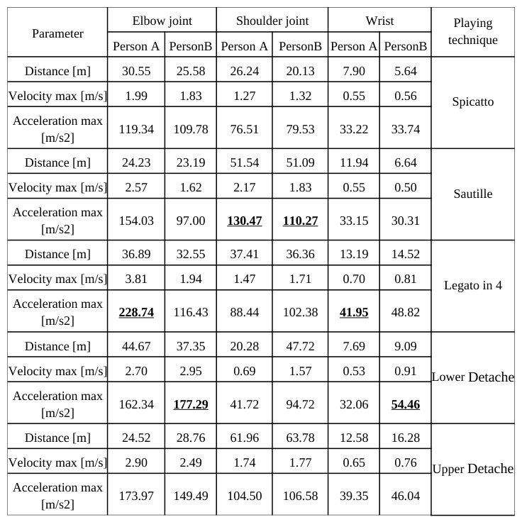
According to the pool results the parameters of movement of the right hand for a Kreutzer’s Etude performed with various techniques were analyzed. The results of the individual parameters are presented in table 1.
Table 1: Comparison of selected parameters of movement of the right hand for a Kreutzer's Etude made by various techniques
The analysis of the play is mainly based on the identification and description of the behavior of fiddler’s hand during the play. An interesting way to understand the difficulty of playing is to determine what distance in a straight line in covered by a hand during such a short time. In the cases analyzed in presented work the distance of the hand movement, for the lowest person (1.58 [m] height - Person A No. 21) at the elbow about 62 [m], while for the highest person (1.83 [m] growth - Person B No. 17) at the elbow about 64 [m].
An interesting fact is that the greatest differences that can be noted do not occur between the same violinists (Figure 3), but in the comparison of various playing techniques (Figure 4). It turns out that the most challenging playing technique in terms of distance covered proved to be lower detache (lower half of the bow), when the "shortest" technique is spiccato (short jerking and bouncing bow). For person A - No. 21 the distance was approximately 30.5 [m], and for person B No. 17, this distance was approximately 25.6 [m].
Figure 3. Rotation of the glenohumeral joint during play of Kreutzer’s Etude executed spiccato technique: a) person A – No. 21, b) person B – No. 17
Figure available in the original PDF paper.
Average speed noted in the analysis of the upper extremity is in the range from 0.26 to 0.65 [m/s]. The maximum speed recorded during the measurement is 3.8 [m/s] was noted for person B No. 21, and in the same technique the hand of person A moves at a speed of 1.9 [m/s]. These speeds occurred in the shoulder joint during playing legato in 4 technique.
A very important parameter for the musicians is the overload related to acceleration. The value of the maximum acceleration measured for the shortest person in the group of respondents (person A) is approximately 228 [m/s2] and for the tallest person (person B) about 177 [m/s2]. Another important parameter from the point of view of ergonomics is to determine the so-called position of the average silhouette of the musician. This will make it possible in future to determine the strains of musicians in relation to the standards indicators. The average position during the performance of a composition for a person A (No. 21) for techniques of spiccato, sautille and lower détaché (Figure 5) was determined.
Figure 4. Rotations of arm joints for person A - No. 21 during playing Kreutzer’s Etude using various techniques: Sustainable Infrastructure (2018)
Figure available in the original PDF paper.
a) Spiccato, b) Sautille, c) Legato on 4, d) upper Détaché
Figure 4. Visualization of average position of hand joints of person A - No. 21 during playing Kreutzer’s Etude using various techniques: a) Spiccato, b) Sautille, c) upper Detache
Figure available in the original PDF paper.
SURFACE ELECTROMYOGRAPHY (SEMG) OF VILONISTS' UPPER EXTREMITY MUSCLES Thanks to 16-channel telemetric sEMG Noraxon device (TeleMyo 2400T G2) the electrical activity of five selected muscles were recorded. The EMG signal is based upon action potentials at the muscle fiber membrane resulting from depolarization and repolarization processes. A raw sEMG signal can range between +/- 5000 microvolts and typically the frequency content ranges from 6 to 500 Hz (Konrad 2005). The surface electrodes, well fixed to previously prepared skin, were used. The electrodes were placed according to the SENIAM indications
(Hermens H., at all, 1999). For the analysis the raw signal was rectified (negative amplitudes are converted to positive amplitudes) and filtered (RMS in 300ms time window).
The analyzed muscles were: trapesius pars descending - bilaterally and deltoideus pars medialis, triceps brachii cap. latteralis, biceps brachii cap. longus - ipsilateral to the hand with the fiddlestick. Every musician played the Etude using five techniques reffered to above.
For the ergonomic assessment and in order to enable comparisons between the fiddlers the amplitude values were normalized to those of Maximal Voluntary Isometric Contraction (Marras W.S., Davis K.G., 2001). The tests were carried out after playing and were preceded by description and training. For each measured muscle there was a task, in which the maximal contraction was held for 3 seconds. After 10s rest the task was repeated twice. All the tests were carried out on the station which consisted of a table and chair with armrests – the resistance was given externally, manually by the therapist.
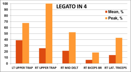
The set of graphs below (Figure 5) presents the results of EMG measurements results for person no.39. It shows mean and peak EMG amplitudes relative to MVC values in five fiddling techniques.
Figure 5. The graphs of mean and peak amplitudes of muscles electric activation relative to MVC in five chosen techniques of person no.39
The most characteristic here is a very high electric activity of both trapezius muscles in every technique. It can be due to the trunk asymmetry of the studied person and habitual tension during violin playing. On the other hand the values for the deltoid muscle are very diverse depending on the technique.
The rate of involvement of the measured muscles varied individually between the tested musicians, even though they all performed the same composition. Nevertheless, the tendency of highest amplitudes was observed in legato in 4 technique for most of muscles in whole group. It is significant that Rodolphe Kreutzer's Etude no. 2 which was played during the experiment, was considered by the examined violinists to be a short and easy composition. When they performed more advanced compositions muscle activity increased.
According to Roman-Liu (Roman-Liu and Konarska, 1997), every work exceeding 15% of MVC should be treated as tiring and heavy. The muscle activity performed by the violinists is of a mixed type – mostly monotype (more than 40 contractions/repetition per minute) and static.
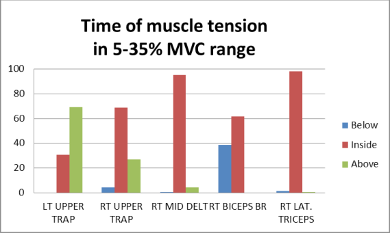
The graph below (Figure 6) shows percentage of time, in a 96s long composition played using legato in 4 technique, for which the amplitude is in the range from 5 to 35% of MVC. This clearly means that for the person no.39 all selected muscles were in nearly constant tension. Such an effort undoubtedly leads to muscle overstrain and injuries
Figure 6. The graph of percentage of time of muscle tension in 5-35% MVC range for Person no.39 during playing legato in 4 technique
THERMOVISION OBSERVATION OF VIOLONISTS Modern thermographic cameras are increasingly often used in medicine, not only to confirm changes in temperature of body surface during various physical therapy procedures, but also in order to make a medical diagnosis. According to Ring (Ring EFJ, 2004): “Today, applications (…) include the quantitative study of inflammation and anti-inflammatory therapies, peripheral circulatory disorders related to connective tissue diseases, and Raynaud's phenomenon, sympathetic disturbances, pain syndromes and locomotor injuries”.
For infrared imagining a thermographic camera (NEC-AVIO TVS-200EX) was used. The camera uses the 8-14µm wave band, temperature resolution better than 0.080C (with sensitivity of 80mK). Such parameters are the most suitable for temperature observation of human skin. The camera was equipped with a high-speed (60 Hz) uncooled FPA 320x240 (HxV) pixels VOx (vanadium oxide) microbolometer. This equipment allows images with spatial resolution of 1.68 mrad and field of view (FOV) 30.60x23.10 (HxV) with using the standard 14mm lenses to be obtained for the visualization of hot spots, from about 2m distance. For analysis of thermal images a specialized program, “Thermography Studio 2007 Professional” was used. To eliminate errors due to the influence of external temperature on a musician’s body temperature and to ensure the adjustment of the recording camera’s temperature to the interior conditions, the measurements began at least 30 minutes after the person had entered the measurement room.
Body temperature of a violinist was observed during a one hour practice session. The musician performed work with Sustainable Infrastructure (2018) an average intensity, similar to his or her everyday practice. Using the thermographic camera the temperature was measured at the beginning of the artist’s work and then every 15 minutes. The examined area included upper extremities, however at the beginning and at the end of the one hour practice also the temperature of the neck, nape of the neck, shoulders and hands was recorded.
Figure 7 presents the distribution of temperatures along the right upper extremity of a violinist (person no. 1) in three selected points of time during the practice.
Figure 7. Distribution of temperature along the upper extremity for person no. 1 in three situations: a) at the beginning of the practice, b) after 30 minutes, c) after 60 minutes – at the end of the practice The highest temperature was noted in the shoulder and elbow areas, where it increases slightly for approx. 45 minutes of the practice, and then decreases. The lowest temperature was noted on the wrist and hand. In majority of participants the distal parts of the hand become colder towards the end of the practice. The study shows a clear difference between temperatures of the upper left and right extremities which is a consequence of a different nature of work of both extremities in violinists. These results may be the basis for the assessment of physiological phenomena in a motor system of a musician and may be related to etiology of ailments experienced by instrumentalists.
Figure available in the original PDF paper.
In many of the examined violinists a significant lowering of the temperature of the right hand was noted after practice – irrespective of the initial temperature (Figure 8).
Figure 8. Change in temperature of hands and fingers: a) before the practice, b) after one hour of practice (person no. 27) The left hand, however, becomes warmer during and at the end of the practice or its temperature is strongly varied (cold fingers, warmer hand and wrist).
Figure available in the original PDF paper.
Some measurements taken with a thermographic camera indicated existing inflammatory focuses, poor blood supply in fingers in case of swelling near a ring during practice, and confirmed painful spots indicated by the musicians.
CONCLUSIONS
The research method presented in this article can help determine the etiology of the most common ailments in string instrumentalists. Such accurate and objectified methodology, biomechanical evaluation of loads of the musician's locomotor system may also pave the way for effective prevention of diseases in this occupational group.
Surface electromyography stands for very useful utility in diagnosis and assessment of muscles tension and overloading. Even though there is great diversity in posture and movement patterns among violin musicians sEMG gives the chance to observe common tendency of muscles hypertension and chronic overload. That can become proof that work of professional musicians should be also treated as physical labor.
The thermographic observations presented in this paper, are an attempt to find an efficient and objective methods for evaluation and observation of physical phenomena occurring at the musicians during their play.
It can be assumed that the results of the multilateral research presented in this paper may help in diagnosis and in the prevention of musculoskeletal disorders of instrumental musicians (violinists).
REFERENCES
Ajidahun A.T., Phillips J. (2013), “Prevalence of Musculoskeletal Disorders Among Instrumental Musicians at a Center for Performing Arts in South Africa”, Medical Problems of Performing Artists, No. 28(2), pp. 96-99. Bejjani F.J., Kaye G.M., Benham M. (1996), “Musculoskeletal and neuromuscular conditions of instrumental musician,. Arch.
Phys. Med Rehabilit., vol. 77, pp.406-416. Bodnar A. (2006), „Schorzenia narzadu ruchu wsrod muzykow instrumentalistow”, Fizjoterapia, Wrocław, No. 14(4), pp.74-78. Brandfonbrener A.G. (2010), “Etiologies of Medical Problems in Performing Artists”, In: Sataloff R.T., Brandfonbrener A.G., Lederman R.J.(eds.), Performing arts medicine, 3rd ed. Science & Medicine, Inc, Narberth, pp. 25-50. Branowski B., Pohl P., Rychlik M., Zabłocki M. (2011), “Integral Model of the Area of Reaches and Forces of Disabled Person
with Dysfunction of Lower Limbs as Tool in Virtual Assessment of Manipulation Possibilities in Selected Work Enviroments”, Proceedings of 14th International Conference on Human - Computer Interaction HCI 2011, Orlando, Florida, USA, Springer-Verlag Berlin Heidelberg 2011 Lecture Notes in Computer Science 6766, pp 12-21. Fotiadis D.G., Fotiadou E.G., Kokaridas D.G., Mylonas A.C. (2013), “Prevalence of Musculoskeletal Disorders in Professional Symphony Orchestra Musicians in Greece: A Pilot Study Concerning Age, Gender, and Instrument-Specific Results, Medical Problems of Performing Artists, vol. 28(2), pp.91-95 Gembris H.,Heye A. (2012), „Älter werden im Orchester“, Lit Verlag, Berlin. Harman S.E. (2010), “The Evolution of Performing Arts Medicine as Seen Through the Literature”, W: Sataloff R.T.,
Brandfonbrener A.G., Lederman R.J.(eds.), Performing arts medicine, 3rd ed. ,Science & Medicine, Inc, Narberth, pp.1-24. Hermens H.J., Freriks B., Marletti R., Stegeman D., Blok J., Rau G., Disselhorst-Klug C., Hagg G. (1999), “SENIAM 8: European Recommendations for Surface ElectroMyoGraphy”, Roessingh Research and Development, Enschede,Netherlands. Konrad P. (2005), “The ABC of EMG. A Practical Introduction to Kinesiological Electromyography”
TECHNOMEX Spółka, Gliwice, Poland. Lederman R.J. (2003), “Neuromuscular and musculoskeletal problems in instrumental musician”, Muscle & Nerve, vol. 27(5), pp. 49-56. Lopez T.M., Martinez J.F. (2013), “Strategies to Promote Health and Prevent Musculoskeletal Injuries in Students from the High
Conservatory of Music of Salamanca, Spain”, Medical Problems of Performing Artists, No. 28(2), pp.100-106. Marras W.S., Davis K.G. (2001), “A non-MVC EMG normalization technique for the trunk musculature: Part 1.
Method development”. Journal of Electromyography and Kinesiology, 11, pp.1–9. Meredith M., Maddock S. (2009), “Motion capture file formats explained”, Department of Computer Science, University of Sheffield 211 Portobello Road, Sheffield, S1 4DP. Pawelec W., Sierszenska-Leraczyk M.:(2013), “Work-related musculoskeletal disorders among string players-questionnaire
results”. Abstract. In: Gesund Musizieren - Wissenschaftliche Tagung mit praxisorientierten Workshops, Tagungsbericht, Wien, pp.50. Ring E.F.J. (2004), „The historical development of thermal imaging in medicine“, Rheumatology, Vol. 34(6), pp.800-802. Roman-Liu D., Konarska M. (1997), „Zasady ergonomii w optymalizacji czynnosci roboczych. In: Koradecka D. (ed.)
Bezpieczenstwo pracy i ergonomia, Centralny Instytut Ochrony Pracy, Warszawa, T.2, pp.893-935. Rychlik M. (2009), “Application of MOCAP systems in medical diagnostic and ergonomic analysis of body movements of disabled persons”, Technologija Kaunas, Biomedical Engineering, Proceedings of 13th International Conference Biomedical Engineering, pp.194-199. Rychlik M. (2010), “Measurements of Body Movements in medical diagnostic and ergonomic analysis of disabled persons”,
Abstracts of the 12th International Interdisciplinary NRW Symposium on Biomaterials and Biomechanics 2010: Fundamentals and Clinical applications; BioMaterialen: Interdisciplinary Journal of Functional Materials, Biomechanics and Tissue Engineering; Verlag Neuer Merkur GmbH, vol. 11 (S1), pp. 76. Rychlik M., Stanowski A. (2012), “Application of advanced reverse engineering and motion capture techniques in modeling of human lower limbs”; in Book Advances in Social and Organizational Factors, editor Peter Vink; CRC Press Taylor & Francis Group USA; pp. 225-234. Strzyzewski W. (2006), „Choroby przeciazeniowe”, W: Marciniak W., Szulc A.(red.) Wiktora Degi Ortopedia i Rehabilitacja,
PZWL, Warszawa, Vol. 2, pp. 218-221. Wagrowska-Koski E. (2008), „Choroby układu ruchu wywołane sposobem wykonywania pracy”, Praca i Zdrowie, No. 7-8, pp.36-37. Zaza Ch. (1998), “Playing-related musculoskeletal disorders in musicians: A systematic review of incidence and prevalence”,
CMAJ: Canadian Medical Association Journal, Vol. 158 (8), s.1019-1029
LISTED IN THE AHFE DIGITAL BOOKSTORE CATALOGUE
Explore the complete digital volume
Advances in Human Factors and Sustainable Infrastructure
Read this individual research paper free through the original publisher, or explore the full digital proceedings book.
ISBN 978-1-4951-2092-3
Explore this ISBN in the AHFE Bookstore ↗Listed in the July 2026 bookstore catalogue; search by ISBN to confirm current availability · Digital book purchase optional