Kartikeya Walia, Kaivalya Raval (2025). Evaluating Clinical Efficacy of Optical Motion Tracking with Real-Time Animation for Rehabilitation Monitoring. In: Healthcare and Medical Devices. AHFE Open Access, vol. 171, pp. 140–150. https://doi.org/10.54941/ahfe1006200
Rehabilitation is a cornerstone for recovering motor function following conditions such as stroke, Parkinson’s disease, aging, or surgery. These conditions often result in musculoskeletal, neurological, or sensory dysfunctions that impair Activities of Daily Living (ADL), delaying recovery. Clinical studies have shown that repeated rehabilitation programs significantly enhance recovery outcomes. Motion capture systems (mocap) provide a robust method to monitor rehabilitation progress, diagnose biomechanical disorders, and adjust treatment plans. This study evaluates the clinical effectiveness of optical motion tracking systems by conducting quantitative kinematic analyses and integrating real-time animation streaming. The study utilized the OptiTrack motion capture system equipped with seven Flex 13 optical cameras and Motive Tracker software to record movement data. Five healthy participants (aged 19–29 years, M=23, SD=3.67; height 170–195 cm, M=181.6, SD=9.63) were recruited to perform three biomechanical tasks: gait, single-leg squat jump, and straight-leg sidewalk. Movements were analysed under normal and braced conditions to investigate key kinematic variables: knee flexion, dorsiflexion/plantar flexion, and hip abduction. Motion capture data was processed in MATLAB for 3D transformation matrix calculations, and the results were streamed into Unity3D to create real-time animations for visualization.Statistical parametric mapping (SPM) paired t-tests (α=0.05) revealed significant differences between normal and braced conditions. During gait, knee flexion range of motion (ROM) was reduced by 32.06° under braced conditions, indicating a limping motion with limited vertical swing-phase movement. In the single-leg jump test, braced conditions resulted in a 7.2° increase in plantar flexion and reduced jump height, while normal conditions demonstrated greater dorsiflexion and knee flexion, indicating compensation. For the sidewalk test, braced conditions showed reduced hip abduction and increased lumbar engagement, with lumbar flexion increasing from 16.82° to 30.89°, suggesting alternate muscle activation patterns. Real-time animation in Unity3D successfully visualized participant biomechanics, offering potential as a recreational and engaging tool for rehabilitation. While the system provided statistically significant data and detailed motion analyses, there are some limitations in the current approach. The rigid-body tracking method required extensive filtering to mitigate inaccuracies caused by marker occlusion and auto-solve algorithms. This approach differs from dynamic skeletal models, which typically integrate lower-body kinetic plugins, and therefore required additional post-processing to accurately calculate joint angles. In real-world applications, factors such as environmental conditions (e.g., lighting and reflective materials), patient behaviour, and movement variability must be considered. Expanding the study to include a diverse range of injury types and larger, more varied sample sizes will be important for understanding how motion capture can be seamlessly integrated into rehabilitation programs. Machine learning could automate the processing of motion data, enabling faster diagnosis and real-time tracking of movement disorders, such as gait abnormalities or Parkinson's symptoms. Furthermore, the integration of virtual reality (VR) with real-time animation could create more immersive rehabilitation experiences, enhancing patient engagement and motivation during therapy. Real-time applications could also enable home-based monitoring, allowing patients to continue their rehabilitation remotely, while clinicians track progress and provide personalized feedback from a distance. Augmented reality (AR) apps could further enrich this experience, guiding patients through exercises with interactive visuals and real-time feedback.This study highlights the growing potential of motion capture systems, combined with advanced computational tools, to transform rehabilitation. By optimizing motion tracking, enabling more personalized treatment, and improving patient engagement, these technologies have the capacity to enhance recovery outcomes and improve the accessibility of rehabilitation programs.
Human locomotion is a complex process requiring coordinated neural activation, joint articulation, muscle control, and balance. Conditions such as stroke, Parkinson’s disease, musculoskeletal injuries, aging, and post- surgical recovery disrupt these functions, leading to impaired mobility and difficulties in Activities of Daily Living (ADLs). ADLs include essential tasks such as ambulation, dressing, and personal hygiene, as well as instrumental activities like transportation, meal preparation, and home maintenance, all of which are critical for independent living. The inability to perform ADLs significantly reduces quality of life and increases dependence on rehabilitation programs and assistive treatments. Aging is one of the primary causes of ADL decline, contributing to reduced bone density, weakened
Evaluating Clinical Efficacy of Optical Motion Tracking With Real-Time Animation 141
neuromuscular function, and decreased sensory perception, which increase the risk of fractures and joint degradation (Edemekong, Bomgaars and Levy, 2023). Stroke is another leading cause of disability, often resulting in partial paralysis and gait disorders due to oxygen deprivation in the brain (American Stroke Association, 2023). Similarly, post-surgical patients, such as athletes recovering from joint surgeries, frequently experience slow recovery due to improper muscle engagement, which delays healing and increases the risk of secondary injuries. Structured rehabilitation programs play a key role in restoring motor function, with studies showing that physical training, technology-assisted therapies, and cognitive interventions significantly improve recovery outcomes. However, effective rehabilitation depends on accurate and continuous monitoring to optimize treatment and ensure long-term progress.
Several motion-tracking technologies have been explored for rehabilitation monitoring, including marker-less motion capture, electromyography (EMG), magnetomyography (MMG), inertial measurement units (IMUs), and pressure-sensing platforms. While marker-less motion capture has been studied for biomechanical assessments, it suffers from inconsistent joint angle measurements, imprecise tracking of multiplanar kinematics, and poor rotation tracking, making it unsuitable for clinical use (Sugiyama, Uno and Matsui, 2023; Wade et al., 2022; Kanko et al., 2023). EMG and MMG, although effective for assessing neuromuscular function, have technical limitations—EMG is affected by skin impedance and electrode placement errors, while MMG is susceptible to environmental magnetic interference (Zuo et al., 2020; Ghahremani Arekhloo et al., 2023). IMUs, while portable, suffer from signal drift, inconsistent sensor placement, and electromagnetic interference, reducing their reliability (Gu et al., 2023; Weygers et al., 2020). Pressure-sensing platforms provide spatiotemporal gait data but fail to capture kinematic movement when the foot is airborne, limiting their application in full-body motion analysis (Hollman et al., 2006; Guaitolini et al., 2021).
Optical motion capture (mocap) systems remain the most reliable tool for precise rehabilitation monitoring, using high-speed cameras and reflective markers to provide accurate kinematic and spatiotemporal data (Ye et al., 2016; Eichelberger et al., 2016). Marker-based mocap systems, such as Vicon and OptiTrack, offer superior precision over IMUs and marker-less methods (Whitting et al., 2013; Tak et al., 2020). However, traditional systems primarily allow offline analysis, limiting their potential for real-time rehabilitation feedback.
This study addresses this gap by integrating optical motion tracking with real-time animation for rehabilitation monitoring. Using an OptiTrack system with seven Flex 13 cameras and Motive Tracker software, this research analyses gait, single-leg squat jump, and straight-leg sidewalk movements under normal and braced conditions to simulate restricted motion. Statistical parametric mapping (SPM) is employed to quantify kinematic differences, while Unity3D enables real-time visualization, creating an interactive platform for movement analysis.
142 Walia and Raval
The novelty of this research lies in real-time biomechanical visualization, offering instant feedback for clinicians and engaging rehabilitation experiences for patients. Unlike previous studies that focus solely on offline motion analysis, this approach enables interactive, data-driven rehabilitation insights. Additionally, this framework could be expanded with machine learning for automated movement analysis and augmented/virtual reality (AR/VR) applications to enhance patient engagement and accessibility. This research paves the way for next-generation rehabilitation technologies, enabling more personalized, effective, and widely accessible rehabilitation solutions.
METHODOLOGY
Experimental Setup
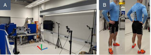
Figure 1: (A) OptiTrack system setup of 7 cameras used for testing. Axis orientation with x in red, y in green and z in blue. (B) Marker placements attached to the participants during the test.
For this study, the OptiTrack motion capture system was employed, featuring a setup of seven Flex 13 cameras operating with Motive Tracker 2.3.7 software to track retroreflective markers and provide real-time motion data. This system facilitated 3D kinetic rigid body tracking, capturing the position and orientation of the body in the global coordinate system. A positional accuracy of ±0.2mm and rotational accuracy of ±0.1 degrees, with an 8.3ms delay and a recording frequency of around 120 Hz (NaturalPoint, 2023) was achieved using the Flex 13 cameras. A world transform-frame was used as a fixed reference (Figure 1A), where the y-axis was oriented upward, the x-axis was set as vertical, and the z-axis was positioned horizontally. This setup allowed the tracking of participant body segments, providing data for individual joints’ kinematics, including flexion, extension, abduction, and adduction (Cloete and Scheffer, 2008). Participants, all healthy males aged 19–29 years (M = 23, SD = 3.67), were selected based on purposive sampling and performed movements under normal and braced conditions. The braced condition simulated injury- related restrictions, allowing for a comparison of kinematic data under two scenarios. Movement tracking utilized 33 retro-reflective markers, with
Evaluating Clinical Efficacy of Optical Motion Tracking With Real-Time Animation 143
specific placements on the right leg, thigh, shank, ankle, and lower back to track joint movements (Figure 1B).
Experimental Conditions Participants were asked to perform three distinct exercises to assess their biomechanics under both normal and braced conditions. The exercises included gait (walking in a straight line), single-leg squat jump on the right leg with a countermovement, and the straight-leg sidewalk movement along the coronal plane. These movements were chosen based on their relevance to assessing lower body joint kinematics and common rehabilitation protocols. In the normal condition, participants performed the exercises without any restrictions, representing typical healthy movements. In the braced condition, specific braces were used to simulate joint restrictions and assess their impact on movement.
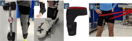
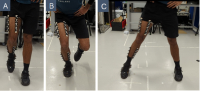
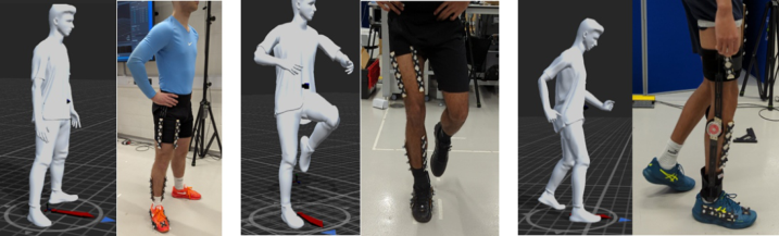
Figure 2: (A) Knee brace while walking in straight line, (B) ankle brace for single leg jump, (C) hip brace, (D) hip brace along with resistance band pulling the hip while performing sidewalk test.Figure 3: (A) Participant walking in straight line during gait test. (B) Single leg jump test. (C) Straight leg sidewalk.
During the gait test (Figure 3A), participants wore a knee brace (Figure 2A), which limited knee flexion. For the single-leg squat jump (Figure 3B), an ankle brace (Figure 2B) was used to restrict ankle mobility. The straight-leg sidewalk test (Figure 3C) involved a hip brace (Figure 2C) to limit hip abduction and additional band resistance (Figure 2D) to simulate further restrictions in mobility. The primary dependent variables analysed were knee flexion angle during the gait test, dorsiflexion angle during the jump test, and hip abduction angle during the sidewalk movement. Additionally, other joint angles and the distance covered were also analysed
144 Walia and Raval
to further understand the differences between normal and braced conditions and provide insights into the underlying causes of changes in the data between the performed tests.
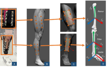
Data Collection and Processing The trajectories of the reflective markers attached to the participant’s body were tracked as they moved through space. Additional computation was required to convert raw data into joint angles, using rigid bodies. Markers were placed on the participants to define distinct body segments, as shown in Figure 4(A) and (B). Each segment was treated as a rigid body, created by manually selecting all relevant markers for that body part, as depicted in Figure 4(C). These rigid bodies were then aligned to anatomical frames, referencing the global coordinate system. Figure 4(D) illustrates the alignment of the femur (fy, fx, fz), tibia (ty, tx, tz), and ankle (ay, ax, az) anatomical frames, with the y-axis along the bone, x-axis as anteroposterior, and z-axis as mediolateral.
Figure 4: (A) Markers attached on the participant. (B) Marker positions captured by the cameras defines femur and tibia into different segments. (C) Femur and tibia segments as rigid bodies. (D) 3 axes realigned where y-axis is along the bone to define the anatomical frames for femur, tibia and ankle.
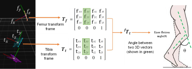
The relationship between two anatomical frames was computed using the Transform Frames. Figure 5 illustrates the calculation of the knee flexion angle between the femur (Tf) and tibia (Tt) by analyzing the rotational matrices along the y-axis, aligned with the bone in both frames. The femur frame served as the reference for the tibia frame. Rotation vectors along the y-axis (green) were employed to compute the angle between the 3D vectors, representing the knee joint angle. This method enabled precise calculation of joint angles for biomechanical analysis, as demonstrated in Figure 5.
The computed joint angle data was analysed using two methods. Quantitative comparison between conditions was performed in MATLAB with Statistical Parametric Mapping (SPM) using the spm1D package. Paired t-tests at a 95% confidence interval and α = 0.05 were conducted for all three tests, analysing joint angle waveforms across all five participants. Additionally, graphical movement analysis plotted joint angles against time
Evaluating Clinical Efficacy of Optical Motion Tracking With Real-Time Animation 145
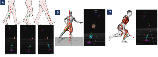
and distance covered in all axes to assess changes in range of motion (ROM) and muscular engagement. The study focused on three movements: gait, single-leg jump, and sidewalk, evaluating knee flexion, dorsiflex- ion/plantar flexion, and hip abduction. Figure 6 illustrates the joint angles analysed.
Figure 5: Anatomical frames captured by the cameras for femur and tibia converted to transform frames. Rotation vectors for y-axis in green. Relation calculated with femur being a reference to tibia. Vector calculation is carried out to compute the knee joint angle.Figure 6: (A) (Top) knee flexion angle examined for the gait test, (bottom) rigid body frames while performing gait test (Themes, 2016). (B) (Left) hip abduction angle analysed during the sidewalk test (DMoose, 2022), (right) frames captured during sidewalk test. (C) (Left) ankle angle computed during single leg jump test to assess plantar flexion and dorsi flexion (Botez, 2023), (right) participant being tracked by the OptiTrack system while performing jump test.
Motion capture data from the OptiTrack system was extracted from Motive and imported into Unity 3D to animate the human model performing the test movements. This real-time animation created a recreational experience for participants and allowed for virtual analysis of normal and braced motions simultaneously, as shown in Figure 7.
146 Walia and Raval
Figure 7: Real time data streaming from OptiTrack to Unity 3D. Integrating rigid body motion of the aligned anatomical frames into dynamic 3D animation.
RESULTS
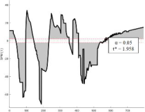
Statistical Parametric Mapping (SPM) Statistical analysis using the spm1D package for paired t-tests revealed significant differences in joint angles between the normal and braced conditions across all three exercises. The SPM analysis for knee, ankle, and hip joint angles showed that the data points outside the significance level (p < 0.05) demonstrated significant changes in joint motion, as seen in Figure 8 (a), (b) and (c). The results indicate that the motion capture system effectively detected biomechanical differences, with the majority of data outside the significance region suggesting restricted movement during the braced condition.
Figure 8: SPM analysis for: (a) knee flexion angle in gait test, (b) ankle angle to assess plantar/dorsi flexion in single leg jump test and (c) hip abduction angle in sidewalk test.
Gait Test The gait test results (Figure 9 (a) and (b)) showed a significant reduction in knee flexion, with the maximum flexion dropping from 43.41
in the normal condition to 23.79
in the braced condition. This reduction in range of motion (ROM) by 32.06
(Table 1) confirmed that participants experienced restricted knee movement and had to adopt a limping gait, dragging their leg during walking.
Evaluating Clinical Efficacy of Optical Motion Tracking With Real-Time Animation 147
(a) (b)
(c) (d)
(e) (f)
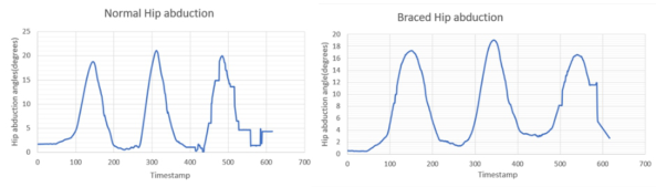
Figure 9: Knee flexion angle vs time graph for one of the 5 participants for (a) normal (a) and (b) braced condition; ankle angle vs time graph for one of the 5 participants for (c) normal and (d) braced condition; hip abduction angle vs timestamp graph for one of the participants for (e) normal and (f) braced conditions.Table 1: Average of knee angle data for the participants under normal and braced conditions.
Table available in the original PDF paper.
In Degrees Normal Condition Braced Condition Average Maximum Flexion 55.52 21.77 Average Maximum Extension 1.94 0.25 Range of Motion 53.58 21.52
Single Leg Squat Jump Test The single-leg squat jump test (Figure 9 (c) and (d)) demonstrated that ankle movement was restricted by the ankle brace, leading to increased plantar flexion (111.54
) compared to dorsiflexion in the normal condition. ROM in the braced condition decreased by 18.7
, and the maximum jump height was reduced from 0.5 m to 0.468 m. Additionally, knee flexion increased in the braced condition, indicating reliance on alternative muscles, such as the quadriceps and hamstrings, to compensate for limited ankle motion.
Straight Leg Sidewalk Test The straight-leg sidewalk test (Figure 9 (e) and (f)) showed a reduction in hip abduction by 7.2
in the braced condition, with participants using more lumbar flexion to compensate. Lumbar flexion increased by 14
, demonstrating greater lower back engagement during movement. However,
148 Walia and Raval
some participants (B and E) showed minimal change in lumbar flexion and instead used more hip abductors, which is typical for strengthening injured muscles during rehabilitation.
The results of this study demonstrate the effectiveness of the motion capture system in quantifying the biomechanical impact of joint restrictions during rehabilitation. Significant changes in joint angles across all three tests (gait, jump, sidewalk) suggest that bracing effectively limits joint motion, resulting in compensatory movement patterns. The observed reductions in range of motion (ROM) and alterations in muscular engagement (e.g., increased lumbar flexion and quadriceps activation) emphasize the system’s potential for assessing rehabilitation progress. These findings offer insights into how joint restrictions influence movement and can inform more targeted rehabilitation strategies to optimize recovery and strengthen specific muscle groups.
DISCUSSION
This pilot study highlights the feasibility and potential benefits of using an optical motion tracking system with real-time animation for rehabilitation monitoring. A key advantage of this method is the provision of immediate visual feedback during exercise, enabling users to self-correct movements in real time and encouraging proper motor pattern development. Real- time animation reflects deviations as they occur, facilitating timely user adjustments and reinforcing correct movement execution. This feedback mechanism supports motor learning and may enhance user engagement and autonomy during rehabilitation. Clinicians also benefit from live motion data, allowing immediate assessment and targeted intervention without relying solely on post-session reviews. This could streamline therapy sessions and support more precise clinical decision-making. While the current study involved only healthy participants with limited or constrained movement tasks, the observed system responsiveness indicates potential value in patient populations with cognitive or neuromuscular impairments who may benefit more from visual guidance than from verbal instruction.
Nonetheless, certain limitations must be recognised. The participant sample in this investigation comprised only healthy individuals, which constrains the extent to which clinical efficacy can be inferred. Accordingly, the present study should be regarded as a feasibility investigation rather than a conclusive clinical evaluation. Future research should incorporate clinical populations and longitudinal study designs to assess the effectiveness of the system in actual therapeutic contexts and to evaluate functional outcomes over time. In summary, while the current findings do not support definitive clinical conclusions, they suggest that real-time visual feedback using optical motion tracking and animation could contribute meaningfully to enhancing rehabilitation engagement and precision. Further clinical validation is warranted to substantiate these preliminary observations.
Evaluating Clinical Efficacy of Optical Motion Tracking With Real-Time Animation 149
CONCLUSION
This study demonstrated the clinical efficacy of optical motion tracking combined with real-time animation for rehabilitation monitoring, providing statistically significant biomechanical insights. By analysing lower-body kinematics under normal and braced conditions, we validated the potential of motion capture technology in detecting movement alterations and compensatory patterns. The results indicated a 32.06
reduction in knee flexion range of motion (ROM) during gait under braced conditions, increased plantar flexion in jump tests, and lumbar engagement shifts in sidewalk movements, confirming the system’s ability to quantify rehabilitation progress effectively. A key contribution of this research is the integration of real-time animation using Unity3D, which enhances visualization and patient engagement in rehabilitation. The ability to stream motion capture data into interactive simulations presents new opportunities for personalized therapy, making rehabilitation more immersive and accessible. The rigid-body approach, while unconventional, successfully captured motion patterns, highlighting its potential for non-invasive rehabilitation tracking.
The findings suggest that machine learning integration could automate motion analysis, allowing faster diagnosis and classification of movement disorders such as Parkinson’s and gait abnormalities. Additionally, virtual reality (VR) applications could further enhance rehabilitation experiences, increasing patient motivation and adherence. This study reinforces the value of optical motion capture for clinical rehabilitation and lays the foundation for future research involving larger, more diverse populations and real- world clinical trials, ultimately advancing technology-assisted rehabilitation solutions.
ACKNOWLEDGEMENT
The authors would like to acknowledge Higher Education Innovation Funding (HEIF) and Nottingham Trent University (NTU) for supporting and part-funding the motion capture equipment, which contributed to the success of this study.
REFERENCES
American Stroke Association (2023). About Stroke. [online] Available at: https:// www.stroke.org/en/about-stroke. Botez, A. (2023). Plyometric Training Program: A Step-by-Step Guide. [online]
Available at: https://fitnessprogramer.com/plyometric-training-program/. Cloete, T. and Scheffer, C., 2008, August. Benchmarking of a full-body inertial motion capture system for clinical gait analysis. In 2008 30th Annual International Conference of the IEEE Engineering in Medicine and Biology Society (pp. 4579–4582). IEEE. DMoose (2022). Cable Hip Abductions. Available at: https://www.dmoose.com/ blogs/quads/cable-hip-abductions.
150 Walia and Raval
Edemekong, P. F., Bomgaars, D. L. and Levy, S. B. (2023). Activities of daily living (ADLs). [online] NCBI. Available at: https://www.ncbi.nlm.nih.gov/books/ NBK470404/. Eichelberger, P., Ferraro, M., Minder, U., Denton, T., Blasimann, A., Krause, F. and
Baur, H., 2016. Analysis of accuracy in optical motion capture–A protocol for laboratory setup evaluation. Journal of biomechanics, 49(10), pp. 2085–2088. Ghahremani Arekhloo, N., Parvizi, H., Zuo, S., Wang, H., Nazarpour, K., Marquetand, J. and Heidari, H., 2023. Alignment of magnetic sensing and clinical magnetomyography. Frontiers in Neuroscience, 17, p. 1154572. Gu, C., Lin, W., He, X., Zhang, L. and Zhang, M., 2023. IMU-based motion
capture system for rehabilitation applications: A systematic review. Biomimetic Intelligence and Robotics, 3(2), p. 100097. Guaitolini, M., Petros, F. E., Prado, A., Sabatini, A. M. and Agrawal, S. K., 2021.
Evaluating the accuracy of virtual reality trackers for computing spatiotemporal gait parameters. Sensors, 21(10), p. 3325. Hollman, J. H., Brey, R. H., Robb, R. A., Bang, T. J. and Kaufman, K. R.,
2006. Spatiotemporal gait deviations in a virtual reality environment. Gait & posture, 23(4), pp. 441–444. Kanko, R. M., Outerleys, J. B., Laende, E. K., Scott Selbie, W. and Deluzio, K. J., 2023.
Comparison of concurrent and asynchronous running kinematics and kinetics from marker-based motion capture and markerless motion capture under two clothing conditions. BioRxiv, pp. 2023–02. NaturalPoint (2023). Motive - In Depth. [online] OptiTrack. Available at: https:// www.optitrack.com/software/motive/. Sugiyama, Y., Uno, K. and Matsui, Y., 2023. Types of anomalies in two-dimensional
video-based gait analysis in uncontrolled environments. PLOS Computational Biology, 19(1), p. e1009989. Tak, I., Wiertz, W. P., Barendrecht, M. and Langhout, R., 2020. Validity of a new 3-D motion analysis tool for the assessment of knee, hip and spine joint angles during the single leg squat. Sensors, 20(16), p. 4539. Themes, U. F. O. (2016). Assessment of Gait. Musculoskeletal Key. Available at:
https://musculoskeletalkey.com/assessment-of-gait/. Wade, L., Needham, L., McGuigan, P. and Bilzon, J., 2022. Applications and limitations of current markerless motion capture methods for clinical gait biomechanics. PeerJ, 10, p. e12995. Weygers, I., Kok, M., De Vroey, H., Verbeerst, T., Versteyhe, M., Hallez, H. and
Claeys, K., 2020. Drift-free inertial sensor-based joint kinematics for long-term arbitrary movements. IEEE Sensors Journal, 20(14), pp. 7969–7979. Whitting, J. W., Meir, R. A., Holding, R. and Crowley-McHattan, Z., 2013.
The use of 3D motion capture to assess back squat technique wearing different footwear: A preliminary investigation. Journal of Australian Strength & Conditioning, 21(suppl. 2), pp. 94–98. Ye, M., Yang, C., Stankovic, V., Stankovic, L. and Kerr, A., 2016. A depth camera motion analysis framework for tele-rehabilitation: Motion capture and person-centric kinematics analysis. IEEE Journal of Selected Topics in Signal Processing, 10(5), pp. 877–887. Zuo, S., Heidari, H., Farina, D. and Nazarpour, K., 2020. Miniaturized magnetic sensors for implantable magnetomyography. Advanced Materials Technologies, 5(6), p. 2000185.
LISTED IN THE AHFE DIGITAL BOOKSTORE CATALOGUE
Explore the complete digital volume
Healthcare and Medical Devices
Read this individual research paper free through the original publisher, or explore the full digital proceedings book.
ISBN 978-1-964867-47-2
Explore this ISBN in the AHFE Bookstore ↗Listed in the July 2026 bookstore catalogue; search by ISBN to confirm current availability · Digital book purchase optional