Alina Vladu, Emilia Visileanu, Alexandra Gabriela Ene, Madalina Georgiana Albu Kaya, Viviana Roman, Carmen Gaidau (2025). Biological evaluation of antimicrobial treated textiles. In: Healthcare and Medical Devices. AHFE Open Access, vol. 171, pp. 89–98. https://doi.org/10.54941/ahfe1006194
1 The National Research and Development Institute for Textiles and Leather, Lucretiu Patrascanu, 030508 Bucharest, Romania ↗
2 The National Research and Development Institute for Textiles and Leather, Lucretiu, Patrascanu, 030508 Bucharest, Romania ↗
3 Division of Leather and Footwear Research Institute, National Research and Development Institute for Textiles and Leather, 93 Ion Minulescu Str., 031215 Bucharest, Romania ↗↗
4 ¸Stefan S. Nicolau Institute of Virology, 285 Mihai Bravu Avenue, District 3, 030304, Bucharest, Romania ↗
Textiles provide a suitable environment for the growth of microorganisms, including fungi and bacteria. Their presence can have detrimental effects on both the fabric and the user. These effects may include unpleasant odors, fabric discoloration, a higher risk of contamination, and a decline in the material's mechanical strength. The transmission of infections through textiles can be mitigated by using antimicrobial fabrics, which either eliminate pathogens on contact or inhibit their reproduction before they spread to another surface or individual. Antimicrobial textiles are created by applying antimicrobial agents to textile substrates or by utilizing fibers that naturally possess antimicrobial properties. This paper is mainly focused on the biological evaluation of antimicrobial treated textiles with doxycycline and collagen hydrolysate. The textile structures were obtained from different fibers such as polyester (PES), cotton/elastane (CO/EL) and cotton/polyester (CO/PES). The antibacterial treatment was carried out by applying the obtained solution on the textile structures using the padding method. The characterization of the treated textiles includes the release profile of active compound, the evaluation of antibacterial activity on two bacterial strains Escherichia coli (E. coli) and Staphylococcus aureus (S. aureus), as well as the assessment of cell viability by MTS and LDH assays. The release kinetics of doxycycline from the textile structures showed a burst release in the first 30 minutes followed by a slow and sustained release until the end of the experiment. The samples presented good results in terms of antimicrobial activity on both bacterial strains, the effect being classified as satisfactory. The viability of HUVEC cells is places between 90.89-95.99%, while the necrosis has low values between 4.01-9.11% suggesting that the antibacterial treated textiles can be considered non-cytotoxic. The obtained results confirm that the textile structures treated with doxycyline and collagen hydrolysate can be used as functional antibacterial textiles in direct contact with human skin.
With increasing public health awareness about the harmful effects of microorganisms, including their role in causing infections, unpleasant odors, and stains, there is a growing demand for antibacterial materials across various applications. These include medical devices, healthcare, hygiene products, water purification systems, hospitals, dental surgery equipment, textiles, food packaging, and storage solutions (Shahidi, 2012). The antimicrobial effectiveness of functional textiles is influenced by various factors, such as the adherence and survival of pathogens on textile surfaces, the type of antimicrobial agents used, and their durability across different textile applications. The primary categories of synthetic and natural antimicrobial agents for textiles include triclosan, metals and their salts, organometallics, phenols, quaternary ammonium compounds (QACs), organosilicons, and essential oils (Vojnits et al., 2024). The incorporation of antimicrobial agents into fabrics depends on the type of textile materials and fibres. Various chemical and physical properties, such as fibre thickness, diameter, and processing conditions, influence the integration methods. Antimicrobial compounds can be incorporated in different ways, including direct addition during fibre synthesis, integration into the fibre sheath during extrusion, or application onto manufactured fibres through techniques like dip coating, polymer coating, spray application, or inclusion in the spin finish. For nonwoven products, antimicrobials can be introduced during bonding or finishing processes. In contrast, knitted and woven textiles are typically treated using the exhaust or pad-dry-cure method (El-Ola, 2008; Gulati et al., 2022). Collagen is the primary structural component of bones, muscles, connective tissues, and skin. Hydrolysed collagen, also known as gelatin, is obtained through the partial hydrolysis of collagen. Previous research has demonstrated the effectiveness of hydrolysed collagen in promoting wound healing and enhancing the health of skin and connective tissues (Bagheri Miyab et al., 2020). Doxycycline hyclate is a water-soluble tetracycline antibiotic that inhibits the growth and eliminates a broad spectrum of gram-positive and gram-negative bacteria. It is commonly used to manage and treat acne, malaria (both prevention and treatment), skin infections, sexually transmitted infections, as well as Lyme disease. Doxycycline’s high lipophilicity, compared to other tetracyclines, enables it to penetrate multiple membranes and effectively reach its target molecules (Cross et al., 2016, Chopra and Roberts, 2001). Doxycycline promotes wound healing with reduced scarring by regulating fibroblast activity and collagen deposition. It suppresses prolidase, an enzyme involved in collagen degradation, thereby inhibiting collagen biosynthesis in human skin fibroblasts. With an IC50 of approximately 150 µg/mL, doxycycline coordinately reduces prolidase activity, collagen production, and gelatinolytic activity. This inhibition may disrupt proline recycling, leading to decreased collagen synthesis (Karna et al., 2001). Doxycycline inhibits prolidase activity and modulates collagen biosynthesis in human skin fibroblasts while also demonstrating antioxidant properties. It enhances the production of type I collagen and elastic fibres, promoting faster skin wound closure (Saliy et al., 2024). The current study
Biological Evaluation of Antimicrobial Treated Textiles 91
is focused on the biological evaluation of three types of textile structures made of different fibres blends and treated with doxycycline and collagen hydrolysed.
EXPERIMENTS Materials and Preparation
First a solution of doxycycline and collagen hydrolysed was prepared for the treatment of textile materials. In the initial solution of 1.5% collagen hydrolysate, 3 g of doxycycline and 30 g of β-cyclodextrin were added (1:10 ratio of doxycycline: β-cyclodextrin). The collagen hydrolysed was supplied by Division of Leather and Footwear Research Institute, National Research and Development Institute for Textiles and Leather (Bucharest, Romania). Doxycycline hyclate of ≥93.5% purity was purchased from Sigma Aldrich (St.Louis, MA, USA) and β-cyclodextrin of ≥99% purity from Fluka (Buchs, Switzerland). All reagents were used without further purification. The obtained solution was applied on three types of textile materials of different fibres blends: PES, CO/EL and CO/PES by padding using a ROACHES machine. The textile structures were passed three times through the treatment solution. Before the treatment the textile materials were washed at a temperature of 30
C with a Kemapon PC/LF solution for 30 min. Then, they were rinsed twice with warm water (30
C) and once with cold water (20
C), then at room temperature.
Methods
Antimicrobial Activity The evaluation of resistance to bacterial action was carried out according to SR EN ISO 20645/2005 (ASRO, 2005), namely resistance to Staphylococcus aureus ATCC 6538 (gram-positive) and Escherichia coli ATCC10536 (gram- negative) bacterial strains. The agarose volume for the lower layer, without bacteria, was first prepared. Then, (10 ± 0.1) mL was dispensed into each sterilized Petri dish and left to solidify. Meanwhile, the agarose for the upper layer was prepared and cooled to 45
C in a water bath. A total of 150 mL of agarose was inoculated with 1 mL of bacterial working solution (1–5 × 10⁸ CFU/mL). The mixture was then vigorously shaken to ensure uniform bacterial distribution, and (5 ± 0.1) mL was added to each Petri dish before allowing it to solidify. Finally, the samples were placed on the surface of the nutrient medium and incubated at 37
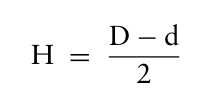
C for 18 to 24 hours. The evaluation is determined by observing the presence or absence of bacterial growth in the contact area between the agar and the sample, as well as the formation of a potential inhibition zone surrounding the sample. The width of the inhibition zone, defined as the bacteria-free area near the sample’s edge, is calculated using the following formula:
Equation reproduced from the original PDF.
2 (1)
where:
H represents the inhibition zone width (mm),
D is the total diameter of the specimen and its inhibition zone (mm),
d is the diameter of the specimen (mm).
Cell Viability The tests were conducted using both untreated textile supports and textile supports treated with doxycycline and collagen hydrolysate. The HUVEC cell line (Primary Umbilical Vein Endothelial Cells; Normal, Human, PCS- 100-010, ATCC, USA) was utilized, cultured in complete DMEM medium supplemented with 10% fetal bovine serum, 1% penicillin/streptomycin solution, and 1% L-glutamine, and incubated at 37
C in a 5% CO2 atmosphere. For testing, the cells were expanded and seeded into 6-well plates at a concentration of 0.5 × 10⁶/mL. After 24 hours of adhesion, the test agents were introduced. To evaluate cell viability, an MTS assay - The CellTiter 96® AQueous One Solution Cell Proliferation Assay kit (Promega, Madison, USA) was used. To asses cell lysis as a form of secondary necrosis, which occurs at the end of the apoptosis process, flow cytometry technique was used. For flow cytometry analysis, the FACScan II cytometer (Becton Dickinson, USA) was employed, along with the BD Pharmingen™Apoptosis Detection Kit (Becton Dickinson, USA). Data analysis was performed using DIVa 6.2 software (Becton Dickinson, USA).
Study of Doxycycline Release Kinetics From Treated Textiles The in vitro release kinetics of doxycycline from the tested textiles were studied using a sandwich device adapted to a dissolution apparatus equipped with paddles (Esadisolver). The pre-weighed antibiotic-impregnated knitwear was placed on the surface of the sandwich setup and then introduced into the release vessels of the dissolution apparatus. The kinetic experiments were conducted at 37
C using phosphate buffer (pH 7.4) as the release medium, stirred at 50 rpm. At predefined intervals over 6 hours, 5 mL samples were withdrawn from the release vessel and replaced with an equal volume of fresh phosphate buffer solution maintained at 37
C ± 0.5
C, ensuring a constant volume. The doxycycline concentration in each sample was determined spectrophotometrically at a wavelength of λ = 348 nm. Kinetic profiles were generated by plotting the cumulative percentage of drug released over time.
Equation reproduced from the original PDF.
To investigate the drug release mechanism, the Power Law model was applied: mt m∞ where:
mt/m∞represents the fraction of antibiotic released at time t,
k is the kinetic constant (1/minn),
n is the release exponent, indicating the drug transport mechanism.
Biological Evaluation of Antimicrobial Treated Textiles 93
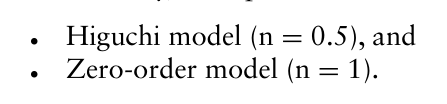
Additionally, two specific cases of the Power Law model were considered:
Equation reproduced from the original PDF.
RESULTS AND DISCUSSIONS The antimicrobial activity of treated textile materials was evaluated using the SR EN ISO 20645/2005 standard – Determination of Antibacterial Activity – Agar Diffusion Plate Test (Table 1). The tests were conducted on Escherichia coli ATCC 10536 (Gram-negative) and Staphylococcus aureus ATCC 6538 (Gram-positive). The results indicate that treated textile structures exhibit significantly higher antimicrobial activity (a satisfactory effect) compared to untreated samples. Cotton-based fabrics demonstrated superior antimicrobial performance compared to polyester fabrics, primarily due to cotton’s higher absorption capacity. Although all treated samples showed good results, DOXY-CO/PES exhibit the highest antimicrobial activity, followed by DOXY-CO/EL and DOXY-PES, most likely because the cotton-polyester blend retains and releases doxycycline in a way that maximizes its antimicrobial potential, whereas the polyester-only and cotton- elastane samples may either retain less of the drug or release it in a less effective manner.
Table 1: Antibacterial efficiency of the treated textile materials according to SR EN ISO 20645/2005.
Table available in the original PDF paper.
Evaluation
Sample Inhibition Zone (mm)
Inhibition Zone (mm) Escherichia Coli Staphylococcus Escherichia Coli Staphylococcus Aureus
Inhibition Zone (mm) Escherichia Coli Staphylococcus Escherichia Coli Staphylococcus Aureus
Equation reproduced from the original PDF.
DOXY-
effect
effect
CO/PES
Equation reproduced from the original PDF.
Satisfactory
DOXY-
effect
effect
CO/EL
Satisfactory
Equation reproduced from the original PDF.
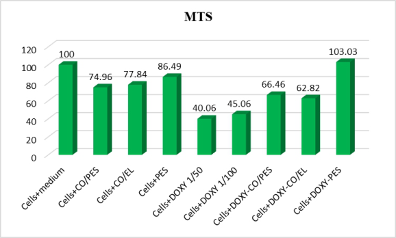
Viability tests were conducted by exposing the specified culture to the samples. The results are shown in Figure 1. The cells were exposed to the treatment solution at final concentrations of 1/50 and 1/100 in the culture medium to assess any potential effects. According to SR EN ISO 10993–5 (ASRO, 2009), cell viability percentages above 80% indicate non-cytotoxicity, while viability between 80% and 60% signifies weak cytotoxicity. Moderate cytotoxicity is classified within the 60%–40% range, and strong cytotoxicity is defined as viability below 40%.
Figure 1: Cell viability – HUVEC cell line.
Biological Evaluation of Antimicrobial Treated Textiles 95
As it can be observed from figure 1, the pure treatment solutions of 1/50 and 1/100 concentration are moderate cytotoxic, as cell viability‘s values are less than 60% (40.06 and 45.06 %). The treatment solutions of doxycycline and collagen hydrolysed appear to reduce the viability of HUVEC cells when compared to controls. The fabrics made of CO/PES and CO/EL treated with the solution can be classified as weak cytotoxic with cell viability values placed between 62.82–66.46%. The highest value of cell viability is recorded for the treated polyester fabric (103,03%), higher than the control, which classifies it as non-cytotoxic.
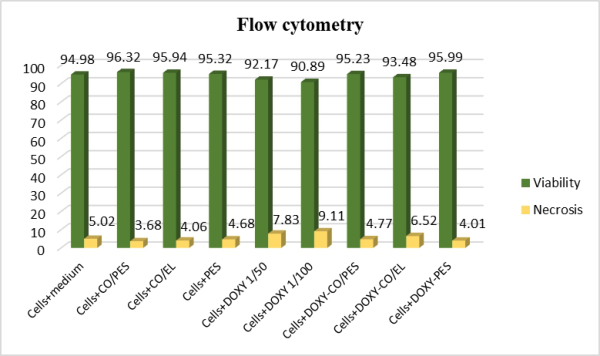
Figure 2: Cell viability and necrosis in HUVEC cell line.
Figure 2 shows the viability and necrosis obtained by flow cytometry for treated and untreated textile supports. From the data analysis, it can be observed that the viability and necrosis levels of antibiotic treated fabrics does not vary significantly when compared to control samples and does not show significant cytotoxicity. Necrotic cells level is very low, between 4.01 – 6.52 % for treated textile materials. Cell viability exceeds 90% for all treated tissue variants, which classifies them as non-cytotoxic.
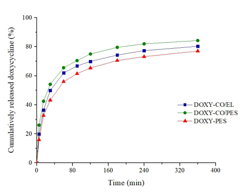
An important aspect in the design and characterization of antibiotic- impregnated textiles is the investigation of the drug release characteristics. Thus, the influence of the type of fabric (CO/PES, CO/EL, PES) impregnated with the drug on the release of doxycycline was analyzed by comparing the kinetic profiles obtained. The cumulative percentage of drug released over time is shown in Figure 3.
Figure 3 shows a similar pattern of drug release kinetic profiles from impregnated textiles. The drug release process was described by two different phases: an initial rapid release in the first 30 minutes, followed by a second, slower and progressive release stage during the next 5.5 hours of the experiment. For topical administration of antibiotics, biphasic kinetic profiles are followed in the formulation of release systems, both for prophylaxis and for the treatment of a skin infection. Thus, the rapid release effect
(“burst release”) is important to control the initial microbial load at the skin level, respectively to prevent bacterial infection that may occur consecutively following skin damage. The most pronounced “burst release” effect was recorded for the CO/PES textile structure (54.14%), followed by the CO/EL fabric (49.96%) and the PES textile (43.20%). The gradual release stage of the drug in the following hours further ensures the prevention of bacterial multiplication or invasion, thus ensuring the acceleration of the healing process. Regarding the cumulative percentage amount of drug released after 6 hours of the experiment, it varies from 77.02 (PES), to 80.33 (CO/EL) and respectively up to 84.29% (CO/PES) (Table 2). The rapid release effect is due to the presence of the drug on the surface of the fabric, while its entrapment in the textile material determines the gradual release over the following hours. To establish the mechanism of doxycycline release from impregnated knitwear, different kinetic models were used. The values of the correlation coefficients for the applied kinetic models are presented in Table 2.
Figure 3: Kinetic release profiles of doxycycline from impregnated textiles.Table 2: Correlation coefficient values for the applied kinetic models.
Table available in the original PDF paper.
Impregnated Textile Structure
Correlation Coefficient (R) Release Exponent, n
Kinetic Constant, k (1/minn)
Release Doxycycline (%) Zero Order Model
Higuchi Model
Power Law Model
DOXY-CO/EL 0,7691 0,9179 0,9806 0,25 20,101 80,33 DOXY-CO/PES 0,7568 0,9101 0,9862 0,23 24,057 84,29 DOXY-PES 0,7977 0,9359 0,9828 0,27 16,373 77,02 The values obtained for the correlation coefficients showed that the drug release was best described by the Power Law model, with R ranging between 0.9806 and 0.9862. The values of the kinetic parameters specific to the Power
Biological Evaluation of Antimicrobial Treated Textiles 97
Law model, the release exponent and the kinetic constant, are shown in Table 2. It is found that the release exponent has values ranging between 0.23 and 0.25, which indicates a non-Fickian drug transport mechanism.
CONCLUSION
The assessment of antibacterial resistance following the SR EN ISO 20645/2005 standard confirmed the effectiveness of treatments with doxycycline and hydrolysed collagen. The results are deemed “satisfactory” as no bacterial multiplication was observed, and the inhibition zones measured between 5 and 11. The cell viability assessment shows that the viability of treated textile samples is between 62.82-103.03% when tested on HUVEC cell line by MTS. However, cell viability assessment by flow cytometry shows that the percent of viable cells is more than 93.48%. The difference in viability results arises because MTS measures metabolic function, which doxycycline can suppress, while flow cytometry assesses cell membrane integrity and counts live cells, which remain unaffected. Also, cell necrosis is very low, with values between 4.01–6.52%. Considering these results the best sample in terms of cell viability is the one made of polyester. Drug release kinetics emphasize an initial burst release within the first 30 minutes, followed by a gradual and sustained release which plays a crucial role in controlling the initial microbial load on the skin and preventing bacterial infections that may arise following skin damage.
ACKNOWLEDGMENT
This research was conducted as part of the Nucleu Programme under the National Research, Development, and Innovation Plan 2022-2027, with support from the Ministry of Research, Innovation, and Digitalization. It was carried out within project no. 6N/2023, PN 23 26 01 02, titled “Intelligent Equipment for Ensuring Combatant Survival in Operational Conditions,” acronym: IRHEM.
REFERENCES
ASRO (2005) SR EN ISO 20645:2005 – Textile fabrics – Determination of antibacterial activity – Agar diffusion plate test. Bucharest: ASRO. ASRO (2009) SR EN ISO 10993-5:2009 – Biological evaluation of medical devices –
Part 5: Tests for in vitro cytotoxicity. Bucharest: ASRO. Bagheri Miyab, K., Alipoor, E., Vaghardoost, R., Saberi Isfeedvajani, M., Yaseri, M., Djafarian, K. & Hosseinzadeh-Attar, M. J. 2020. The effect of a hydrolyzed collagen-based supplement on wound healing in patients with burn: A randomized double-blind pilot clinical trial. Burns, 46, 156–163. Chopra, I. & Roberts, M. 2001. Tetracycline antibiotics: Mode of action, applications, molecular biology, and epidemiology of bacterial resistance. Microbiol Mol Biol Rev, 65, 232–60; second page, table of contents. Cross, R., Ling, C., Day, N. P., McGready, R. & Paris, D. H. 2016. Revisiting
doxycycline in pregnancy and early childhood–time to rebuild its reputation? Expert Opin Drug Saf, 15, 367–82.
El-Ola, S. M. A. 2008. Recent Developments in Finishing of Synthetic Fibers for Medical Applications. Designed Monomers and Polymers, 11, 483–533. Gulati, R., Sharma, S. & Sharma, R. K. 2022. Antimicrobial textile: Recent
developments and functional perspective. Polymer Bulletin, 79, 5747–5771. Karna, E., Pałka, J. & Wołczy´nski, S. 2001. Doxycycline-induced inhibition of prolidase activity in human skin fibroblasts and its involvement in impaired collagen biosynthesis. European Journal of Pharmacology, 430, 25–31. Kumar, M., Banerjee, P., Das, A., Singh, K., Guith, T., Kacar, S., Gourishetti, K.,
Sen, C. K., Roy, S. & Khanna, S. 2024. Hydrolyzed Collagen Powder Dressing Improves Wound Inflammation, Perfusion, and Breaking Strength of Repaired Tissue. Adv Wound Care (New Rochelle), 13, 70–82. Saliy, O., Popova, M., Tarasenko, H. & Getalo, O. 2024. Development strategy of novel drug formulations for the delivery of doxycycline in the treatment of wounds of various etiologies. European Journal of Pharmaceutical Sciences, 195, 106636. Shahidi 2012. Antibacterial Agents in Textile Industry. In: Bobbarala, V. (ed.)
Antimicrobial Agents. Rijeka: IntechOpen. Vojnits, K., Mohseni, M., Parvinzadeh Gashti, M., Nadaraja, A. V., Karimianghadim, R., Crowther, B., Field, B., Golovin, K. & Pakpour, S. 2024. Advancing Antimicrobial Textiles: A Comprehensive Study on Combating ESKAPE Pathogens and Ensuring User Safety. Materials [Online], 17.
LISTED IN THE AHFE DIGITAL BOOKSTORE CATALOGUE
Explore the complete digital volume
Healthcare and Medical Devices
Read this individual research paper free through the original publisher, or explore the full digital proceedings book.
ISBN 978-1-964867-47-2
Explore this ISBN in the AHFE Bookstore ↗Listed in the July 2026 bookstore catalogue; search by ISBN to confirm current availability · Digital book purchase optional