Yingwei Li, Qianxiang Zhou, Zhongqi Liu, Mengmeng Jin (2026). A Chest-Worn Quad-Modal Cardiac Monitoring Device Combining ECG, PCG, SCG, and GCG with Cross-Modal Motion Artifact Suppression. In: Healthcare and Medical Devices. AHFE Open Access, vol. 211, pp. 261–271. https://doi.org/10.54941/ahfe1007489
Continuous monitoring of both electrical and mechanical cardiac activity is essential for early detection and management of cardiovascular diseases in real-life environments. This paper presents the design and preliminary evaluation of a chest-worn, Holter-like device that enables 24-hour quad-modal cardiac monitoring by synchronously acquiring electrocardiogram (ECG), phonocardiogram (PCG), seismocardiogram (SCG), and gyrocardiogram (GCG) signals. The main unit is attached to the chest and integrates a heart sound sensor, a 6-axis inertial measurement unit (IMU), data acquisition and storage circuits, and a battery into a single compact housing, while four limb leads (RA, RL, LA, LL) are extended from the device to record ECG. All cardiac signals are sampled at 10 kHz under a shared hardware clock, ensuring absolute temporal synchronization across modalities.Building on the IMU, SCG (chest wall micro-acceleration) and GCG (chest wall micro-rotation) are treated not only as auxiliary motion references, but also as cardio-mechanical signals that are jointly analyzed with ECG. A cross-modal motion artifact suppression framework is proposed, in which ECG, SCG, and GCG mutually constrain each other: motion-dominated components are identified via their inconsistent morphology across modalities, while cardiac components exhibit stable beat-synchronous patterns. The denoised ECG then serves as a temporal reference to perform ECG-guided heart sound segmentation on the PCG, enabling robust extraction of the first to fourth heart sounds (S1–S4). A custom desktop software platform supports synchronized visualization, beat-level quality assessment, and batch analysis of 24-hour recordings.Preliminary tests on healthy subjects during daily activities (resting, walking, posture changes) show that the proposed quad-modal system effectively reduces motion-induced artifacts, improves the morphological consistency of ECG, SCG, and GCG, and achieves reliable multi–heart sound segmentation under ambulatory conditions.The chest-worn, integrated design and cross-modal processing pipeline demonstrate strong potential as a user-friendly and low-cost solution for continuous, multi-dimensional cardiovascular monitoring in clinical and home settings.
Continuous cardiovascular monitoring outside the clinic is increasingly important for early detection, long-term management, and home-based follow-up. Wearable sensing supports this shift, but real-life use introduces substantial variability in posture, motion intensity, and sensor coupling, which can degrade signal quality and clinical interpretability (Xie et al., 2025).
Electrocardiography (ECG) is the most established wearable modality for capturing cardiac electrical activity, yet it provides limited direct information about cardiac mechanical events. Therefore, complementary cardiomechanical modalities, including phonocardiography (PCG), seismocardiography (SCG), and gyrocardiography (GCG), have attracted growing interest. Prior studies and reviews have shown that SCG and GCG can capture beat-synchronous mechanical patterns and clinically relevant timing cues when measurements are sufficiently stable (Jafari Tadi et al., 2017; Sieciński et al., 2020; Rai et al., 2021). In parallel, ECG-assisted approaches are widely used to provide temporal anchors for PCG segmentation in noisy recordings (Prasad et al., 2020).
A key challenge for ambulatory cardiomechanical monitoring is motion contamination. Daily activities such as walking and posture transitions can introduce artifacts that overlap with cardiac components in SCG/GCG and also degrade wearable ECG. Practical artifact reduction therefore remains an active topic, especially under walking conditions where signal morphology can change rapidly (Javaid et al., 2017; Castaño Usuga et al., 2022). Another limitation of many existing prototype systems is their reliance on multi-device acquisition or weak synchronization, which complicates cross- modal fusion and beat-level timing analysis. Recently released datasets with truly simultaneous mechanical signals, PCG and ECG further highlight the importance of tight synchronization for developing and benchmarking multimodal methods (Parlato et al., 2025).
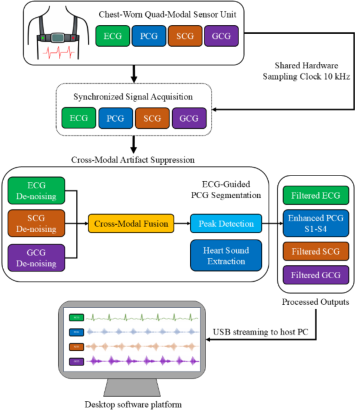
To address these challenges, this work presents a chest-worn, Holter-like quad-modal system that synchronously acquires ECG, PCG, SCG and GCG under a shared hardware clock at 10 kHz, enabling absolute temporal alignment across modalities. Unlike prior studies that typically investigate individual modalities or limited combinations, the proposed system integrates all four sensing modalities within a single wearable platform and combines hardware-level synchronization with a unified cross-modal processing pipeline. Specifically, we propose a cross-modal motion artifact suppression framework in which ECG, SCG and GCG mutually constrain one another to preserve beat-synchronous cardiac components under motion, while the denoised ECG is further used to guide PCG segmentation and support robust extraction of S1–S4 under ambulatory conditions (Prasad et al., 2020). A desktop software platform is also developed for synchronized visualization, beat-level quality assessment, and batch analysis of long-duration recordings. Figure 1 summarizes the overall system architecture and data flow. The main contributions of this work are fourfold: (1) a chest-worn wearable platform integrating ECG, PCG, SCG, and GCG within a single hardware system; (2) a shared-clock acquisition architecture at 10 kHz for strict hardware-level synchronization and elimination of inter-device drift; (3) a cross-modal motion artifact suppression framework
A Chest-Worn Quad-Modal Cardiac Monitoring Device 263 for preserving beat-synchronous cardiac components under motion; and (4) ECG-guided PCG segmentation for more stable extraction of heart sound events in ambulatory recordings.
Figure 1: Overview of the chest-worn quad-modal cardiac monitoring system.
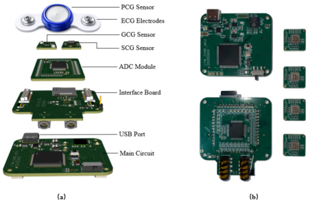
SYSTEM DESIGN AND IMPLEMENTATION Following the system overview in Fig. 1, this section describes the hardware and software implementation of the proposed synchronized quad-modal system. The prototype integrates multiple analog sensing chains, a shared digitization stage, and an FPGA-based control and streaming unit into a compact main module for chest-worn operation. To illustrate the physical realization beyond the abstract workflow, Fig. 2 presents the structural layout of the main unit together with the corresponding printed circuit board used in this study.
Figure 2: Physical structure and hardware implementation of the system: (a) schematic illustration of the main unit structure; (b) photograph of the corresponding printed circuit board.
The system simultaneously acquires four cardiac-related modalities— ECG, PCG, SCG, and GCG—all in analog form. ECG is implemented using the CN121 single-lead ECG chip (Xichen Semiconductor, China), which outputs an analog signal suitable for direct digitization. Heart sounds are captured by the HKY-06E full-frequency heart–lung sound sensor (Hefei Huake Electronic Technology Research Institute, China). Cardiomechanical vibrations are measured using inertial sensors with analog outputs: SCG is acquired via the tri-axial ADXL354 accelerometer (Analog Devices), while GCG is obtained using three orthogonally arranged ADXRS649BBGZ single-axis gyroscopes (Analog Devices). In total, eight analog channels are digitized (ECG ×1, PCG ×1, SCG ×3, and GCG ×3), enabling consistent multi-modal analysis and beat-level alignment.
All analog channels are digitized by a shared multi-channel ADC (AD7606) at a unified sampling rate of 10 kHz, providing a single time base for ECG, PCG, SCG, and GCG and eliminating inter-device clock drift. Acquisition timing, sample buffering, and data framing are coordinated by a Cyclone IV FPGA (EP4CE10F17V8), which ensures deterministic control of synchronized sampling and organizes the multi-channel data stream into fixed-format frames.
The synchronized data stream is transmitted to a host computer via USB for high-throughput streaming of raw multi-channel waveforms. On the host side, a desktop software platform reconstructs time-aligned channels for real-time visualization and storage, supports long-duration recording management, optional event annotation, and batch export of raw and processed data for offline analysis. This software layer completes the acquisition–analysis loop depicted in Fig. 1 and serves as the entry point for the signal processing methods described in Section 3. Table 1 summarizes the key hardware components and acquisition settings of the proposed system.
Table 1: Key components and acquisition settings of the synchronized quad- modal system.
Sampling Rate 10 kHz (All Channels) Controller Cyclone IV EP4CE10F17V8
Communication Interface USB (High-Throughput Streaming)
METHODS
This section describes the host-side signal processing pipeline operating on the synchronized quad-modal recordings acquired by the proposed system. The processing is designed around two practical requirements for ambulatory
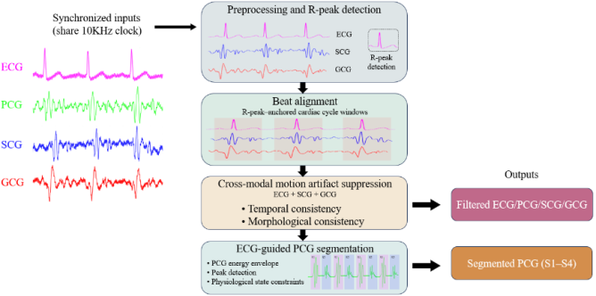
A Chest-Worn Quad-Modal Cardiac Monitoring Device 265 monitoring: (i) all modalities must be analyzed on a shared time base to support beat-level fusion, and (ii) motion artifacts should be attenuated while preserving beat-synchronous cardiac components. Figure 3 provides an overview of the complete processing workflow from synchronized inputs to final outputs.
Figure 3: Overview of the signal processing pipeline, including preprocessing and beat alignment, cross-modal motion artifact suppression (ECG+SCG+GCG), and ECG- guided PCG segmentation (S1–S4).
All recorded channels are first preprocessed using modality-specific band- limiting filters to suppress baseline drift and high-frequency noise while preserving cardiac-related components. For ECG, filtering enhances QRS complexes and enables reliable R-peak detection, which serves as the temporal anchor for beat-level analysis. For SCG and GCG, filtering retains frequency bands associated with cardiomechanical vibrations while attenuating slow motion trends and high-frequency disturbances. PCG signals are similarly filtered to emphasize heart sound components prior to envelope extraction.
Using detected R-peaks, ECG, PCG, SCG, and GCG signals are segmented into fixed-length cardiac-cycle windows. Because all modalities are acquired under a shared hardware clock, beat alignment across channels is preserved by design and no post-hoc resynchronization is required, providing a consistent temporal framework for subsequent cross-modal analysis.
Motion artifacts induced by daily activities may affect electrical and mechanical signals simultaneously, but their manifestations are generally non- synchronous and vary across modalities, whereas true cardiac components remain beat-synchronous and morphologically stable over adjacent beats. Based on this observation, a cross-modal motion artifact suppression strategy is applied in which ECG, SCG, and GCG mutually constrain each other. For each beat-aligned segment, components exhibiting coherent timing relative to the ECG R-peak and consistent morphology across SCG and GCG axes are retained, while non-synchronous or rapidly varying components are treated as motion-dominated artifacts. This electrical–inertial consistency principle allows SCG and GCG to act as motion references for ECG denoising,
while ECG timing simultaneously constrains the selection of meaningful mechanical components, resulting in filtered ECG, SCG, and GCG signals with attenuated motion disturbances.
For heart sound analysis, the denoised ECG is further used as a temporal anchor to guide PCG segmentation. R-peaks define the approximate onset of each cardiac cycle, within which a PCG energy envelope is computed to emphasize acoustic events. Candidate peaks are detected using adaptive thresholding, and simple physiological state constraints, such as event ordering and plausible systolic and diastolic durations, are applied to assign peaks to heart sound states, enabling robust extraction of S1–S4 when identifiable.
Overall, the proposed method operates on strictly synchronized quad-modal data and follows a modular workflow: ECG-based beat alignment establishes a common temporal reference; cross-modal suppression attenuates motion artifacts in ECG, SCG, and GCG; and ECG-guided segmentation improves the stability of PCG state detection. The resulting outputs include cleaned electrical and cardiomechanical signals, segmented heart sound states, and beat-level markers for downstream visualization and quantitative evaluation.
EXPERIMENTAL SETUP The experimental setup was designed to evaluate the proposed synchronized quad-modal system and its signal processing pipeline under practical operating conditions, focusing on synchronized acquisition stability, cross-modal motion artifact suppression for ECG, SCG, and GCG, and the robustness of ECG-guided PCG segmentation in the presence of motion.
All experiments were conducted using the chest-worn prototype described in Section 2. ECG, PCG, SCG, and GCG signals were simultaneously recorded through the shared digitization and timing architecture at a unified sampling rate of 10 kHz and streamed to a host computer via USB for real- time monitoring and storage. Continuous recordings were collected across multiple sessions to verify long-duration operation and data integrity under sustained throughput.
Data acquisition was performed under representative daily-use conditions to introduce realistic motion disturbances, including static postures (e.g., standing or sitting) and mild dynamic activities involving natural body movements and posture transitions. No external synchronization devices or reference sensors were used; all modalities relied exclusively on the shared hardware clock of the proposed system to ensure temporal alignment.
All recorded data were processed offline using the host-side software platform with fixed preprocessing and analysis parameters across all sessions. ECG signals were filtered to enhance QRS complexes for reliable R-peak detection, SCG and GCG signals were filtered to preserve cardiomechanical vibration components while attenuating motion-related trends and noise, and PCG signals were filtered to emphasize heart sound components prior to envelope extraction. No recording-specific parameter tuning was applied.
A Chest-Worn Quad-Modal Cardiac Monitoring Device 267 ECG-guided PCG segmentation was performed automatically using R-peaks from the denoised ECG as temporal anchors. Within each cardiac cycle window, PCG energy envelopes were computed and candidate acoustic events were detected via adaptive thresholding, followed by physiological state constraints to assign events to heart sound states (S1, S2, and S3/S4 when identifiable), without manual intervention.
Evaluation combined qualitative review with fixed quantitative metrics defined consistently across recordings. Inter-channel skew was reported as the maximum absolute temporal offset observed between simultaneously acquired channels under the shared hardware clock. ECG beat-to-beat timing jitter was computed from variability in detected R-peak timing across consecutive aligned beats, and the ECG non-synchronous component ratio was defined as the proportion of beat-segment energy not explained by the beat-synchronous component after beat alignment. For SCG and GCG, a beat-level consistency index was calculated as the mean normalized correlation between individual beat segments and the corresponding session template, so that larger values indicate more repeatable cardiomechanical morphology. For PCG, S1 and S2 timing variability versus the ECG anchor was measured as the standard deviation of the delays from detected heart-sound events to the corresponding ECG R-peaks. All statistics in Table 2 are reported as mean +/− SD across recordings processed with the same parameter set, so that the observed improvements reflect method robustness rather than recording-specific tuning.
RESULTS
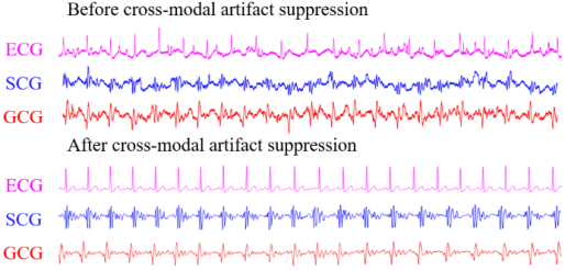
The proposed synchronized quad-modal system was evaluated in terms of signal quality, robustness to motion disturbances, and the stability of downstream processing results. Representative ECG, SCG, and GCG waveforms before and after cross-modal motion artifact suppression are shown in Fig. 4. In raw recordings, motion-induced disturbances introduced non-synchronous fluctuations and amplitude variations across modalities, particularly during posture transitions and mild body movements. After cross-modal suppression, beat-synchronous cardiac components became more prominent, while non-synchronous artifacts were attenuated across all three modalities.
Figure 4: Representative ECG, SCG, and GCG waveforms before and after cross-modal motion artifact suppression.
For ECG, denoised signals exhibited improved waveform continuity and more stable QRS morphology across consecutive beats, without distortion of underlying electrical timing. For SCG and GCG, filtered signals showed clearer beat-aligned mechanical patterns with reduced baseline wandering and fewer high-amplitude motion spikes. Owing to the shared hardware clock, beat-level alignment across ECG, SCG, and GCG was preserved by design, enabling reliable comparison of corresponding cardiac cycles without post-hoc correction.
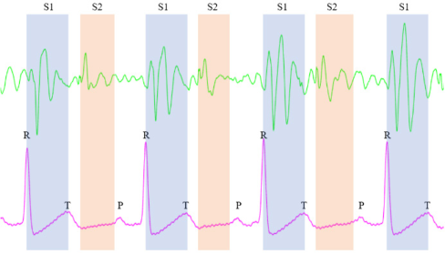
Results of ECG-guided PCG segmentation are illustrated in Fig. 5. In raw PCG signals, heart sound components were often obscured by motion and environmental noise, leading to unstable acoustic peaks. After ECG-guided segmentation, detected heart sound events exhibited consistent temporal relationships with the ECG cardiac cycle, and repeated S1–S2 patterns were observed across consecutive beats. When identifiable, additional acoustic states were also detected in a temporally consistent manner. Across recordings, segmentation maintained stable ordering and improved repeatability under both static and mildly dynamic conditions.
Figure 5: Example of ECG-guided PCG segmentation showing detected heart sound events aligned to ECG-derived cardiac cycles.
Quantitative evaluation results are summarized in Table 2. Metrics related to beat-level consistency and non-synchronous component reduction confirmed the qualitative improvements observed for ECG, SCG, and GCG after cross-modal artifact suppression. For PCG, timing variability of detected heart sound events relative to ECG markers was reduced following ECG-guided segmentation. All results were obtained using fixed processing parameters across sessions, without recording-specific tuning.
Table 2: Summary of quantitative evaluation results for synchronized acquisition, cross-modal artifact suppression, and ECG-guided PCG segmentation.
PCG S2 timing variability vs. ECG anchor (ms) Overall, the results demonstrate that the proposed system achieves stable synchronized acquisition of ECG, PCG, SCG, and GCG and supports robust downstream processing under practical operating conditions. Hardware- level synchronization combined with cross-modal signal processing enables effective attenuation of motion artifacts while preserving beat-synchronous cardiac information across modalities. The quantitative metrics supporting these observations include inter-channel skew under the shared hardware clock, ECG beat-to-beat timing jitter, non-synchronous component ratio, beat-level consistency indices for SCG and GCG, and PCG event timing variability relative to ECG-derived cardiac cycle anchors.
DISCUSSION
The experimental results demonstrate that strict hardware-level synchronization combined with cross-modal signal processing provides a practical foundation for robust ambulatory cardiac monitoring. By enforcing a shared sampling clock across ECG, PCG, SCG, and GCG, the proposed system avoids inter-device drift and post-hoc resynchronization, simplifying downstream processing and enabling reliable beat-level fusion across electrical, acoustic, and inertial modalities.
The proposed cross-modal motion artifact suppression strategy exploits the complementary characteristics of electrical and inertial signals. While motion disturbances manifest differently across modalities, true cardiac components remain beat-synchronous and temporally constrained. Leveraging this consistency enables attenuation of motion-dominated components without aggressive filtering or modality-specific heuristics, and the use of a fixed processing configuration across all recordings highlights the general robustness of the approach.
ECG-guided PCG segmentation further illustrates the benefit of synchronized multi-modal acquisition. In ambulatory conditions, purely acoustic segmentation is vulnerable to motion and environmental noise, whereas constraining heart sound detection within ECG-defined cardiac cycles improves segmentation stability while maintaining flexibility in
acoustic feature extraction. This strategy avoids complex model training and is well suited for practical monitoring systems.
Importantly, the contribution of the present study lies in the end-to-end integration of these elements within one wearable system. Rather than presenting only a new sensor combination or only a post-processing method, the work links chest-worn hardware integration, strict synchronization, cross- modal denoising, and ECG-guided PCG analysis in a single experimentally validated pipeline.
Overall, the results indicate that combining synchronized hardware design with cross-modal processing can substantially improve signal stability and interpretability in wearable multi-modal cardiac monitoring systems under motion-prone conditions.
CONCLUSION
This paper presented an original chest-worn quad-modal cardiac monitoring system that synchronously acquires ECG, PCG, SCG, and GCG under a shared hardware clock. A unified digitization and FPGA-controlled streaming architecture was developed to ensure strict temporal alignment across modalities, enabling reliable beat-level fusion without post-hoc synchronization. Building on this hardware foundation, the paper further introduced a host-side processing pipeline that combines cross-modal motion artifact suppression for ECG, SCG, and GCG with ECG-guided PCG segmentation for robust heart sound analysis.
Experimental results under practical operating conditions demonstrated stable synchronized acquisition and effective attenuation of motion-induced disturbances across modalities. The combination of electrical timing and inertial sensing improved robustness in both cardiomechanical signals and heart sound segmentation, highlighting the benefits of integrated multi- modal design for ambulatory monitoring scenarios.
The proposed system provides a flexible platform for further investigation of multi-modal cardiac sensing and processing strategies. Future work may focus on real-time implementation, extended activity scenarios, and integration of additional analysis modules. The presented design and evaluation contribute toward practical, synchronized multi-modal wearable systems for continuous cardiac monitoring.
ACKNOWLEDGMENT
This work was supported by the National Key R&D Program of China (Grant No. 2023YFF0615904). The authors would like to thank this supporter and all participants for their cooperation in the experiments.
REFERENCES
Castaño Usuga, F.A., Gissel, C. and Hernández, A.M. (2022). Motion Artifact Reduction in Electrocardiogram Signals Through a Redundant Denoising Independent Component Analysis Method for Wearable Health Care Monitoring Systems: Algorithm Development and Validation. JMIR Medical Informatics 10(11), e40826. doi: 10.2196/40826.
A Chest-Worn Quad-Modal Cardiac Monitoring Device 271 Jafari Tadi, M., Lehtonen, E., Saraste, A., Tuominen, J., Koskinen, J., Teräs, M.,
Airaksinen, J., Pänkäälä, M. and Koivisto, T. (2017). Gyrocardiography: A New Non-invasive Monitoring Method for the Assessment of Cardiac Mechanics and the Estimation of Hemodynamic Variables. Scientific Reports 7, 6823. doi: 10.1038/s41598-017-07248-y. Javaid, A.Q., Ashouri, H., Dorier, A., Etemadi, M., Heller, J.A., Roy, S. and Inan, O.T. (2017). Quantifying and Reducing Motion Artifacts in Wearable Seismocardiogram Measurements During Walking to Assess Left Ventricular Health. IEEE Transactions on Biomedical Engineering 64(6), 1277–1286. doi: 10.1109/TBME.2016.2600945. Parlato, S., Centracchio, J., Cinotti, E., Manzi, M.V., Canciello, G., Prastaro, M.,
Lembo, M., Brandwood, B.M., Gargiulo, G.D., Bifulco, P., Esposito, G., Izzo, R. and Andreozzi, E. (2025). A Forcecardiography dataset with simultaneous SCG, Heart Sounds, ECG, and Respiratory signals. Scientific Data 12, 1370. doi: 10.1038/s41597-025-05694-2. Prasad, R., Yilmaz, G., Chételat, O. and Magimai.-Doss, M. (2020). Detection of S1 and S2 Locations in Phonocardiogram Signals Using Zero Frequency Filter. In: Proceedings of the IEEE International Conference on Acoustics, Speech and Signal Processing (ICASSP 2020), pp. 1254–1258. doi: 10.1109/ ICASSP40776.2020.9053155. Rai, D., Thakkar, H.K., Rajput, S.S., Santamaria, J., Bhatt, C. and Roca, F. (2021).
A Comprehensive Review on Seismocardiogram: Current Advancements on Acquisition, Annotation, and Applications. Mathematics 9(18), 2243. doi: 10.3390/math9182243. Sieciński, S., Kostka, P.S. and Tkacz, E.J. (2020). Gyrocardiography: A Review of the Definition, History, Waveform Description, and Applications. Sensors 20(22), 6675. doi: 10.3390/s20226675. Xie, H., Yang, L., Jiang, B., Huang, Z. and Lin, Y. (2025). State-of-the-art wearable
sensors for cardiovascular health: a review. npj Cardiovascular Health 2, 90. doi: 10.1038/s44325-025-00090-6.
LISTED IN THE AHFE DIGITAL BOOKSTORE CATALOGUE
Explore the complete digital volume
Healthcare and Medical Devices
Read this individual research paper free through the original publisher, or explore the full digital proceedings book.
ISBN 978-1-964867-87-8
Explore this ISBN in the AHFE Bookstore ↗Listed in the July 2026 bookstore catalogue; search by ISBN to confirm current availability · Digital book purchase optional