Akisue Kuramoto, Masaya Noguchi, Motomu Nakashima (2024). Biomechanical modeling of subjective fatigue during high-frequency repetitive manual-handling tasks. In: Human Factors in Design, Engineering, and Computing. AHFE Open Access, vol. 159, pp. 1521–1529. https://doi.org/10.54941/ahfe1005720
Department of Systems and Control Engineering, School of Engineering, Institute of Science Tokyo (Tokyo Institute of Technology), Meguro, Tokyo 1580082, Japan ↗
Accumulation of muscle fatigue and subjective fatigue are significant causes of decline in individual performance. Those fatigues can also lead to work errors and the associated musculoskeletal disorders. Thus, a quantitative evaluation of fatigue accumulation during work is required to manage the risk of industrial accidents. Most of the current assessment methods of workload are based on observing and scoring the range of joint motion and work frequency at a point in time. In other words, these assessment methods do not fully consider the continuous accumulation of fatigue. However, even while repeating the same task, muscle fatigue-recovery states and work movements change over time. Therefore, risk management of industrial accidents is important to objectively evaluate the subjective sense of strain and muscle fatigue from work movement data. This study aims to biomechanically model muscle fatigue and subjective fatigue during high-frequency repetitive manual-handling tasks. In an experiment, participants were asked to repeatedly lift a bottle weighing approximately 1 kg, containing salt as ballast, from a chest-height shelf to an eye-level shelf every two seconds for ten minutes. Both start and end points were set at the point approximately 80% of the upper limb length from shoulders at those heights in the midsagittal plane. During the experiment, whole-body motion was measured using an inertial sensor-based motion capture system. In addition to the body motion, electromyograms and subjective evaluations based on the Borg-CR10 scale of the upper limb were recorded. The measured body motion data were applied to a human musculoskeletal model to simulate muscle activity at each sampling time in the experiment. The results were then applied to the Xia and Frey-Law muscle fatigue model to simulate each muscle's residual capacity and fatigue at each sampling time during the experiment. The ratio of the simulated muscle activity to the simulated residual capacity was defined as the substantial muscle activity rate (SMAR). Changes in the SMAR during the experiment were compared with the changes in subjective fatigue and EMG median frequency. Throughout the task, slight abduction and forward flexion were kept in the upper arm. Therefore, we focused our discussion on the deltoid muscle, which might be the most heavily loaded during the experiment. The frequency analysis result of electromyograms indicated that the frequency power spectrum in the medial deltoid shifted to a lower frequency band in the first few minutes and was generally constant in the rest. The residual capacity of the medial deltoid simulated by the muscle fatigue model declined nonlinearly in the first few minutes and was almost constant after that. These results indicate that the muscle fatigue model sufficiently represented the fatigue at the medial deltoid. The muscle activity rate simulated by the musculoskeletal model was almost the same throughout the experiment. On the other hand, the SMAR declined in the first few minutes and continued at a higher range than the muscle activity rate. This changing trend of the SMAR was similar to the time change of the subjective fatigue of the shoulder.
Workload management with considering accumulated fatigue is very important in occupational perspective of health. It is said that accumulation of muscle fatigue and subjective fatigue causes of decline in individual performance (Aryal, 2017). Excessive physical fatigue can lead to long-term health problems, such as work-related musculoskeletal disorders (Valero, 2017; Gallagher, 2016). Therefore, from the occupational perspective of health, various methods have been tried to use to manage fatigue. For example, subjective exertion level is one of the physical fatigue evaluation items in the actual workplace. This aims to evaluate the magnitude of physical strain through ratings of perceived exertion. However, it is difficult to quantitatively evaluate actual muscle fatigue and the resulting risk of musculoskeletal disorders from subjective evaluation alone.
Objective workload and work frequency evaluation methods, such as RULA, REBA, OWAS, etc., are currently used to find potential risks of musculoskeletal disorders in actual work environments. Those observation methods score the range of joint motion and work frequency during a specific period. However, the challenge with those methods is that they cannot quantitatively consider the accumulation of fatigue caused by continued work.
Muscle fatigue-recovery state and physical movements change over time while repeating the same work. In order to consider the changes, the change in relative strength (the ratio of absolute muscle strength at a specific time to baseline strength) has been widely accepted as an objective indicator of cumulative muscle fatigue. It has already been reported that a moderate correlation exists between the decrease in relative strength and the increase in subjective strain (Vahedi, 2023). However, it is not practical to ask workers to measure MVIC to record relative strength or to rate subjective exertion while continuing to perform tasks in the workplace. If there are some alternative methods to evaluate the decline in relative muscle strength, it would be useful for estimating subjective exertion.
Accumulation of muscle fatigue indices asymmetric posture (Penedo, 2021) and changes in physical movements (Cortes, 2014). In other words, it results in changes of working posture and movements (Davidson, 2024; Fischer, 2015). This indicates that physical movements change to use muscles with lesser fatigue as muscle fatigue accumulates. Therefore, if we could objectively and quantitatively evaluate muscle fatigue of each part of the body from physical movements during work, it would be possible to estimate the subjective strain, which is expected to be very useful in managing the risk of workplace accidents.
A conventional indicator of physical muscle load is the muscle activation rate, that is, the ratio of the number of activated muscle units to the total number of units. However, accumulation of muscle fatigue reduces residual capacity (the number of available units). This means that the accumulation of muscle fatigue may increase the subjective exertion level. When the same muscle force is continuously required, the actual muscle activity rate can increase along with decreasing the residual capacity, whereas the conventional index do not change. In other words, this substantial muscle activation rate is expected to be an objective indicator of the subjective exertion level that changes over time during work. This study aims to confirm this expectation. This paper proposes a new estimation method of subjective exertion level biomechanically using musculoskeletal simulation, considering a muscle fatigue model. An experiment on high-frequency repetitive manual handling tasks was conducted. The applicability of the proposed method was verified by comparing the simulation results to the results with the frequency spectrum analysis of electromyograms and actual subjective evaluation results.
Biomechanical Modelling of Subjective Fatigue During High-Frequency 1523
MATERIALS AND METHODS
Experimental Protocol
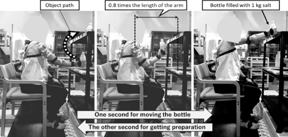
Figure 1 shows the experimental task environment. This simulates repetitive manual handling tasks like picking work in front of a shelf. The work object was bottle weighing approximately 1 kg, containing salt as ballast. In an experiment, a participant was asked to repeatedly lift the bottle from a chest- height shelf to an eye-level shelf every two seconds for ten minutes. Both start and end points were at points approximately 80% of the upper limb length from shoulders at those heights in the midsagittal plane. Here, the task pace was set to once every two seconds to induce changes in body movements and muscle usage by making the participants more tired in a short period. This experiment was performed in time with a metronome that sounded once per second.
To assess perceived fatigue, a participant was asked about subjective exertion level in each of five body parts (waist, back, shoulder, upper arm, and forearm) on a Borg CR-10 scale (Borg, 1982) every minute of the experiment. A table for the Borg CR 10 scale was displayed in front of the participant during the experiment.
Measurements Throughout the experiment, three-dimensional upper body motion was recorded at 100 Hz using an IMU-based motion capture system (Perception Neuron Studio, Noitom Inc, US).
Six electromyography (EMG) sensors (Surface EMG Sensor SX230, Biometrics Ltd, UK) were used to measure muscle activity and fatigue from the anterior deltoid, intermedius deltoid, biceps brachii, triceps brachii, brachioradialis, and pronator teres on the right upper limb used for work. The EMG sensors were fixed to the skin over the muscles after skin treatment to reduce impedance affections. EMG data was recorded with a sampling rate of 2000 Hz. After fixing the EMG sensors, the subject was asked to produce maximal voluntary contractions (MVCs) for 5–10 s each. Then, the participant rested for about 10 minutes while the experimenter fixed the IMU sensors on the participant’s body.
Figure 1: Experimental task environment and 2-seconds working cycle sequence.
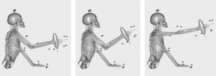
Musculoskeletal Simulation Musculoskeletal simulation for simulating muscle activity rates at each sampling time was performed using AnyBody Modeling System (Version 7.4.4, AnyBody Technology, Aalborg, Denmark). Measured body motion data during the experiment was input into a human musculoskeletal model as shown in Fig. 2. The muscle force distribution was determined by optimization calculations performed with the objective function of minimizing the sum of the cubes of muscle stress (Crowninshild, 1981), in terms of a compromise between minimal fatigue and energy efficiency.
Figure 2: Musculoskeletal simulation. Body movements during the task, measured using an IMU-based motion capture system, was recorded as time series data of joint angles of a stick figure model considering body dimensions. Additional nodes were manually defined to the musculoskeletal and stick-figure models at locations corresponding to identifiable points. Joint angles of the human musculoskeletal model was determined to minimize the squared error between virtual markers on the stick figure and on the musculoskeletal model.
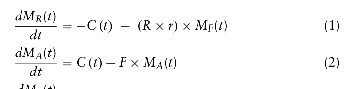
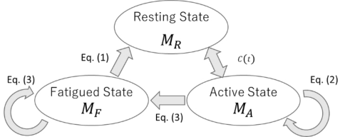
Muscle Fatigue Simulation To estimate task-related muscle fatigue, we use the three-component muscle fatigue model (Xia and Frey-law, 2008). As shown in Fig. 3, this model assumes that muscle motor units can be classified into following three states at each time: active state, recovery state, and fatigued state. Hereinafter, the percentages of muscle motor units in active, resting, and fatigued states at time t are denoted as MA (t), MR (t), and MF (t), respectively. The sum of M (t)A, MR (t), and MF (t) equals 100. The time derivatives of M (t)A, MR (t), and MF (t) are defined as follows,
Equation reproduced from the original PDF.
Biomechanical Modelling of Subjective Fatigue During High-Frequency 1525
where F and R are fatigue and recovery coefficients. The values of F and R were set to 0.1 and 0.02 (Frey-Law, 2012), respectively. The r is 1 if the target load TL(t) > 0 otherwise 0, and C (t) is a value corresponding to TL(t) applied to the muscle at time t. The C (t) is defined as follows, C (t) = TL(t) −MA(t), if (MA(t) < TL(t) and MR(t) < (TL(t) −MA(t)) MR(t), otherwise.
(4) Here, TL(t) is the percentage of muscle activity at time t and can be defined as follows,
TL(t) = F(t) F0 × 100, (5) where F(t) is the force exerted on the muscle at time t, and F0 is maximum exertion force. In this study, TL(t) was calculated by multiplying the muscle activity rate of each muscle obtained by musculoskeletal simulation by 100.
From the above, the residual muscle capacity RC (t) at time t can be expressed by the following equation: RC(t) = 100 −MF(t). (6)
Substantial Muscle Activity Rate (SMAR) Smaller RC (t) indicates that muscle capacity is decreased. This would lead that the subjective exertion increase according to RC (t) decreases even if target load is constant. Therefore, we focus on the ratio of the simulated muscle activity to the simulated residual capacity was defined as the substantial muscle activity rate (SMAR). In order to predict the subjective exertion when RC (t) changes, the SMAR(t) was defined as follows,
Equation reproduced from the original PDF.Figure 3: Schematic diagram of the flow of the motor unit in the three-component muscle fatigue model. To show the reduction to its own state elements, the authors have modified the diagram from the original one in the reference paper (Xia and Frey-law, 2008).
EMG Data Analysis A sliding window Fast-Fourier Transform technique was applied to the raw EMG data for power spectrum analysis. Time window width and time step were 6.0 sec and 2.0 sec, respectively. The window width was for including several task motions in a window, and the time step was for down-sampling.
RESULTS AND DISCUSSION
Throughout the task, slight abduction and forward flexion were kept in the upper arm. Therefore, we focused our discussion on the deltoid muscle, which might be the most heavily loaded during the experiment.
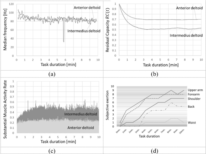
It has been reported that the higher the muscle exertion rate, the more rapidly the median power frequency (MPF) decreases (Nanthavanij, 1989). Figure 4(a) shows that the MPF in the deltoid muscles decreased in the first few minutes and was almost constant in the rest. The results show a similar trend to that report, indicating that this experimental task required a high muscle exertion rate to decrease the deltoid muscle MPF.
Figure 4(b) shows the time change of RC (t) of the deltoid muscles estimated based on the musculoskeletal simulation. In both the anterior and the intermedius deltoid, the RC (t) decreased rapidly in the first few minutes, but remained almost constant after that. The change trend of RC (t) is similar to that of MPF.
Figure 4(c) shows the time change of SMAR (t). In the intermedius deltoid, SMAR (t) increased by about 0.25 in the first few minutes and then remained almost constant. On the other hand, SMAR (t) of the anterior deltoid increased by about 0.05 in the first few minutes, but did not show as large a change as that of the intermedius deltoid throughout the experiment. From these results, it is expected that the subjective sense of shoulder strain increased in the first few minutes of the experiment.
On the other hand, the subjective exertion level shown in Fig. 4(d) increased earlier in the upper arm and forearm than in the shoulder. After the experiment, we confirmed what the participant thought of as the shoulder and upper arm to him. As a result, it was found that he thought of the trapezius muscle and scapula as the shoulder area, and the shoulder joint and elbow joint as the upper arm area. Therefore, the subjective sense of strain around the deltoid muscle was thought to be recorded as that of the upper arm. This difference is due to a lack of explicit confirmation of which body parts were referred to as “shoulder” or “upper arm” before the experiment began. Considering this, it is indicated that the subjective exertion level around the deltoid muscles increased throughout the first half of the experiment and then remained almost constant. From the above, it was found that the changes in subjective exertion level based on the Borg CR-10 during the experiment were similar to those of SMAR (t).
One issue that needs to be investigated in the future is the difference in magnitude between SMAR (t) and subjective exertion level based on the Borg CR-10 scale. The SMAR (t) was lower than the subjective exertion level throughout the experiment. Ideally, their magnitudes should be similar because the Borg CR-10 level is designed to be close to 10 times the %MVC,
Biomechanical Modelling of Subjective Fatigue During High-Frequency 1527
or muscle activation rate. One reason for this might be the values of the fatigue model parameters, F and R. The values used in this study were the average of reported values for several muscles in each part of the body (Frey-Law, 2012) because obtaining accurate subject-specific human models is very complex (Michaud, 2023). Even with such a rough parameter setting, the estimated subjective fatigue index proposed in this study may be sufficient to recognize the signs of fatigue. For assessing the progression of fatigue in more detail, there is still room to discuss methodologies to determine the individual fatigue parameters more precisely.
Figure 4: Time trends of each analysis result of the deltoid muscles. (a) Median frequency of EMG in the 6-seconds time window. (b) Simulated residual capacity. (c) Substantial muscle activity rate (SMAR). (d) Subjective exertion level in the Borg CR-10 scale.
CONCLUSION
This study proposed a methodology to model subjective fatigue during high- frequency repetitive manual-handling tasks by considering muscle fatigue and activity biomechanically. The residual capacity of the medial deltoid simulated by the muscle fatigue model declined nonlinearly in the first few minutes and was almost constant after that. These results indicate that the muscle fatigue model sufficiently represented the fatigue at the medial deltoid. The muscle activity rate simulated by the musculoskeletal model was almost the same throughout the experiment. On the other hand, the SMAR declined
in the first few minutes and continued at a higher range than the muscle activity rate. This changing trend of the SMAR was similar to the time change of the subjective fatigue of the shoulder.
ACKNOWLEDGMENT
A part of this study was supported by JKA Foundation (Grant No. 2024M-389).
REFERENCES
Aryal, A., Ghahramani, A., Becerik-Gerber, B. (2017). Monitoring fatigue in construction workers using physiological measurements. Autom. Constr., Volumne 82, pp. 154–165 Borg, Gunnar A. (1982). Psychophysical bases of perceived exertion. Medicine and science in sports and exercise, Volume 14, No. 5, pp. 377–381. Cortes, N., Onate, J., Morrison, S. (2014). Differential effects of fatigue on movement
variability. Gait & posture, Volume 39, No. 3, pp. 888–893. Crowninshield, Roy D., Brand, Richard A. (1981). A physiologically based criterion of muscle force prediction in locomotion, Journal of Biomechanics, Volume 14, No. 11, pp. 793–801. Davidson, J. B., Cashaback, J. G., Fischer, S. L. (2024). Exploring optimal objective
function weightings to predict lifting postures under unfatigued and fatigued states. Human Factors, Volume 66, No. 2, pp. 510–527. Fischer, S. L., Greene, H. P., Hampton, R. H., Cochran, M. G., Albert, W. J. (2015).
Gender-Based Differences in Trunk and Shoulder Biomechanical Changes Caused by Prolonged Repetitive Symmetrical Lifting. IIE Transactions on Occupational Ergonomics and Human Factors, Volume 3, No. 3–4, pp. 165–176. Frey-Law, Laura A., Looft, John M., Heitsman, Jesse. (2012). A three-compartment muscle fatigue model accurately predicts joint-specific maximum endurance times for sustained isometric tasks. Journal of biomechanics, Volume 45, No. 10, pp. 1803–1808. Gallagher, S., Schall, M. C. (2016) Musculoskeletal disorders as a fatigue failure
process: evidence, implications and research needs. Ergonomics, Volume 60, No. 2, pp. 255–269 Michaud, F., Frey-Law, L. A., Lugrís, U., Cuadrado, L., Figueroa-Rodríguez, J., Cuadrado, J. (2023). Applying a muscle fatigue model when optimizing load- sharing between muscles for short-duration high-intensity exercise: A preliminary study. Frontiers in physiology, Volume 14, Paper ID 1167748. Nanthavanij, Suebsak., Subramaniam, Deivanayagam. (1989). On the assessment of
muscle fatigue rate via various EMG frequency spectral parameters. International Journal of Industrial Ergonomics, Volume 4, No. 3, pp. 213–224. Penedo, T., Polastri, P. F., Rodrigues, S. T., Santinelli, F. B., Costa, E. D. C., Imaizumi, L. F. I., Barbieri, F. A. (2021). Motor strategy during postural control is not muscle fatigue joint-dependent, but muscle fatigue increases postural asymmetry. PLoS One, Volume 16, No. 2, Paper ID: e0247395. Vahedi, Z., Kazemi Kheiri, S., Hajifar, S., Ragani Lamooki, S., Sun, H., Megahed,
F. M., Cavuoto, L. A. (2023). The relationship between ratings of perceived exertion (RPE) and relative strength for a fatiguing dynamic upper extremity task: A consideration of multiple cycles and conditions. Journal of Occupational and Environmental Hygiene. Volume 20, No. 3–4, pp. 136–142.
Biomechanical Modelling of Subjective Fatigue During High-Frequency 1529
Valero, E., Sivanathan, A., Bosche, F., Abdel-Wahab, M., Bosché, F., Abdel-Wahab, M. (2017). Analysis of construction trade worker body motions using a wearable and wireless motion sensor network. Automation in Construction, Volume 83, pp. 48–55. Xia, Ting., Frey-Law, Laura A. (2008). A theoretical approach for modeling
peripheral muscle fatigue and recovery, Journal of biomechanics, Volume 41, No. 14. pp. 3046–3052.
LISTED IN THE AHFE DIGITAL BOOKSTORE CATALOGUE
Explore the complete digital volume
Human Factors in Design, Engineering, and Computing
Read this individual research paper free through the original publisher, or explore the full digital proceedings book.
ISBN 978-1-964867-35-9
Explore this ISBN in the AHFE Bookstore ↗Listed in the July 2026 bookstore catalogue; search by ISBN to confirm current availability · Digital book purchase optional